UHOD
Prognostic Factors Affecting Survival in Patients
with Ovarian Cancer: A 5-Year Experience in an
University Hospital
Gül PINAR1*, Tevfik PINAR2*, Ayşe DURUKAN3, Ali AYHAN3
1 Yıldırım Beyazıt University, Faculty of Health Sciences, Department of Nursing 2 Hacettepe University, Institute of Public Health
3 Baskent University Ankara Hospital, GyneOncologic Clinic, Ankara, TURKEY
ABSTRACT
The aim if this study is to explore the impact of the various prognostic factors on overall survival in women with ovarian cancer (OC). Patients diagnosed with gynecologic malignancy in Gynecology Clinic of Baskent University Medicine Faculty between 2010 and 2015 included to study. Patients with ovarian (n= 112) cancers examined retrospectively. Kaplan-Meier, Univariate and Multivariate Cox re-gression model were performed to estimate for associations of potential variables with survival factors. The mean patient age was 56.4 y, range 20-80. The overall survival was 94.3%, 83.4%, 66.4%, 54.7% and 42.8% at 1, 2 3, 4 and 5 years respectively (60.5 months, range 43-68). 3-year disease-free survival was 25.3% (18.3 months, range 17-20). Multivariate analysis of patients indicated that stage, histology, grade, age at diagnosis, comorbidity, recurrence, BMI, menopausal status and regional distance were independent prognos-tic factors on survival (p< 0.05). In conclusion, these results will presents a framework to identify fundamental causes in survival for OC. Keywords: Ovarian cancer, 5-year survival, Prognostic factors
ÖZET
Over Kanserli Hastalarda Sağkalımı Etkileyen Prognostik Faktörler: Bir Üniversite Hastanesinin 5 Yıllık Deneyimi
Bu çalışmanın amacı, over kanseri (OK) hastalarında sağkalımı etkileyen prognostik faktörleri araştırmaktır. Çalışmada, 2010 ile 2015 yılları arasında Başkent Üniversitesi Tıp Fakültesi Hastanesi Jine-Onkoloji Bölümü’nde tanı alan 112 over kanserli hasta ret-rospektif olarak incelenmiştir. Sağkalım süreleri hesaplanırken Kaplan-Meier metodu ve sağkalımı etkileyen faktörlerin ilişkisini ke-stirmek amacıyla Çok yönlü ve Tek yönlü Cox Regression modeli kullanılmıştır. Hastaların ortalama yaşı 56.4 (20-80)’dür. Genel sağkalım oranları sırasıyla; 1 yıllık-%94.3, 2 yıllık-%83.4, 3 yıllık- %66.4, 4 yıllık-%54.7 ve 5 yıllık- %42.8 (65.5±6.2 months)’dir. 3 yıllık hastalıksız sağkalım oranı %25.3 (18.3±3.94 months)’dir. Çok değişkenli analize göre, hastaların tanı aldığı yaş, BKİ, komorbidite, evre, grade, histoloji, rekürrens, menopoz durumu ve bölgesel uzaklığın OK hastalarının sağkalımı üzerinde etkili olduğu bulundu (p< 0.05). Bu çalışmanın sonucunda elde edilen bulgular, OK sağkalımının temel nedenlerini belirlemede önemli bir çerçeve sunacaktır.
Anahtar Kelimeler: Over kanseri, 5 yıllık sağkalım, Prognostic faktörler
INTRODUCTION
Ovarian cancer (OC) is the deadliest gynecologic cancer worldwide, with nearly 225.000 new cases
diagnosed each year and 140.000 deaths annu
-ally.Lifetime risk of OC in women is one in 71,
and the risk of dying from the disease is 1 in 95.1
Based on GLOBOCAN estimates, the incidence and mortality of OC varies in different regions of the world; Scandinavian (14.9/100000), USA
(13.3/100000), UK (11.7/100000), Russian Fed
-eration (11.3/100000), and Japan (4.8/100000).2
In Turkey (6.6/10000), OC is also a major health problem in women following breast cancer. This is
higher than that of the previous reports in Turkey.3
Regrettably, identifying this disease is difficult. There is no routine, screening test to accurately and reliably detect OC in the general population so diagnosis of OC in the advanced stages leads to this cancer being considered as a fatal disease.
Although the incidence and mortality of this dis
-ease is high, its prognostic factors are still not com
-pletely understood especially in low-income coun
-tries.4,5 In the literature, survival depends on many
factors such as patient’s family history, multiple
comorbidities, postoperative complications, chem
-otherapy toxicities, increasing age, postmenopau
-sal period, recurrence, presence of positive lymph nodes, advanced stage and grade, carcinosarcomas subtype, ascites, never having been pregnant and having never taken oral contraceptives, as well as
lifestyle factors such as performance status, smok
-ing and obesity.6-11
Statistics show that survival from all cancers in women was 64.2% while it was approximately 45.0% for OC globally and the 5-year survival rate has improved in OC by only 9% since 1975 even
in high-income countries.2 A 5-year survival rate
is about 43% in Turkey (1970s-37%, 1980s-40%,
and 2000s-45%).3 Despite the recently improve
-ments such as advances in the diagnosis, staging,
and treatment of OC, and provided significant ad
-vancement of patients survival, major challenges related to the prognostic implications of its clinic-pathologic characteristics remain controversial in the management of OC. Most women with OC
are likely living with the disease rather than liv
-ing cancer-free and some oncologists consider OC
a chronic disease.9,12 Relatively, limited compre
-hensive national information describing clinical and nonclinical factors associated with survival is available in Turkish women with OC. Therefore, estimating survival is remarkable that provide an understanding of the disease course and better prognosis for OC.
PATIENTS AND METHODS
The aim of this study was to investigate determi
-nant factors associated with survival of women
with OC over the course of five years. The popula
-tion of the study consisted of all patients with OC in years 2010-2015 who were actively followed-up
and age 18 years or older were included at the Gy
-necologic Oncology Clinic, Faculty of Medicine, Baskent University Hospital (n= 112). Patients
with unknown treatment data, or missing infor
-mation were excluded. In addition, patients who survived less than 12 months after diagnosis were excluded to limit bias. Patients were scheduled for follow-up every two months in the first year, every three months during the second year and every six months thereafter. Follow-up data such as date of last visit and disease status at the time of the last contact were also noted. This study was conducted
at a multidisciplinary tertiary care center. The re
-search was approved by the University of Baskent Institutional Review Board and was conducted in compliance with principles of Helsinki declaration.
The data were collected by using three differ
-ent forms between April 2016 and January 2016
through hospital-based cancer registry; 1) The Pa
-tient Information Form, 2) The Charlson Comor
-bidity Index (CCI), and 3) The Eastern Coopera
-tive Oncology Group (ECOG) Performance Scale.
1. The Patient Information Form
This form includes 21 questions to define individ
-ual and medical characteristics of the women with OC such as patient age at diagnosis, educational level, occupation, marital status, family income,
smoking history, alcohol use history, family his
-tory, distance of patient travel, residence, survival
time information, clinic-pathological character
re-hospitalization and readmission, primary surgery,
treatment modality, treatment toxicities, preopera
-tive CA125, postopera-tive and intraopera-tive com
-plications). Histology was classified based on the
International Classification of Diseases for Oncol
-ogy (ICD-O) as serous, mucinous, endometrioid, clear cell; and tumor grade as well differentiated, moderately differentiated or poorly differentiated. Stage categories were based on the International Federation of Gynecology and Obstetrics (FIGO) stages I, II III and IV. Survival time was calculated from date of diagnosis until death or censoring. The overall survival (OS) was defined as the time
from first diagnosis to death or the date of last fol
-low up. Disease-free survival (DFS) is the time elapsed from PE to first recurrence and OS is the time from PE to the last follow-up date or death.
BMI values of patients were classified as under
-weight (BMI <18.5 kg/m2), normal (BMI
18.5-24.9 kg/m2), overweight (BMI 25-29.9 kg/m2), and
obese (BMI ≥30 kg/m2) according to World Health
Organization (WHO) criteria.13
2. The Charlson Comorbidity Index (CCI)
Comorbidity was measured for each patient using
the Charlson Comorbidity Index (CCI), catego
-rized as 0, 1, or ≥2, with a higher score indicating
a larger number or greater severity of comorbidi
-ties.14 The CCI, ranging from 0 to 29, consists of
a weighted sum of 17 major illnesses (e.g., myo
-cardial infarction, stroke, diabetes, liver disease, dementia, renal disease). HT was included in the list of possible comorbid illnesses.
3. European Cooperative Oncology Group (ECOG) Performance Scale
This scale was developed by the Eastern Coopera
-tive Oncology Group (ECOG) in 1982 to classify a patient according to their functional impairment, compare the effectiveness of therapies, and assess
the prognosis of a patient (from 0 to 5, with 0 de
-noting perfect health and 5 death).15
Statistical Analysis
Descriptive statistical analyses were performed with SPSS version 18 (SPSS inc., Chicago, IL).
Categorical variables were compared using Pear
-son chi-square test and continuous variables using the Wilcoxon–Mann–Whitney and Kruskal–Wallis tests. Survival was estimated using the Kaplan– Meier method. Univariate and multivariate Cox Regression model (95% CIs) were used to evaluate
the effects of multiple prognostic factors on sur
-vival.16 The significance level was p< 0.05.
RESULTS
There were 112 patients who met inclusion crite
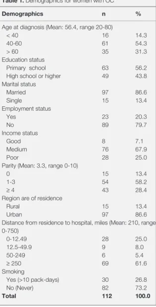
-ria. The patients’ general characteristics are shown in Table 1. The median age at diagnosis was 56.4
years + 11.3 (min: 20 - max: 80). Findings deter
-mined that 56.2% were primary school graduates,
Table 1. Demographics for women with OC
Demographics n %
Age at diagnosis (Mean: 56.4, range 20-80)
< 40 16 14.3
40-60 61 54.3
> 60 35 31.3
Education status
Primary school 63 56.2
High school or higher 49 43.8
Marital status Married 97 86.6 Single 15 13.4 Employment status Yes 23 20.3 No 89 79.7 Income status Good 8 7.1 Medium 76 67.9 Poor 28 25.0
Parity (Mean: 3.3, range 0-10)
0 15 13.4
1-3 54 58.2
≥ 4 43 28.4
Region are of residence
Rural 15 13.4
Urban 97 86.6
Distance from residence to hospital, miles (Mean: 210, range 0-750) 0-12.49 28 25.0 12.5-49.9 9 8.0 50-249 6 5.4 ≥ 250 69 61.6 Smoking Yes (>10 pack-days) 30 26.8 No (Never) 82 73.2 Total 112 100.0
79.7% were housewives, 86.6% were married, 67.9% had their income equal to expenditure, and 86.6% were urban dwellers. The mean parity was 3.35, ranging from 0 to 10. All of the women had health insurance, 73.2% did not smoke (99.5% did not drink alcohol). Distance from residence to center, miles (range) was 210±10.2 (0-750) (Table 1).
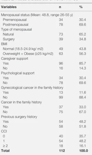
Seventy percent of women were diagnosed during
menopause (the rate of surgically induced meno
-pause was 34.8%). More than half of women were
obese (56.3%). The majority of women had a car
-egiver (85.7%). One in three received psychologi
-cal support for their illnesses. Thirty-three percent of women had cancer stories in their families and
Table 2. Medical characteristic for women with OC
Variables n %
Menopausal status (Mean: 48.8, range 26-55 y)
Premenopausal 34 30.4 Postmenopausal 78 69.6 Type of menopausal Natural 73 65.2 Surgery 39 34.8 BMI Normal (18.5-24.9 kg/ m2) 49 43.8 Overweight + Obese (≥25 kg/m2) 63 56.3 Caregiver support Yes 96 85.7 No 16 14.3 Psychological support Yes 34 30.4 No 78 69.6
Gynecological cancer in the family history
Yes 13 11.6
No 99 88.4
Cancer in the family history
Yes 37 33.0
No 75 67.0
Previous surgery history
Yes 54 48.2 No 58 51.8 CCI 0 40 35.7 1 54 48.2 ≥ 2 18 16.1 Total 112 100.0
Abbreviations: BMI: Body mass index, CCI: Charlson Comorbidity Index
Table 3. Cancer and treatment characteristic for women with OC
Variables n %
Initial symptoms at first admission
Abdominal-pelvic pain 22 19.6
Vaginal bleeding-irregular menstrual cycle 40 35.7
Abdominal distension 50 44.6 Stage 1-2 30 17.8 3-4 92 82.2 Grade 1/2 38 33.9 ≥ 3 72 66.1 Subtype Serous 85 75.9 Endometrioid 16 13.2 Mucinous 6 6.4 Clear cell 5 4.5 Primary surgery USO + BPPLND + Omentectomy 9 8.0 TAHBSO 12 10.7 TAHBSO + BPPLND + 91 81.3 Omentectomy Other treatments CT 88 78.6 RT 23 20.5 CT+RT 11 9.9 CT+RT Toxicities Yes 96 85.7 No 33 29.4 ECOG 0-1 74 66.1 2-3 38 33.9 Preop CA12-5 35-500 85 75.9 ≥ 500 27 24.1 Recurrence 1 71 63.4 >1 41 36.6 Postoperative complication Yes 21 18.7 No 91 81.3 Intraoperative complication Yes 8 6.2 No 104 92.8
Unplanned readmission at least one (hospitalized -76%)
Yes 69 61.6
No 43 38.4
Abbreviations: CA125: Serum cancer antigen 125, CT: Chemothera-py, RT: RadiotheraChemothera-py, USO: Unilateral Salpingo-Oophorectomy, TAH-BASO: Total Abdominal Hysterectomy and Bilateral Salpingo-Oopho-rectomy, BPPLND: Bilateral pelvic and paraaortic lymphadenectomy
eleven-one percent of women had gynecological cancer stories in their families. Among patients with OC, 35.7% had an CCI- 0, and 48.2% had an CCI- 1, (28.2%) had an CCI -2 (16.1%), had an
CCI-3 (8.5%) (Hypertension also has been identi
-fied as 48.6%). %48.2 patients had previous sur
-gery (Cesarean section 29.5%, cholecystectomy 14.3%, and thyroidectomy 6.3%) (Table 2). Primary surgery was performed on 112 patients during this 5-year period (secondary cytoreductive surgery was performed on all of the patients and
nearly half of the patients underwent tertiary cytro
-ductive surgery) and was removed all visible tu
-mor. 91 patients; TAHBSO + BPPLND + omentec
-tomy, 12 patients; only TAHBSO, 9 patients; USO + BPPLND + omentectomy (as additional surgery;
41 patients appendectomy; 8 patients cholecystec
-tomy; 12 patients splenec-tomy; 6 patients colon resection + colostomy. Hospital length of stay after
surgery was 13.14±4.64. Postoperative complica
-tions were seen in 21 patients (18.7%) (within 4 weeks after surgery). These complications were
fewer (18.6%), wound evisceration (4.5%), res
-piratory complication (3.3%), ileus (11.3%), DVT (16.3%), lymphocele (3.2%). Rate of intraoperative
complication was 8.4% (defects in the bowel se
-rosa during surgery). Readmission rate was 61.6%. Patterns of unplanned readmission in patients were deep vein thrombosis-DVT (19.6%), pyrexia
(60.7%), acute abdomen (29.6%), genital tract in
-fection (22.3%), pain and weakness (56.3%), dysp
-nea (7.4%) and ileus (22.3%) (Table 3).
Most (82.2%) had stage III or IV cancer (all of them were epithelial type OC). More than half the cancers (75.9%) were serous subtype and
66.1% were high-grade (≥ 3). All of patients un
-derwent primary surgery (The mean operative time was 120 minutes). 85.7% of patients received CT (median cure was 6.2+3.3) (platinum-taxane
-paclitaxel+carboplatin 88.3%, cisplatin/ gemcit
-abine 8% and doxorubicin + carboplatin 6%) and only 20.5% RT. ECOG performance status was
“0-1” (66.1%), “≥2” (33.9%) in patients’ last hospital
-izations (Table 3). Hematologic and non-hemato
-Table 4. Distributions of OS and DFS scores
Years (y) Overall survival Disease-free survival (OS) (DFS) 1 y 94.3% 68.7% 2 y 83.4% 33.4% 3 y 66.4% 15.3% 4 y 54.7% -5 y 42.8%
-Median 60.5 months 18.3 months
(%95 GA) (range 43-68) (range 10-27)
(95%CI) (95%CI)
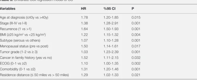
Table 5. Univariate Cox regression model of OS
Variables HR %95 CI P
Age at diagnosis (≤40y vs >40y) 1.78 1.20-1.85 0.015
Stage (III-IV vs I-II) 1.38 1.28-2.91 0.001
Recurrence (1 vs >1) 1.64 1.50-1.93 0.001
BMI (≥25 kg/m2 vs <25 kg/m2) 1.22 1.15-1.32 0.004
Subtype (serous vs others) 1.07 1.10-1.28 0.001
Menopausal status (pre vs post) 1.50 1.14-1.61 0.017
Tumor grade (1-2 vs ≥ 3) 1.03 1.23-2.39 0.001
Cancer in family history (yes vs no) 1.52 1.11-2.15 0.032
ECOG (0-1 vs ≥2) 1.10 1.00-1.35 0.002
Comorbidity (0-1 vs ≥2) 1.31 1.20-1.46 0.001
Residence distance (≤ 50 miles vs > 50 miles) 1.29 1.02-1.33 0.021
logic toxicity of the CT and RT comprised anemia (29.7%), neutropenia (28.6%), thrombocytopenia (26.3%), pain (33.4%), alopecia (48.9), diarrhea
(18.4%), nausea/ vomiting (38.4%), and oral mu
-cositis (13.7%). No patient was lost to follow-up.
The OS for patients was 60.5 months and the medi
-an DFS rate was 18.3 months. The 1-year survival rate was 94.3%, 2-year survival rate was 83.4%, 3-year 66.4%, 4-year 54.7%, and 5- year 42.8%. DFS scores were 1-year 68.7%, 2-year 33.4%,
and 3-year 15.3 % (Table 4). Univariate model re
-vealed that the menopausal status, age at diagnosis,
the FIGO stage, grade, histological type, perfor
-mance status, recurrence, BMI, residence distance, cancer in the family history and the comorbidity were related with OS significantly (Table 5). On
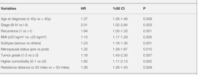
multivariate analysis, advanced age was signifi
-cantly associated with worse OS (HR, 1.3 [95% CI,
1.2-1.4] than younger women. Mucinous, endome
-trioid and clear cell subtypes were associated with worse OS than serous cancers (HR, 1.2 [95% CI, 1.1-1.3]). Postmenopausal period (HR, 1.2 [95% CI, 1.2–1.8), obesity (HR, 1.1 [95% CI, 1.1-1.2), presence of recurrence (HR, 1.4 [95% CI, 1.0–1.3), and higher comorbidity scores (≥ 2), higher grade (HR, 1.1 [95% CI, 1.4-2.3) and stage (HR, 2.0 [95% CI, 1.0-2.8) had significantly poorer OS rates
(HR, 1.6 [95% CI, 1.1-2.1). Regional–remote resi
-dence were also associated with poorer OS (HR, 1.3 [95% CI, 1.2-1.4]) (Table 6).
Compared to good prognosis women, poor progno
-sis women were less educated, and income level,
had before surgery, high toxicity, and re-hospital
-ization. However, there was no significant differ
-ence in OS between the groups (p> 0.05). There were no major differences in distribution of other prognostic factors (p> 0.05).
DISCUSSION
OC is often called the silent killer; women often ignore early signs because there are no clearly identifiable initial symptoms often confused with
complaints of common benign gastrointestinal dis
-orders such as abdominal distension and pain due to acidity. Because of diagnostic challenges only
one fifth of OC patients are detected at the local
-ized stage. Frequently, women are medically man
-aged for indigestion or other complaints without
having a pelvic examination, thus significant de
-lays before diagnosis are very common.4,5
In our study abdominal distension was seen in
44.6% of patients, which was followed by abdomi
-nal/pelvic pain with a rate of 19.6%, and vaginal bleeding/irregular menstrual cycles was found in
35.7%. In accordance with our study Oge et al7
also reported abdominal swelling as the most com
-mon complaint (59.1%), and the second most fre
-quent complaint was reported as abdominal pain
Table 6. Multivariate Cox regression model of OS
Variables HR %95 CI P
Age at diagnosis (≤ 40y vs > 40y) 1.37 1.26-1.46 0.009
Stage (III-IV vs I-II) 2.01 1.02-2.84 0.003
Recurrence (1 vs >1) 1.84 1.05-1.33 0.001
BMI (≥25 kg/m2 vs <25 kg/m2) 1.15 1.17-1.29 0.005
Subtype (serous vs others) 1.23 1.16-1.30 0.001
Menopausal status (pre vs post) 1.32 1.28-1.87 0.010
Tumor grade (1-2 vs ≥ 3) 1.16 1.43-2.30 0.007
Higher comorbidity (0-1 vs ≥2) 1.65 1.11-2.13 0.002
Residence distance (≤ 50 miles vs > 50 miles) 1.36 1.26-1.43 0.009
(20.8%). Physician and nurses recommendations
regarding alarming symptoms and screening pro
-grams in this population are a vital investment on survival for OC. In addition there is a real need for new, more effective screening options for women with OC.
Cancer statistics often use 5-year OS rate to pre
-sent a better idea of the longer-term outlook for
people with cancer.5 We found that the OS for OC
was 94.3%, 66.4%, and 42.8% at 1, 3, and 5 years, respectively. Also, the median OS time was 60.5 months. The slope of decline in OS was reduced during the first years after diagnosis. Most patients diagnosed with OC, survival is theorized to be
100%, who are still alive after 5-years, are presum
-ably living with the disease rather than living dis
-ease-free.5 Our analysis demonstrated that the DFS
was 18.3 months (10-27). The Surveillance, Epi
-demiology and End Results-SEER database from
1995 to 2007 with epithelial OC who were ac
-tively followed-up and age 20 years or older were included for analyzing OS in the United States (40.692 patients) and OS was 65%, 44%, and 36%
at 2, 5, and 10 years, respectively.10 Bailey et al.17
evaluated the 5-year outcome of women (n= 361) with advanced OC in the South West of England as stage III was 16% and with stage IV was 10%.
Recent prospective and randomized trials demon
-strated that patients with advanced OC (n= 718)
from 1998 through 2006 at 59 institutions in Cana
-da have similar survival (OS: 29-30 months, DFS:
12 months).18 In a study the median DFS and OS
for patients with OC were 35.0 months and 54.0
months respectively.19 In different study done by
Anuradha et al in 2014, 1192 Australian women di
-agnosed with invasive epithelial OC in 2005 were identified through state-based cancer registries and
the 5-year OS was 53%.20 A Swedish study pub
-lished in 2009 of 682 patients with epithelial OC found a 10-year relative survival rate of 38.4%, the median OS time was 81 months (52-109 months)
in OC patients.6 Larger population-based analyses
of 10-year OS in OC have not been published in the Turkish populations. In a study performed in Turkey, according to the surgical stages I, II, III and IV median survival was 78.5 months, 60.1 months, 33.9 months and 16.1 months respectively
and significantly different.7 In a study by Karaca21
study, a number of 13.590 women (from 9 nation
-wide cancer registry centers) were evaluated who got gynecological cancer diagnosis (31.2% OC) in Turkey between 2004 and 2011, overall 5-year observed OC survival rate was 46%. Buldanlı et al reported that OS was 51.6% in Western Turkey. When we compare the all of results with national or international studies, the 5-year survival rate in our study did not differ from the 5-year survival published from the general population.
Previous studies have shown that the survival of
OC is affected by personal history, and genetic fac
-tors including low socioeconomic status, and life
style.18,22-26 We scrutinized the analyzing related to
various prognostic factors for OC. In the present study younger women, those diagnosed before
the age of 40, had significantly better survival af
-ter five years than women diagnosed at older ages
(p< 0.05). Older age at diagnosis was often asso
-ciated with comorbidities and functional deficits that impact their survival. Gershenson et al27 re
-ported on a cohort of 112 patients with stage II-IV low-grade serous carcinoma from M.D. Anderson Cancer Center in 2006, and noted age older than 45 years at diagnosis was associated with longer survival. Some studies reported that increasing
age was most strongly associated with poor sur
-vival.9,10,18-20,22 However, in the other studies of Oge
et al7 and Buldanlı et al11 revealed that age was not
was not effected by the age of the patients in OC. Recently, treatments for cancer has resulted in
increasingly complex care patterns and individu
-alization of care by multidisciplinary teams. Lack
of access to a qualified region for proper oncol
-ogy center may impact outcome and contribute to disparities in early stage cancer. Access to on site
appropriately qualified healthcare center is essen
-tial to decrease distance barriers and to improve survival.22 Conversely, Shylasree et al28 analyzed
that there were no significant differences survival between women managed in the cancer center and those managed in the peripheral units. In our study,
the patients are referred to the study hospital be
-cause they do not have a high-quality healthcare setting in the periphery. In our study, patients who traveled less than 50 miles for their cancer care
were more often favorable prognosis than patients who had traveled more than 50 miles (p< 0.05).
Similarly, Anuradha et al20 reported that regional
distance (HR, 1.2 [95% CI, 1.0-1.4]) was also asso
-ciated with poorer OS. A Cochrane Review by Woo
et al24 in 2012 identified five studies (total 62.987
women with gynecological cancer), and concluded that women with advanced OC may have improved outcomes if treated in specialist centers (in a two-fold). In the light of this information, these results would be important for future studies to assess the
survival associated with centralization of this can
-cer care.
Beyond some demographic and medical features, relatively little is known about reproductive factors
that may influence survival after OC diagnosis.5 In
our study revealed women with postmenopausal
had poorer OS scores than women with premeno
-pausal (p< 0.05). OC may be diagnosed earlier in premenopausal women than in postmenopausal
women, because one major symptom used to iden
-tify OC is a change in menstruation. Premenopau
-sal women are also more likely to develop types of tumors that are easier to detect and therefore,
the chances of survival are higher.18,22,23 Our results
were similar previously reported in the literature but inconsistent with a population-based cohort
study of Australian women that found no associa
-tion.20
Comorbidities such as diabetes, heart disease and hypertension may impact this treatment disparity. The majority of studies show that comorbidities
were found as the prognostic factors that influ
-ence the survival.6,18,28,29 In a different study20, a
high comorbidity score of ≥2 was associated with a decreased OS rates (HR, 1.5 [95% CI, 1.1-2.1]). When we performed our analyses, we either did found a relationship, higher comorbidity score was found to have poorer OS. The results of our study are consistent with the literature.
Due to the prolonged OC course, obesity as poten
-tially modifiable risk factors may alter the survival.
Suh et al30 reported that survival of OC was more
influenced by the obese environment. In a different
study25, obesity was found significant predictors of
OS. In this study similarly, overweight/obesity de
-fined as a BMI ≥25 kg/m2, demonstrated associa
-tion with worse OS. The role of obesity on survival
both in the literature and in our results provides ev
-idence for women diagnosed with OC. Therefore, avoiding obesity in relation to treatment practices is the main strategy to improve the survival, as the
health provider is expected to address obesity fac
-tors in developing a plan of care.
Some types of OC have a more favorable prog
-nosis. As reported in several studies, mucinous or clear-cell histology was associated with a worse
OS compared with serous carcinomas.6,9,11,18,20
Similar to others, we found that women with clear cell, endrometrioid and mucinous type had the poorest survival, possibly reflecting aggressive tumor biology or less sensitivity to routinely used
chemotherapy. However, Benvito et23 and Shysas
-ree et al28 were not found of survival differences
between subtypes and survival. Akhtar-Danesh et
al22 reported that the worst survival observed for
serous tumors.
In our study, low-grade tumor was associated with a more favorable prognosis than high-grade (p<
0.05). Bodurka et al31 analyzed 378 women with
low-grade tumors had significantly longer median DFS values than those with high-grade carcinomas (45.0 vs 19.8 months; p< 0.001). Similarly, other
studies found that higher grade remained signifi
-cantly associated with survival.6,9,11,17,18,20,23 A mul
-tivariate analysis showed that, for OC, the histo
-logical grade was a significant prognostic factor
for DFS but not for OS.19 As reported in several
studies, grade as important prognostic factors is still controversial.
Matz et al8 analyzed data from 60 countries for
695.932 women with OC during 2005-2009. 5-year
OS ranged from 40 to 70% for type I localized epi
-thelial OC, for type II advanced epi-thelial tumors was much lower (20-45%). In a different study, the survival rates were higher for women with earlier
stage cancers.9 We found that stage was the most
important prognostic factor with regard to survival. Consistent with other work, we also revealed that high stage had significantly effect on OS (p< 0.05).
In contrast, Shylasree et al28 analyzed the outcomes
of 250 women with OC and there were no signifi
Currently, there is no recommendation for rou
-tine OC screening from any national organization. Routinely checking markers and CT scans do not result in early detection or longer survival in either
the general or high-risk populations.5 An elevated
CA125 was not determinant factors of OS in our study (p> 0.05) in our study. Similar to the study
of Whoo et al24 CA125 level was not influence on
OS for OC. However we found that presence of recurrence had significant effect on survival in OC
(p> 0.05). As mentioned in several studies the pa
-tients with recurrent had significantly shorter sur
-vival.24,29
Conclusion
In the result of this study, the 5-year survival for OC was 42.8%. The time of OS for patients was 60.5 months and the median DFS rate was 18.3
months, clearly reinforcing the need for preven
-tion, early detection and better treatments. Ad
-vanced age, subtype, recurrence, comorbidities, obesity, distance to residence, postmenopausal period, stage and grade were also associated with
OS. Ideally, any prospective studies should be per
-formed on large number of patients to accurately establish whether geographic and socioeconomic differences, and medical features relate to survival. Until better screening tools are available, patient
education and close follow-up remain the most im
-portant intervention for prevention of OC. Also, in
-dividualized healthcare may be able to determinant impact on survival for these women.
Limitations
All of data from were a single hospital registry, thus not representative of the general population.
However, this study was a maiden attempt to re
-veal that substantial prognostic factors of OC. Also, none of the studies looked at risk of some social factors such as regional distance or caregiver
support, which would be important to those com
-missioning healthcare services.
REFERENCES
1. Ferlay J, Shin HR, Bray F, et al. Estimates of worldwide burden of cancer in 2008: GLOBOCAN 2008. International journal of cancer. Int J Cancer 127: 2893-28917, 2010.
2. Sankaranarayanan R, Ferlay J. Worldwide burden of gyneco-logical cancer: the size of the problem. Best Prac Res Clin Obstet Gynaecol 20: 207-225, 2006.
3. Republic of Turkey Ministry of Health (2009-2015) Department of Cancer Control National Cancer Program, Ministry Publica-tion No: 760.
4. Chan JK, Kapp DS, Shin JY, et al. Influence of the gynecologic oncologist on the survival of ovarian cancer patients. Obstet Gynecol 109 : 1342-1350, 2007.
5. Chabner BA, Lynch TJ, Longo DL. Ovarian Cancer. Harrison’s Manual of Oncology. 2nd Ed., New York, McGraw-Hill 2014: 485-502.
6. Akeson M, Jakobsen AM, Zetterqvist BM, et al. A population-based 5-year cohort study including all cases of epithelial ovarian cancer in western Sweden: 10-year survival and prog-nostic factors. Int J Gynecol Cancer 19: 116 -123, 2009. 7. Oge T, Ozalp S, Yalcin OT. Prognostic Factors In Epithelial
Ovarian Carcinoma: A Reference Institution Experience. J Turk Soc Obstet Gynecol 8: 51-56, 2011.
8. Matz M, Coleman MP, Carreira H, et al. Worldwide compari-son of ovarian cancer survival: Histological group and stage at diagnosis (CONCORD-2). Gynecol Oncol 144: 396-404, 2017.
9. Winter WE III, Maxwell GL, Tian C, et al. Prognostic factors for stage III epithelial ovarian cancer: a Gynecologic Oncology Group Study. J Clin Oncol 25: 3621-3627, 2007.
10. Lauren AB, Bin H, Rachel WM, et al. Ten-Year Relative Sur-vival for Epithelial Ovarian Cancer. Obstet Gynecol 120: 612-618, 2012.
11. Buldanli N, Uslu T, Saygili U, et al. Jinekolojik Tümörlerde Sag-kalim ve Buna Etki Eden Faktörler: DEJOG Serisi. TJOD 9: 67-74, 2006.
12. Crawford R, Greenberg D. Improvements in survival of gy-necological cancer in the Anglia region of England: are these an effect of centralization of care and use of multidisciplinary management? BJOG 119: 160-165, 2012.
13. Charlson ME, Pompei P, Ales KL, et al. A new method of clas-sifying prognostic comorbidity in longitudinal studies: develop-ment and validation. J Chronic Dis 40: 373-83, 1987. 14. WHO (World Health Organization). Obesity: Preventing and
managing the global epidemic. Report of a WHO consultation. World Health Organization technical report series. Report No: 894, Geneva 2000.
15. Oken M, Creech R, Tormey D, et al. Toxicity and response criteria of the Eastern Cooperative Oncology Group. Am J Clin Oncol 5: 649-655, 1982.
16. Cox DR. Regression models and life-tables. JR Stat Soc 34: 187-220, 1972.
17. Bailey J, Murdoch J, Anderson R, et al. Stage III and IV ovar-ian cancer in the South West of England: five-year outcome analysis for cases treated in 1998. Int J Gynecol Cancer 16: 25-29, 2006.
18. Vergote I, Trope CG, Amant F, et al. Neoadjuvant chemothera-py or primary surgery in stage IIIC or IV ovarian cancer. N Engl J Med 363: 943-953, 2010.
19. Chen M, Jin Y, Yalan B, et al. A survival analysis comparing women with ovarian low-grade serous carcinoma to those with high-grade histology. Onco Targets Ther 7: 1891-1899, 2014.
20. Anuradha S, Web PM, Blomfield P, et al. Survival of Australian women with invasive epithelial ovarian cancer: a population-based study. Med J Aust 201: 283-288, 2014.
21. Karaca MZ. Gynecological cancer trends and 5-year survival in Turkey: Analysis ff 13.590 cancer patients. ESGO-eAcademy 0761. Abstract Oct 24, 2015.
22. Akhtar-Danesh N, Elit L, Lytwyn A. Temporal trends in the rela-tive survival among patients diagnosed with ovarian cancer in Canada 1992-2005: A population-based study. Gynecol On-col 123: 192-195, 2011.
23. Benito V. What types of gynecological cancer are expected in women under the age of 40? Long term follow-up study on 461 patients. ESGO eAcademy Abstract No: 37857, October 2013.
24. Woo YL, Kyrgiou M, Bryant A, et al. Centralization of services for gynecological cancer. Cochrane Database of Systematic Reviews 2012, Issue 3. Art. No: CD007945.
25. Previs RA, Kilgore J, Craven R, et al. Obesity is associated with worse overall survival in women with low-grade papillary serous epithelial ovarian cancer. Int J Gynecol Cancer 24: 670-675, 2014.
26. Tracey E, Hacker NF, Young J, Armstrong BK. Effects of ac-cess to and treatment in specialist facilities on survival from epithelial ovarian cancer in Australian women: a data linkage study. Onco Targets Ther 7: 1891-1899, 2014.
27. Gershenson D, Sun C, Lu K, et al. Clinical behavior of stage II-IV low-grade serous carcinoma of the ovary. Obstet Gynecol 108: 361-368, 2006.
28. Shylasree TS, Howells RE, Lim K, et al. Survival in ovarian can-cer in Wales: Prior to introduction of all Wales guidelines. Int J Gynecol Cancer 16: 1770-1776, 2006.
29. Chi DS, Eisenhauer EL, Zivanovic O, et al. Improved progres-sion-free and overall survival in advanced ovarian cancer as a result of a change in surgical paradigm. Gynecol Oncol 114: 26-31, 2009.
30. Suh DH, Kim HS, Chung HH, et al. Body mass index and sur-vival in patients with epithelial ovarian cancer. J Obstet Gynae-col Res 38: 70-76, 2012.
31. Bodurka DC, Deavers MT, Tian C, et al. Reclassification of serous ovarian carcinoma by a 2-tier system: a Gynecologic Oncology Group Study. Cancer 118: 3087-3094, 2012.
Correspondence: Dr. Gül PINAR 318. Cadde No: 11 / 2 CANKAYA / ANKARA e-meail: gpinar_1@hotmail.com