BIOINSPIRED MATRICES
FOR IN VITRO HEPATIC
DIFFERENTIATION
LIISA KANNINEN
UNIVERSITY OF HELSINKI FACULTY OF PHARMACY HELSINKI 2016 ISBN 978-951-51-2053-3 LI IS A K AN NIN EN B IO IN SP IRE D M AT RIC ES FO R IN VIT RO HE PA TIC DIF FE RE NT IA TIO NCentre for Drug Research Division of Pharmaceutical Biosciences
Faculty of Pharmacy University of Helsinki
Finland
Bioinspired matrices for in vitro
hepatic differentiation
Liisa Kanninen
ACADEMIC DISSERTATION
To be presented, with the permission of the Faculty of Pharmacy of the University of Helsinki, for public examination in Auditorium 1041, Biocenter 2,
on 27 May 2016, at 12 noon. Helsinki 2016
© Liisa Kanninen Cover: Erkki Kanninen
ISBN 978-951-51-2053-3 (pbk.) ISBN 978-951-51-2054-0 (PDF) https://ethesis.helsinki.fi/
Hansaprint, Helsinki, Finland 2016 Professor Marjo Yliperttula
Division of Pharmaceutical Biosciences Faculty of Pharmacy
University of Helsinki Finland
Members of steering group Ph.D. Timo Tuuri
Department of Obstetrics and Gynecology
University of Helsinki and
Helsinki University Central Hospital Finland
Pre-examiners Professor Outi Hovatta
Division of Obstetrics and Gynecology Department of Clinical Science, Intervention and Technology Karolinska Institute and Karolinska University Hospital
Sweden Opponent Professor Hanry Yu Department of Physiology Yong Loo Lin School of Medicine National University of Singapore The Republic of Singapore Custos
Professor Marjo Yliperttula
Professor Tuula Heinonen
Finnish Centre for Alternative Methods School of Medicine
University of Tampere Finland
Ph.D. Mikko Koskinen
Chemistry and Safety Sciences, R&D Orion Corporation
Finland
Professor Timo Otonkoski
Research Programs Unit, Molecular Neurology and Children’s Hospital University of Helsinki
Finland Supervisors
Ph.D. Yan-Ru Lou
Division of Pharmaceutical Biosciences Faculty of Pharmacy
University of Helsinki Finland
Abstract
Standard two-dimensional (2D) in vitro cell culture systems do not mimic the complexity found in the liver as three-dimensional (3D) cell-cell and cell-matrix interactions are missing. Although the concept of cell culturing was established over 100 years ago the currently used culture techniques are not yet ideal. In the field of pharmacy especially, the need of physiologically-relevant models to characterize biotransformation pathways during drug development is urgent. Hepatocytes, the main cell type of the liver, are essential components in these in vitro models. Liver cell lines and derivation of hepatocyte-like cells from stem cells are alternative sources to primary isolations for obtaining hepatocytes.
In the liver, hepatocytes are in continuous interaction with other cells and surrounding extracellular matrix (ECM). Moreover, liver functions are strictly dependent on correct tissue architecture. One approach to improve the standard cell culture systems is to mimic the hepatocytes’ natural microenvironment and organization by culturing the cells within biomaterial matrices. Matrix-based culture systems for hepatocytes have been developed from natural, synthetic and hybrid biomaterials and the cells can be grown in 2D or 3D configuration. The aim of this thesis was to find new defined culture matrices for in vitro hepatic differentiation.
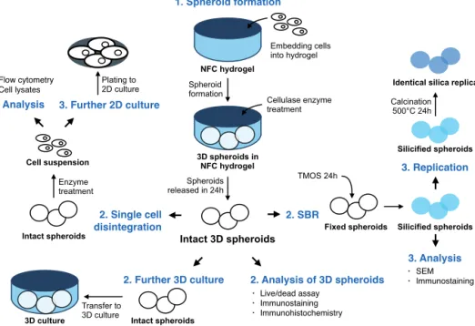
First, we studied two biomaterials, nanofibrillar cellulose (NFC) hydrogel and hyaluronic acid-gelatin (HG) hydrogel, to construct functional liver 3D organoids. Both of the studied hydrogels supported 3D spheroid organization of human liver progenitor HepaRG cells and their functional polarization. The 3D culture systems promoted hepatic differentiation of progenitor cells faster than the standard 2D culture. However, the 3D hydrogels did not enhance hepatocyte-like properties if the HepaRG cells were pre-differentiated to hepatocyte-like cells in advance. Subsequently, we showed that NFC hydrogel culture can be combined with high-resolution imaging since the intact spheroids can be enzymatically released from the matrix. This was not possible with the HG hydrogel. We demonstrated that silica bioreplication preserved the 3D spheroid structure with its fine details and cellular antigens and allowed detailed morphological analysis of the spheroids cultured in NFC hydrogel.
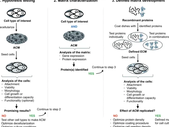
Next, we developed a xeno-free matrix for hepatic specification of human pluripotent stem cell-derived definite endoderm (DE) cells using a three-step approach. We first proved our hypothesis that a liver progenitor-like matrix, HepaRG-derived acellular matrix (ACM), supports hepatic lineage differentiation of DE cells. Then, we characterized the ECM proteins secreted by HepaRG cells, and finally we showed that the identified proteins, laminin-511 and laminin-521, can replicate the effect of HepaRG-ACM. The human pluripotent stem cell-derived hepatic cells expressed mature hepatocyte-like functions but the phenotype of the cells was eventually closer to fetal hepatocytes than mature cells. Thus, hepatic maturation should be further studied. In conclusion, this thesis describes new biomaterials for hepatic differentiation, a protocol to form 3D spheroids and to transfer intact spheroids to high-resolution imaging, and that the described three-step approach can guide the identification of new defined matrices.
Acknowledgements
This thesis work was carried out in the Centre for Drug Research (CDR), Division of Pharmaceutical Biosciences of the Faculty of Pharmacy at the University of Helsinki in Finland. Part of the experiments were performed in the Center for Micro-Engineered Materials at the University of New Mexico in Albuquerque in the USA. Ph.D. training was also received in the Finnish Centre of Alternative Methods (FICAM) in the School of Medicine at the University of Tampere. My Ph.D. work and conference trips were financially supported by the University of Helsinki, the University of Tampere, Doctoral Programme in Materials Research and Nanosciences (MATRENA), National Doctoral Programme in Nanoscience (NGS-NANO), Orion-Farmos Research Foundation, The Finnish Pharmacists’ Society, ACT Germany and the German Foundation SET, and The Finnish Concordia Fund.
First of all, I would like to thank the following persons for providing research facilities and educational opportunities: Professor Jouni Hirvonen, the dean of the Faculty of Pharmacy, Professor Heikki Vuorela, head of the Division of Pharmaceutical Biosciences, and Professor Arto Urtti, the head of CDR and the former head of the Division of Pharmaceutical Biosciences. I wish to warmly thank Professor Kai Nordlund, the former leader of MATRENA, and Dr. Alma Kartal-Hodzic, the coordinator of MATRENA, a dear colleague and a dear friend.
I am honored that Professor Hanry Yu has accepted the invitation to be my opponent in the public defense of this thesis. I would like to thank Professors Outi Hovatta and Timo Otonkoski for reviewing this thesis. Drs. Timo Tuuri and Mikko Koskinen are acknowledged for being part of my Ph.D. steering group.
I would like to express my sincere gratitude to my supervisor Dr. Yan-Ru Lou for your endless support and wise advice during these years. I am very grateful to my other supervisors Professors Marjo Yliperttula and Tuula Heinonen. Thank you Marjo for always inspiring and believing in me. Tuula, I am really thankful that I had a chance to work in your group in Tampere and learn the GLP spirit.
I am very grateful to all my co-authors for their contribution which has made this work possible. Above all, I wish to thank Dr. Melina Malinen for sharing her knowledge in liver cell research and being an important role model for me. I want to thank the current and old members in Yan-Ru’s team, especially Riina, Johanna, Mariia, Tuuli, Tomáš, and Ara, for fruitful scientific discussions and making each day at work more fun. I would like to express my thanks to all the present and former members at the Division of Pharmaceutical Biosciences and all other great people at the Faculty of Pharmacy. I am so happy that I got to share my Ph.D. journey with these amazing colleagues: Otto, Alma, Elisa, Dominique, Noora, Ansku, Aniket, Mecki, Cris, Lukasz, Mari, Mariangela, Susanna, Sofia, Victor, Feng, Marco, Heidi, Gloria, Polina, Astrid, Jaakko, Tatu R and L, Heli, Patrick, Katja-Emilia, Viviana, Leena K, Teemu, Leena P, Erja, Timo, Krista and all others!
Making this Ph.D. would not have been possible without the support from my friends and family. I am blessed for having so many wonderful friends in my life. You all mean so much to me. I can not thank my parents, Heli and Heikki, enough for all your love and encouragement. My dearest siblings, Tiina, Kalle, and Erkki, tykkään teistä niin että halkeen! Haluan erityisesti kiittää isovanhempiani, Leenaa, Taunoa, Salmea ja Heikkiä, kaikista viisaista elämänohjeista.
Finally, I want to express my deepest gratitude to Petri, my future husband, for your neverending support. You have made me bolder and taught me to dream big. We are the best team, L&P, ever.
Helsinki, April 2016 Liisa Kanninen
Contents
Abstract 3 Acknowledgements 4
List of original publications 8
Author’s contribution 9
Abbreviations 10
1 Introduction 12
2 Review of the literature 14
2.1 Liver tissue environment 14
2.1.1 Liver structure and function 14
2.1.2 Embryonic liver development and intrahepatic lineage maturation 16
2.1.3 Extracellular matrix chemistry 18
2.2. Liver cells sources for in vitro culture 20
2.3 Matrix-guided liver cell cultures 21
2.3.1 Liver-derived biomaterials 22
2.3.2 Liver ECM component-based matrices 25
2.3.3 Artificial liver mimicking matrices 29
3 Aims of the study 34
4 Materials and methods 35
4.1. Cell culture matrices 36
4.1.1 Two-dimensional matrices 36
4.1.2 Three-dimensional matrices 36
4.2 Cell cultures 37
4.2.1 Human liver cell lines 37
4.2.2 Human primary hepatocytes and liver tissue 37
4.2.3 Human pluripotent stem cells 37
4.2.4 Hepatic differentiation of human pluripotent stem cells 38
4.2.5 Three-dimensional cell culturing 38
4.3 Analysis methods 39 4.3.1 Viability assays 39 4.3.2 Gene expression 39 4.3.3 Protein expression 40 4.3.4 Cell functionality 41 4.3.5 Silica bioreplication 42 4.3.6 Imaging 42 4.3.7 Statistical analysis 43 5 Results 44
5.1. Three-dimensional matrices promote hepatic differentiation and NFC
hydrogel enables high-resolution imaging of 3D spheroids 44
5.2. Laminin-511 and laminin-521-based matrices support hepatic specification
of definite endoderm cells 47
6 Discussion 51
6.1 Mimicking liver tissue in a dish with bioinspired matrices 51
6.1.1. Three-dimensional hydrogels induce differentiation of liver cells 52
6.1.2 Lineage-specific matrix guides efficient hepatic differentiation 53
6.2 Multidimensional cell cultures require advanced analysis techniques 56
7 Conclusions 58
List of original publications
This thesis is based on the following publications:
The publications are referred to in the text by their roman numerals (I-IV). Reprinted with the permission of the publishers.
Malinen MM, Kanninen L, Corlu A, Isoniemi H, Lou Y-R, Yliperttula M, Urtti A. Differentiation of liver progenitor cell line to functional organotypic cultures in nanofibrillar cellulose and hyalyronan-gelatin hydrogels. Biomaterials 35: 5110-5121, 2014.
Lou Y-R*, Kanninen L*, Kaehr B, Towson J.L, Niklander J, Harjumäki R, Brinker C.J, Yliperttula M. Silica bioreplication preserves three-dimensional spheroid structures of human pluripotent stem cells and HepG2 cells. Scientific reports 5: 1-9, 2015. *Equal contribution
Kanninen L, Porola P, Niklander J, Malinen MM, Corlu A, Guguen-Guillouzo C, Urtti A, Yliperttula ML, Lou Y-R. Hepatic differentiation of human pluripotent stem cells on human liver progenitor HepaRG-derived acellular matrix. Experimental Cell Research 341: 207-217, 2016.
Kanninen L, Harjumäki R, Peltoniemi P, Porola P, Niklander J, Smutný T, Urtti A, Yliperttula M, Lou Y-R. Laminin-511 and laminin-521 based matrices for efficient hepatic specification of human pluripotent stem cells. Manuscript.
I
II
III
Author’s contribution
Publication IThe author helped to design the experiments, performed 2D and 3D cell culturing, phase contrast microscopy, alamarBlue viability assay, CYP3A4 activity, CYP3A4 induction, and whole mount immunostaining together with M.Sc. (later Ph.D.) Melina Malinen. M.Sc. Malinen performed live/dead viability assay, RT-PCR, genomic DNA quantification, immunohistochemistry, functional polarity studies, and analyzed the data. The author commented on the paper.
Publication II
The author designed the experiments with Ph.D. Yan-Ru Lou and the co-authors. The author performed HepG2 cell culturing and whole mount immunostaining. The phase contrast microscopy, immunohistochemistry, enzymatic removal of the hydrogels, silica bioreplication, and calcination were done together with co-authors. Flow cytometry of 3D cultured stem cells was done together with undergraduate (later M.Sc.) Riina Harjumäki. The scanning electron microscopy was carried out by Ph.D. Bryan Kaehr. The stem cell cultures were performed by Ph.D. Lou, undergraduate Johanna Niklander, and Ms. Harjumäki. The author analyzed the data with co-authors and wrote the paper with Ph.D. Lou.
Publication III
The experiments were designed by the author and Ph.D. Lou. The author carried out the cell culture experiments with WA07 and iPS(IMR90)-4 cell lines and the assays related to their analysis with the help of Ms. Niklander and Ph.D. Lou. M.Sc. (later Ph.D.) Pauliina Porola performed H9-GFP cell cultures and the related analysis with Ph.D. Lou. The author analyzed the data and wrote the paper together with Ph.D. Lou.
Publication IV
The experiments were designed by the author and Ph.D. Lou. The cell culturing and analysis of H9-GFP cell line was done by undergraduate (later M.Sc.) Pasi Peltoniemi and Ph.D Lou. The characterization of the HepaRG-ACM by conventional PCR was done by Ms. Porola, Mr. Peltoniemi and Ph.D. Lou and by immunostaining by the author, Mr. Peltoniemi and Ph.D. Lou. The cell culturing of WA07 and iPS(IMR90)-4 cell lines and the assays related to their analysis were performed with the help of co-authors. The author analyzed the data and wrote the paper together with Ph.D. Lou.
Abbreviations
2D Two-dimensional 3D Three-dimensional
ACM Acellular matrix
ActA Activin A
AAT Alpha-1 antitrypsin
AFP Alpha-fetoprotein
ALB Albumin
BMP Bone morphogenetic protein
CK Cytokeratin
CV Central vein
CXCR-4 Chemokine receptor type 4
CYP Cytochrome P450
DPBS Dulbecco’s modified phosphate salt buffer
DE Definite endoderm
DEX Dexamethasone
DMSO Dimethyl sulfoxide
DNA Deoxyribonucleic acid
ECM Extracellular matrix
EHS Engelbreth–Holm–Swarm mouse sarcoma
F-actin Filamentous actin
FBS Fetal bovine serum
FGF Fibroblast growth factor
FN Fibronectin
EpCAM Epithelial cell adhesion molecule
GAG Glycosaminoglycan
GFP Green fluorescent protein
HA Hyaluronic acid, also known as hyaluronan
HCM Hepatocyte culture medium
hESC Human embryonic stem cell
HG Hyaluronan-gelatin
HGF Hepatocyte growth factor
HNF Hepatocyte nuclear factor
hPSC Human pluripotent stem cell
hiPSC Human induced pluripotent stem cell
ICAM Intercellular adhesion molecule
LN Laminin
MSC Mesenchymal stem cell
MDR Multidrug resistance protein
mRNA Messenger ribonucleic acid
MRP Multidrug resistance-associated protein
NaBut Sodium butyrate
NCAM Neural cell adhesion molecule
NFC Nanofibrillar cellulose
NR1I2 Nuclear receptor subfamily 1 group I member 2, also known as
NR3C1 Nuclear receptor subfamily 3 group C member 1, also known as glucocorticoid receptor (GR)
OCT4 Octamer-binding transcription factor 4
OSM Oncostatin M
PCL Poly-ε-caprolactone
PEG Polyethylene glycol
PEGDA Polyethylene glycol diacrylate
PFA Paraformaldehyde
PT Portal triad
qPCR Quantitative polymerase chain reaction
RGDS Arginine-glycine-asparagine-serine
rhEGF Recombinant human epidermal growth factor
RNA Ribonucleic acid
RT-PCR Real time polymerase chain reaction
SBR Silica bioreplication
SEM Scanning electron microscopy
SSEA Stage-specific embryonic antigen
TMOS Tetraethyl orthosilicate
1 Introduction
The approximated cost of bringing a new molecular entity, a novel drug, to a market is 1.8 billion US dollars, and is constantly rising (Paul et al. 2010). Only over 10 years ago the respective price was estimated to be 0.8 billion US dollars (Dickson and Gagnon 2004). The high costs are primarily due to high failure related to unaccepted efficacy (56%) and toxicity in human (28%) (Arrowsmith and Miller 2013). Currently less than one in ten drugs that enter into the clinical phase will eventually get market approval (M. Hay et al. 2014). Additionally, withdrawals from the market are still seen: 19 drugs were withdrawn in the EU during 2002-2011 (McNaughton et al. 2014). For economic and regulatory reasons, the researchers in the academia and industry are evermore looking for tools to better predict the effects of new molecules in humans (LeCluyse et al. 2012). Importantly, in 21% of the cases hepatic disorders was the reason for withdrawn drugs in the EU in 2002-2011 (McNaughton et al. 2014).
Currently, there are satisfactory pharmacokinetic in vitro models to study induction and inhibition of liver enzymes and many assays have been proposed for studying hepatotoxicity (LeCluyse et al. 2012). However, there is an urgent need for advanced physiologically-relevant models to characterize biotransformation pathways. Hepatocytes, the main cell type of the liver, are essential components in these in vitro models. Subcellular systems such as liver microsomes can be exploited for metabolic profiling but only living cells can replicate all the metabolic processes occurring in vivo (Guillouzo and Guguen-Guillouzo 2008; A.P. Li 2007). Viable hepatocytes can be isolated from human liver, and they continue to serve the golden standard cell type for in vitro tests even though there are certain limitations in their use (Guillouzo and Guguen-Guillouzo 2008; FDA 2012). One of the reasons hampering their use is their limited life-span in culture. To overcome this, significant improvements in maintaining their phenotype have been achieved when using in vivo mimicking culture matrices (Bissell et al. 1987; Michalopoulos and Pitot 1975; Uygun et al. 2010; Sellaro et al. 2010). Alternative source for obtaining viable human hepatocytes are human pluripotent stem cells (hPSC). Both human embryonic stem cells (hESC) and human induced pluripotent stem cells (hiPSC) have the ability to form any cell type of the human body (Thomson et al. 1998; Yu et al. 2007; K. Takahashi et al. 2007) and thus they are broadly studied for various biomedical applications, as well as for drug testing. The concept of culturing cells was established over 100 years ago but the currently used culture techniques are not yet ideal. Cell culturing can be seen to have started in 1885 when cells were kept alive outside a body for a few days (Wall 2015). In 1907 cells were successfully grown in a laboratory for the first time (Harrison et al. 1907). Thereafter, numerous cell culture systems, reagents, and devices have been reported. Still, cell cultures are routinely performed with very simple techniques in two-dimensional (2D) configuration (Breslin and O’Driscoll 2013). Standard 2D monolayer cultures on flat, typically plastic surfaces do not simulate the complexity found in in vivo tissue as cell-matrix and multidimensional cell-cell interactions are missing (Owen and Shoichet 2010; Breslin and O’Driscoll 2013). Mimicking the cells’ natural microenvironment, extracellular matrix (ECM), in vitro with biomaterials has been shown to be a successful strategy to improve tissue-like functions of the
cultured cells (Tibbitt and Anseth 2009; Breslin and O’Driscoll 2013; Owen and Shoichet 2010). Development of bioinspired materials has two approaches. The materials are either extracted or derived from nature and then modified, or the idea for a new material is from nature but the product is manufactured synthetically (Smitthipong et al. 2014). Thus, bioinspired materials can be classified as materials originating from nature or materials mimicking the nature. Developing liver mimicking culture systems for hepatocytes should start by learning from the liver.
The shifting trend from the standard 2D cultures to more advanced systems, often using the third culture dimension, challenges the basic analysis techniques. In more complex culture formats visualization of the sample or data collection can be problematic. For example, in microfluidic devices the cell number and the culture volume can be so low that detection of an analyte can cause issues, or in case of three-dimensional (3D) culture system, the matrix or a thick sample itself can hinder microscoping of the specimen (Pampaloni et al. 2007; Esch et al. 2015). Consequently, it is important that the innovations in analysis techniques keep up with the development of advanced culture systems. This thesis describes first structure, functions, development, and ECM proteins of the liver and reviews matrix-based cell culture systems for hepatocytes. After the literature review, the aims and materials and methods of this thesis are introduced. Finally, the obtained results are briefly presented and the findings are discussed.
2 Review of the literature
2.1 Liver tissue environment
During the last decades hepatic research has significantly advanced and increased the knowledge in liver structure, function, and hepatic lineage maturation (Abdel-Misih and Bloomston 2010; Si-Tayeb et al. 2010a; L. Zhang et al. 2008). New molecular biology technologies have provided deeper understanding in regulation of gene expression, proliferation, cell cycle, and DNA repair in hepatocytes (Si-Tayeb et al. 2010a). This chapter gives an overview of the liver anatomy and function, development, describes recently proposed hepatic lineage maturation process, and illustrates the extracellular matrix chemistry in the liver.
2.1.1 Liver structure and function
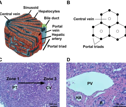
In adults, liver is the largest gland and it weighs approximately 1.5 kg comprising two to three percent of body weight (Si-Tayeb et al. 2010a). The liver has various functions which are strictly dependent on correct liver tissue architecture (Figure 1A). A liver lobule is the basic architectural unit consisting of plates of hepatocytes with thickness of one to two cells (Schiff, et al. 2011; Si-Tayeb et al. 2010a). The hexagonal-shaped lobule is lined by a portal triad vessels composed of portal vein, bile duct, and hepatic artery (Figure 1B, D). The portal vein and hepatic artery supply blood to the lobule from each of the lobule’s six corners and a network of sinusoidal capillaries carry the oxygen and nutrients to a central vein in the middle of the lobule. Indeed, a highly vascularized liver receives more blood than any other organ in the body (Abdel-Misih and Bloomston 2010). The periportal area close to the portal triad is also called zone 1 and the pericentral area next to the central vein is called zone 3.
Hepatocytes are the major cell type in the liver representing approximately 80% of the liver volume and 60% of all liver cells (Malarkey et al. 2005). These polyhedral-shaped cells are highly polarized, having a specialized canalicular region at their apical membrane and plenty of microvilli on the basolateral surface towards the space of Disse, the area between the hepatocytes and sinusoidal endothelial cells (Schiff et al. 2011). The canalicular regions of two neighboring hepatocytes are connected with tight junctions and form bile canaliculi that drains to the bile duct. Hepatocytes are responsible for most of the liver’s biosynthesis and biomolecules’ storage functions (Juza and Pauli 2014). They exhibit functional gradients over the lobule from zone 1 to zone 3 (Malarkey et al. 2005) (Figure 1C). Hepatocytes in zone 1, also called periportal hepatocytes, receive abundant blood supply rich in oxygen and nutrients and thus show higher activity in gluconeogenesis, amino acid catabolism, ureagenesis, cholesterol synthesis, and bile acid secretion compared to the cells close to the central vein, also called centrilobular cells (Malarkey et al. 2005; Schiff et al. 2011). In zone 3, the hepatocytes are responsible for glycolysis, lipogenesis, and biotransformation of endogenous and exogenous compounds.
In addition to hepatocytes, at least 14 other cells types can be found in a normal liver (Malarkey et al. 2005). Out of these, cholangiocytes, endothelial and sinusoidal endothelial cells, Kupffer cells, and hepatic stellate cells are closely cooperating with hepatocytes (Si-Tayeb et al. 2010a). Cholangiocytes, also called biliary epithelial cells, transport bile and maintain its pH (Tietz and Larusso 2006). Kupffer cells are liver macrophages with immunological and phagocytic functions and reside in the sinusoidal vessels (Malarkey et al. 2005; Schiff et al. 2011; Si-Tayeb et al. 2010a). Hepatic stellate cells, located in the spaces of Disse, are normally quiescent but when activated by various cytokines they function as the principal hepatic fibroblasts (Malarkey et al. 2005; Schiff et al. 2011; Martinez-Hernandez and Amenta 1995).
Figure 1 Architecture of the liver. A) The main cell type of the liver are hepatocytes which are organized in plates between portal triad and central vein. B) A liver lobule, the smallest architectural unit of the liver, is lined by portal triads. C) The hepatocytes exhibit functional gradients over the lobule from portal triad (PT), zone 1, to central vein (CV), zone 3. D) The portal triad is composed of portal vein (PV), hepatic artery (HA), and bile duct (B). Picture A is modified from Si-Tayeb et al. 2010a, picture B is inspired by LeCluyse et al. 2012, and pictures C and D are from hematoxylin and eosin stained human liver sections (Lou et al. unpublished).
B
PV HA B CV PT Zone 1 Zone 3 Central vein Sinusoid Hepatocytes Bile duct Portal vein Hepatic artery Portal triadC
A
Central vein Portal triadsD
2.1.2 Embryonic liver development and intrahepatic lineage maturation
Processes occurring during liver development can be categorized into two main chains of events: differentiation of all the cell types in liver from their embryonic progenitors and the arrangement of the derived cells into highly organized structures (Zong and Friedman 2014). In human, gastrulation takes place on approximately day 16 of gestation, after which the embryo is composed of ectodermal, mesodermal and endodermal germ layers. Liver epithelial cells, hepatocytes and cholangiocytes, are derived from the endoderm which also gives rise to pancreas, lung, thyroid, and gastrointestinal tract. The next step in liver development is hepatic specification during which developing cardiac mesoderm and the septum transversum mesenchyme secrete inductive signals, fibroblast growth factor (FGF) and bone morphogenic proteins (BMPs), to endoderm cells that differentiate to hepatoblasts (Gualdi et al. 1996; Zong and Friedman 2014).
The formed hepatoblasts are proliferating and form a liver bud, anatomical outgrowth from the ventral wall of the foregut endoderm (Zhao and Duncan 2005). Immediately after the bud being formed, endothelial cells or angioblasts envelop the bud and play important role in the bud’s expansion and separate it from the septum transversum mesenchyme (Matsumoto et al 2001; Zhao and Duncan 2005). Next, the hepatoblasts begin to invade into surrounding septum transversum mesenchyme as cords, and simultaneously, the hepatic vasculature is being developed. The hepatoblast population is rapidly proliferating and as a bipotent cell type they differentiate into hepatocytic and cholangiocytic lineages (Schiff et al. 2011; Cardinale et al. 2011). In concert, other liver cell types are being differentiated, biliary tract is developed, and the ECM is formed (Zhao and Duncan 2005).
Multiple transcription factors, including hepatocyte nuclear factors (HNFs), have shown to be crucial for the hepatocyte differentiation (Zhao and Duncan 2005). In addition, oncostatin M (OSM), secreted by hematopoietic cells, is essential in controlling the late stage hepatocyte differentiation. The maturation of functional hepatocytes is gradual and also continues after birth. During fetal and neonatal periods, the metabolizing enzyme and transporter expressions occur in several patterns and vary for each enzyme subfamily and isoform (Moscovitz and Aleksunes 2013). The ECM proteins present in the liver are discussed below in chapter 2.1.3. At birth, the liver comprises approximately four percent of the newborn’s body weight (Suchy 2014).
In a healthy adult liver, hepatocytes are proliferatively quiescent and their estimated life-span is over one year (Roskams 2006; Sell 2001). However, in the case of partial hepatectomy, surgical removal of liver mass, or in acute injury the lost liver mass is rapidly replaced although the native liver architecture is not reconstituted (Fausto and Campbell 2003). The liver has enormous regenerative capacity but results from studies on how liver regeneration occurs have been controversial. It has been shown that after a partial hepatectomy, hepatocytes resting at G0 phase re-enter the cell-cycle and start to replicate (Taub 2004). However, after toxin-mediated injuries hepatocytes have decreased capacity to proliferate. Some of the findings support the assumption that liver stem/progenitor cells (LPCs) participate in repopulating the liver after a severe injury in concert with proliferating hepatocytes (Forbes et al. 2002). In addition, a recent study reported that hepatocytes can be converted into cholangiocytes in vivo which might play a role in restoring functions and architecture
of the liver after injuries (Yanger et al. 2013).
The research on liver stem cells does not have explicit history. The early observations and speculations on LPCs were made before the 19th century and the characterization of oval cells, small epithelial cells with oval nuclei, in the 1950s started their broad examination (Fausto and Campbell 2003; Farber 1956). LPCs are classified as cells that have the ability to differentiate into hepatocytes and cholangiocytes during liver regeneration and cellular turnover (Tanimizu and Mitaka 2014). Even though the role and origin of LPCs is still not unambiguous their importance in liver biology has been proven (Forbes et al. 2002). The LPCs have been suggested to derive from periductular cells, hematopoietic stem cells, or the quiescent hepatocytes themselves have been suggested to be the stem cells of the liver as their differentiation can be activated (Fausto and Campbell 2003; Sell 2001; Y. Zhang et al. 2003). Later, Reid and co-workers have reported that both hepatic stem cells and hepatic progenitors are found in liver of all donor ages (L. Zhang et al. 2008; Turner et al. 2011; Cardinale et al. 2011).
The stem cells are located in a unique microenvironment called a niche which regulates self-renewal and differentiation of the stem cells (Scadden 2006). In pediatric and adult livers, the hepatic stem cell niches are found in canals of Hering and within the biliary tree (Theise et al. 1999; Kuwahara et al. 2008; Turner et al. 2011). It has been recently shown that peribiliary glands throughout the biliary tree host multipotent stem/progenitor cells which give rise to cholangiocytes, hepatocytes and pancreatic committed progenitors (Cardinale et al. 2011; Y. Wang et al. 2013). These biliary tree stem/progenitor cells express typical endodermal markers but only low-levels or no lineage markers of liver or endocrine pancreas (Cardinale et al. 2011). Whether the liver is a classical stem cell lineage system or not is a divisive question in the field of hepatology (Sigal et al. 1992; Fausto and Campbell 2003). Intrahepatic lineage maturation, as suggested by Reid and co-workers, consists of eight stages proceeding from zone 1 to zone 3 in the liver lobule (Turner et al. 2011; Furth et al. 2013) (Figure 2). Multipotent hepatic stem cells represent approximately 0.5-2% of the liver cells in donors of all ages (Turner et al. 2011). The hepatic stem cells co-express epithelial cell adhesion molecule (EpCAM), neural adhesion molecule (NCAM), cytokeratin-19 (CK-19), low levels of albumin (ALB) and very low levels or no alpha-fetoprotein (AFP) (L. Zhang et al. 2008; Furth et al. 2013; Schmelzer et al. 2006). In the next stage, the hepatic stem cells differentiate into hepatoblasts which can give rise both to hepatocytic and cholangiocytic lineages. Hepatoblasts express intercellular adhesion molecule-1, (ICAM-1), EpCAM, early cytochrome P450s, and are highly positive for AFP (Turner et al. 2011; Furth et al. 2013; Schmelzer et al. 2006).
Next, the hepatoblasts are committed to hepatic progenitors which, according to this classification, are unipotent cells and can differentiate only to hepatocytes (Turner et al. 2011). Hepatic progenitors express ALB but lack CK-19 and AFP (L. Zhang et al. 2008; Furth et al. 2013). In the liver lobule, they are located in the beginning of hepatocyte plates. During the later stages of lineage maturation the hepatocytes grow in size, become binucleated, and express mature liver markers such as CYP3A4, glutathione S-transferases, and high levels of ALB (Turner et al. 2011; Furth et al. 2013). Finally, the apoptotic hepatocytes at zone 3 have been reported to produce hepatocyte growth factor that simulates the expansion of stem cells and/or progenitors which works as positive loop signaling.
Lineage stage
(viable cells) 1 2 3 4 5 6 7
Parenchymal
cells stem cellsHepatic Hepato-blasts progenitorsComitted Diploid adult hepatocytes pericentral Tetraploid hepatocytes Cell size 7-10 µm 10-12 µm 12-15 µm 15-20 µm 22-25 µm 25-30 µm >30 µm Cell growth Maximum (complete cell division) Intermediate
(complete cell division) Negligble Gene
expression EpCAMNCAM CK-19 AFP-ICAM CYP3A7 AFP+++ ALB+ ALB++ AFP-Glycogen <<< Gradient >>> ALB+++ CYP3A4 Glutathione S-transferases
Figure 2 Hepatic lineage maturation from hepatic stem cells to apoptotic
hepatocytes is suggested to consist of eight steps (Turner et al. 2011; Furth et al. 2013). The liver lobule exhibits structural and functional gradients from zone 1 to zone 3 (L. Zhang et al. 2008; Y. Wang et al. 2011; Turner et al. 2011; Furth et al. 2013). AFP, alpha-fetoprotein; ALB, albumin; CK-19, cytokeratin-19; CV, central vein; CYP, cytochrome P450 enzyme; EpCAM, epithelial cell adhesion molecule; ICAM-1, intercellular adhesion molecule-1; NCAM, neural adhesion molecule; PT, portal triad. Figure is modified from Turner et al. 2011 and LeCluyse et al. 2012.
2.1.3 Extracellular matrix chemistry
In liver, hepatocytes are in a continuous intereaction with an ECM. The ECM is a mixture of molecules which by their structure and function provide essential cues for cell proliferation, migration and differentiation, and maintenance of tissue homeostasis (Faulk et al. 2014). In turn, dysfunction of ECM dynamics leads to unregulated cell proliferation and differentiation causing severe pathological events such as fibrosis and cancer (Rozario and DeSimone 2010; Lu et al. 2011). The secretion of the ECM starts already at the embryonic stage and its remodeling is an important mechanism by which tissue formation is regulated (Rozario and DeSimone 2010; Lu et al. 2011). The ECM is modified and degraded by enzymes secreted by the cells. The most important enzymes in ECM remodeling are matrix metalloproteinases (Lu et al. 2011).
Zone 1 Zone 2 Zone 3
Metabolism
Oxygen
CV PT
ECM proteins are functionally diverse; both rigid, elastic, wet, and sticky proteins are needed in the tissue formation and maintenance (Mecham 2001). Biochemically, ECM components can be divided into proteins, proteoglycans, and glycoproteins (Rozario and DeSimone 2010; Lu et al. 2011). Fibrillar collagens and elastin form fibrils and determine the viscoelasticity and tensile strength of the tissue. Fibronectin and laminins contribute as building blocks of the matrix network and connecting proteins. ECM occupies only a very limited part of a healthy liver and is restricted to portal triads, sinusoids, and central veins (Bedossa and Paradis 2003). In the plates of hepatocytes, the ECM is located in spaces of Disse (Martinez-Hernandez and Amenta 1995).
The most abundant ECM proteins the liver are collagens which also are the most frequently found proteins in a human body compromising approximately 30% of the total protein mass (Bedossa and Paradis 2003; Ricard-Blum 2011). Out of the described 28 members of the collagen family, fibrillar collagen, types I, III, and V, and basement membrane collagen, types IV and VI, have been described in the liver (Y. Wang et al. 2011; Martinez-Hernandez and Amenta 1995). In addition to collagens, other matrix components found in the liver are glycoproteins, including laminins, fibronectin, and tenascin, and proteoglycans, such as hyaluronic acid, also called as hyaluronan, heparan, and chondroitin sulfate (Bedossa and Paradis 2003). Even though several groups have examined the liver ECM proteins, more studies are still needed to understand their relative quantities in different locations.
Laminins (LNs) are present predominantly in the basement membrane in most tissues in human and they bind to cell surface via integrin and nonintegrin receptors (Durbeej 2010; Malinda and Kleinman 1996). These multidomain heterotrimers are named on the basis of their α, β, and γ chains (Aumailley et al. 2005) and different subtypes can serve distinct biological functions (Colognato and Yurchenco 2000). By now, 18 laminin isoforms have been described in the literature (Durbeej 2010). Out of these, at least LN-211/221, LN-411/421, and LN-511/521 are found in adult human liver (Kikkawa et al. 2008; Liétard et al. 1998). According to Kikkawa and co-workers, LN-511 and LN-521 are the main laminin isoforms in the liver (2008). Presence of LN-111 in the adult liver is uncertain. Expression of laminin α1 chain mRNA in human liver has been reported (Liétard et al. 1998) but in other studies the α1 chain protein was not found (Virtanen et al. 2000; Kikkawa et al. 2008). In general, distribution of laminin isoforms in the human liver has not yet been broadly studied and thus a complete picture on their localization can not be made.
Fibronectin is a multidomain protein that binds to cell surface receptors, collagens, and other fibronectin molecules (Schwarzbauer and DeSimone 2011). Fibronectin is expressed in most human tissues (Stenman 1978), and it is abundant in liver as well (Martinez-Hernandez and Amenta 1995). Proteoglycans are a heterogenous group of macomolecules composing of glycosaminoglycans (GAGs), linear polysaccharides, bound to a core protein. Proteoglycans are categorized based on their GAG chain and size. Degree of sulfation of GAGs and proteoglycans is believed to play an important role in intahepatic differentiation processes (Y. Wang et al. 2010).
The ECM chemistry changes during the development, and in the adult liver expression of the matrix proteins vary between the zones. (McClelland et al. 2008; Y. Wang et al. 2010). In periportal areas, zone 1, the matrix is composed of laminins, collagen types I, III, IV, and V, fibronectin, hyalyronan, entactin, perlecan, and
chondroitin sulfate proteoglycans as shown in animal and human studies (Kikkawa et al. 2008; Seebacher et al. 1997; McClelland et al. 2008; Turner et al. 2011; Y. Wang et al. 2010; Mazza et al. 2015; Roskams et al. 1995). Kikkawa and co-workers (2008) reported that LN-511 is abundant in the bile duct where fibronectin expression has been reported to be low or absent (Hahn et al. 1980). Fibronectin, in turn, is the most abundant ECM protein in the space of Disse (Martinez-Hernandez and Amenta 1995). Other ECM components localized in spaces of Disse include all types of collagen found in the liver and perlecan (Martinez-Hernandez and Amenta 1995; Y. Wang et al. 2011; Mazza et al. 2015). Laminin is not present in the sinusoids of a healthy liver (Martinez-Hernandez and Amenta 1995; Kikkawa et al. 2008). Elastin is expressed in the whole liver lobule (Y. Wang et al. 2010; Turner et al. 2011; Van Eyken et al. 1990)
The matrix close to the central vein, zone 3, contains collagens type I, IV, and VI and a weak expression of collagen type III, fibronectin, syndecans, and highly sulfated heparan proteoglycans (McClelland et al. 2008; Y. Wang et al. 2011; Mazza et al. 2015). Zone 3 matrix differs from typical epithelial basement membrane by almost total absence of laminins and entactin, but contains large amounts of tenascin (Van Eyken et al. 1990; Y. Wang et al. 2010; Turner et al. 2011). Low or absent expression of laminin isoforms in zone 3 was confirmed by Kikkawa et al. (2008).
The matrix chemistry within intrahepatic stem cell niche is only partially characterized (Furth et al. 2013). The suggested ECM components include hyalyronan, collagen type III, α6β4 integrin-binding form of laminin, and chondroitin sulfate proteoglycan with minimal sulfation (Furth et al. 2013; Seebacher et al. 1997; L. Zhang et al. 2008; Y. Wang et al. 2010; Turner et al. 2011). As stem cells migrate from the niche, the surrounding GAGs and more extensively sulfated proteoglycans together with growth factors direct the differentiation process (Y. Wang et al. 2010). The microenvironment of hepatoblasts already differs from the stem cell niche consisting collagen types III, IV and V, α3β1 integrin-binding form of laminin, normally sulfated chondroitin sulfate proteoglycan, and heparan sulfate proteoglycans as proposed by Y. Wang and co-workers (2010).
2.2. Liver cells sources for in vitro culture
Human liver cell cultures have multiple applications both as in vitro test systems and in tissue engineering. Of the liver cells, hepatocytes are needed in drug development for metabolic profiling of drug candidates (FDA 2012). Primary hepatocytes serve as the gold standard for in vitro drug biotransformation and liver toxicity testing even though their limitations in terms of availability, rapid loss of metabolizing capacity, and batch-to-batch variation are well recognized (LeCluyse and Alexandre 2010). Contradictory to liver tissue, the isolated primary hepatocytes have low proliferation capacity in vitro which hampers their use (Fausto and Campbell 2003; Ramboer et al. 2014). After recognizing that hepatic functions of primary hepatocytes decrease rapidly during culture (Bissell 1973) numerous culture improvements by using various matrices, devices, and flow-based systems have been described (Bissell et al. 1987; Michalopoulos and Pitot 1975; Uygun et al. 2010; Sellaro et al. 2010; C. Lin et al. 2015).
Hepatocyte cell lines can be obtained from tumors or by oncogenic immortalization and thus have an unlimited lifespan (Guillouzo and Guguen-Guillouzo 2008; Rodríguez-Antona et al. 2002). Compared to primary hepatocytes, hepatic cell lines exhibit a more stable phenotype, are easily available, and they can be genetically manipulated (Gomez-Lechon et al. 2010). However, their major limitation is their relatively weak metabolizing capacity. Only a few hepatic cell lines express some liver-specific functions (Gomez-Lechon et al. 2010; Guillouzo and Guguen-Guillouzo 2008).
In addition to primary isolations and immortalized cell lines, hepatocyte-like cells can be obtained from PSCs. In 1998 Thomson and co-workers established the first hESC lines. Cells derived from the inner cell mass of a blastocyst maintained capacity to self-renew and their pluripotency, capability to form all three germ layers, when expanded in vitro (Thomson et al. 1998). Only less than ten years later, a method for reprogramming pluripotent stem cells from somatic cells was described (K. Takahashi and Yamanaka 2006; Yu et al. 2007; K. Takahashi et al. 2007). These cells, named as induced pluripotent stem cells, were produced by delivering key transcription factors related to pluripotency via viral transduction into the cells in vitro. Thereafter, hiPSCs have been reprogrammed from various somatic cell types and by several alternative methods such as non-integrating vectors, small molecules, and microRNA (Okita et al. 2008; Shi et al. 2008; Anokye-Danso et al. 2011). Generally, hiPSCs exhibit similar properties to those of hESCs but do not share the same ethical concerns. As the hPSCs have an ability to form all cell types of an adult body they are highly interesting sources of cells for various human cell-based applications including disease modelling, drug screening, and cell therapies. Hepatic differentiation of the hPSCs has already been broadly studied (Cai et al. 2007; Duan et al. 2010; Hay et al. 2008; Si-Tayeb et al. 2010b). Still, obtaining fully mature, in vivo counterparts mimicking hepatocytes from hPSCs is challenging and none of the current protocols entirely fulfil this task. Alternative stem cell sources for hepatic differentiation are hepatic stem cells and mesenchymal stem cells (MSC) (X. Zhang and Dong 2015; Y. Wang et al. 2011). In addition, direct reprogramming of fibroblasts to hepatocytes has been recently described (Huang et al. 2014).
2.3 Matrix-guided liver cell cultures
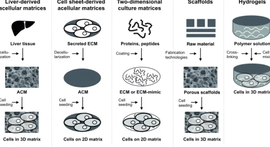
It has been stated that hepatocytes lose their phenotypic functions once isolated from liver because the cell-matrix interactions are disrupted (Kikkawa et al. 2011). Liver environment can be simulated in vitro by using several approaches. Liver tissue can be decellularized and used as a native scaffold for reseeding cells in vitro. ECM proteins, individually or in combinations, provide cues from the liver matrix to cultured cells. Alternatively, the liver microenvironment can be mimicked with biologically inactive biomaterials to resemble mechanical properties of the liver. This chapter describes how different matrices, both 2D and 3D systems, have been used to culture human liver cells, including primary isolations and liver cell lines, or facilitate the hepatic differentiation of stem cells in vitro. The materials discussed are categorized into three groups: liver tissue-derived matrices, liver ECM-based matrices, and artificial liver mimicking matrices. Due to the high number of published matrices this review can only give examples from each matrix category.
The non-matrix-based culturing techniques, such as suspension cultures or culture models built only on co-culture setup or flow systems, are not included in this review. Figure 3 gives an overview of the used matrix-guided liver cell culture approaches.
Figure 3 Different approaches to mimic liver tissue in a dish. Liver-derived acellular matrices (ACM) have native liver structure and matrix chemistry. Cell sheet– derived ACMs can be made from cell lines. Two-dimensional (2D) culture can be performed on liver extracellular proteins (ECM) or synthetic polymers. Liver structure or stiffness can be resembled with scaffolds made from natural or synthetic biomaterials. Hepatic cells can be embedded in hydrogels, which mimic liver stiffness or matrix chemistry, to form three-dimensional (3D) spheroids. Picture is inspired by Chan and Leong 2008.
2.3.1 Liver-derived biomaterials
Native liver tissue can be used for in vitro cell culturing after decellularization, a process which aims to remove all cellular materials from the tissue. The obtained acellular matrices (ACM) can be used as such or they can be further processed into powders and gels (Uygun et al. 2010; Y. Wang et al. 2011; Sellaro et al. 2010). After first being described in 1948, techniques for decellularization and reseeding the cells have greatly improved and currently ACMs are promising culture materials for many biomedical applications (Hinderer et al. 2016).
2.3.1.1. Whole liver acellular matrices
Decellularized whole organs are the most natural, in vivo simulating scaffolds since tissue macroarchitecture and matrix microarchitecture can be maintained after the decellularization process (Hoshiba et al. 2010). The structure, chemical composition and functions of a matrix are tissue specific but not species specific (Y. Wang et al. 2011). Though, differences in ECM component quantities between
modified from Eur Spine J 2008 (17)
Secreted ECM Decellu- larization ACM Cell seeding Cells on 2D matrix Cell sheet-derived acellular matrices Liver-derived acellular matrices ACM Decellu- larization Cell seeding Cells in 3D matrix Liver tissue Two-dimensional culture matrices ECM or ECM-mimic Proteins, peptides Coating Cell seeding Cells on 2D matrix Hydrogels Polymer solution Cells in 3D matrix Cross-
linking Cell mixing
Scaffolds Raw material Porous scaffolds Cell seeding Cells in 3D matrix Fabrication technologies
species have been reported (Dahms et al. 1998). Animal-derived liver ACMs can be used for culturing human cells because ECM proteins are well-conserved among species and xenogeneic matrices are tolerated (Hoshiba et al. 2010).
Both whole liver and liver slices have been used to create liver structure mimicking cell culture scaffolds. The aim in tissue decellularization is to remove a whole cell population and to restore the complete tissue matrix without affecting its 3D structure, surface topology, and biological activity (Faulk et al. 2014). However, it is accepted that removal of cells from their integrin-bound anchors and intercellular adhesion sites while maintaining surface topography and resident ligands of the ECM intact is demanding (Badylak et al. 2012).
The decellularization can be performed by using enzymatic, physical, chemical and ionic methods (Badylak et al. 2012). In case of whole liver, the decellularization is preferably done by perfusion via the organ’s vasculature, recent innovation that preserves the 3D structure of an organ entirely (He and Callanan 2013). The liver ACM slices have been prepared by decellularizing liver tissue sheets or sectioning the decellularized whole liver (P. Lin et al. 2004; Y. Wang et al. 2011). Before reseeding the cells into acellular liver the matrix has to be sterilized which might have a harmful effect on the mechanical properties of the scaffold (Badylak et al. 2015). The sterilization techniques include gamma irradiation, electron beam irradiation, and treatment with acid or ethylene oxide (Badylak et al. 2015; Badylak 2002; Uygun et al. 2010).
The recently described decellularization by perfusion is an attractive method to produce bioarticifial liver scaffolds. Wang et al. (2011) reported a four-step perfusion decellularization which maintains the collagen content high and the remaining growth factors and cytokines were at physiological level. Uygun and co-workers (2010) demonstrated the decullarization of rat and bovine livers by portal vein perfusion with increasing concentrations of sodium dodecyl sulfate (SDS) and sterilized with peracetatic acid. After decellularization, collagen types I and IV, fibronectin and laminins, were maintained. The acellular liver scaffold was then reseeded with rat primary hepatocytes, which showed similar ALB protein expression compared to normal liver. However, ALB and urea production were at the similar level as in the cells cultured in between collagen layers, called as sandwich culture, a much simpler technique compared to liver ACM. The concept of culturing primary hepatocytes in whole liver ACM has also been proven with mouse hepatocytes in rat liver ACM (Soto-Gutierrez et al. 2011).
Wang et al. (2011) used slices made from rat liver ACM for differentiating human hepatic stem cells to mature liver cells. Decellularization was performed with four-step protocol by portal vein perfusion including delipidation, washing four-step with high salt concentration to maintain the insoluble collagens, eliminating nucleic acids with nucleases, and finally washing the matrix with cell culture medium to remove salts and detergents and to equilibrate the ECM components. After freezing, the tissue matrix was sectioned, sterilized and placed to cell culture plates. Gene and protein expression data and functionality studies showed that hepatic stem cells were able to differentiate to adult liver cell types and liver specific functions were higher compared to the cells cultured on collagen type I matrix. Rat and human primary hepatocytes have also been cultured on liver ACM slices prepared from porcine tissue sheets (P. Lin et al. 2004; Lang et al. 2011). Recently, Mazza et al. (2015) described the first decellularization of a human liver. By repopulating
cubes cut from the liver ACM with three liver cell types they showed that the ACM supported homing of the different cell types into their natural locations. Liver-like functions of the recellularized ACM were not examined.
The acellular scaffolds are promising for regenerative medicine, especially for end-stage organ failures (Badylak et al. 2012). So far, transplantation of recellularized liver ACM has been tested only in animal studies (Uygun et al. 2010; Barakat et al. 2012; Mazza et al. 2015). To scale up the whole liver ACM approach, to achieve human-sized organ, researchers have engineered scaffolds from porcine and human livers (Baptista et al. 2011; Barakat et al. 2012; Mazza et al. 2015). In vitro models based on liver ACM cultures could provide a deep understanding of the biotransformation routes and mechanisms of a drug candidate. However, they will not be applicable for high-throughput drug testing, at least in the near future, due to demanding decellularization and repopulation processes using perfusion systems. 2.3.1.2 Modified liver acellular matrices
Liver ACMs can be further processed to create culture systems with less batch-to-batch variation compared to native whole tissue ACM. Already in 1980, fibrils obtained from homogenized and decellularized rat liver was used for long-term culturing of rat hepatocytes (Rojkind 1980). After that, liver ACMs have been used to create liver ECM mimicking gels and powders both for 2D and 3D cell culturing (J. S. Lee et al. 2014; Sellaro et al. 2010; Y. Wang et al. 2011).
Sellaro et al. (2010) prepared liver ACM gel from porcine liver to culture human primary hepatocytes. The hepatocytes were cultured in a sandwich configuration in between liver ACM gel layers. ALB secretion remained stable for 10 days in the cells cultured in a liver ACM gel system. However, the liver-like functions of the cells cultured in ACM gel were not improved compared to Matrigel (described in chapter 2.3.2.2) sandwich culture. Wang et al. (2011) reported exceptionally long functional stability of human primary hepatocytes cultured on pulverized bovine liver ACM. Decellularized liver matrix was milled with the presence of liquid nitrogen, mixed with culture medium and dispersed to culture wells. The human hepatocytes on ACM powder were functional for eight weeks while the cells cultured on collagen type I started to die after three weeks. A 3D hydrogel derived from rat liver ACM has been used to culture rat primary hepatocytes (J. S. Lee et al. 2014). Lyophilized ACM containing collage type I, laminins, and fibronectin was cross-linked by mixing with PBS and incubated in neutral conditions at 37°C and used to encapsulate the cells. The hepatocytes were viable for a longer period of time in 3D ACM-hydrogel culture compared to 3D collagen type I hydrogel. The cells secreted ALB and urea over one week but other hepatic functions in vitro were not characterized.
Liver ACM-derived gel has also been shown to support hepatic differentiation of mouse adipose-derived MSCs (X. Zhang and Dong 2015). The cells were plated on an ACM gel made from pulverized rat liver ACM and cultured with and without the presence of hepatic differentiation inducing reagents. Compared to other tested coating agents, collagen, fibronectin and Matrigel, the liver ACM gel supported the hepatic differentiation best. The cells treated with inducing agents showed higher hepatic functions than the non-treated cells as shown by the secretion of ALB and urea. However, the culture of stem cells without soluble factors showed that the
culture matrix alone supported induction of the MSCs towards hepatic lineage. Compared to the whole liver ACM, the advantage of the modified liver ACM culture systems is their flexibility and possibility to decrease batch-to-batch variation by pooling several tissues together. However, the 3D structure of native liver tissue cannot be resembled with processed ACM culture systems. Nonetheless, the cells embedded in suspended or gelified ACMs can be easily transplanted in vivo with minimally invasive techniques offering a promising tool for tissue engineering (Badylak et al. 2015).
2.3.2 Liver ECM component-based matrices
The chemistry of liver tissue can be mimicked by using ECM proteins in culture systems. The ECM proteins have been used as culture substrata for liver cells since the 1970s when hepatocytes were cultured on collagen membranes (Michalopoulos and Pitot 1975). Thereafter, ECM proteins, mixtures or individual components, have been used as 2D matrices and materials for creating 3D culture systems. The ECM component-based matrices are introduced in the three following chapters: cell sheet-derived acellular matrices, ECM protein mixtures, and defined ECM proteins. 2.3.2.1 Cell sheet-derived acellular matrices
Compared to whole organ ACMs, decellularized cultured cells offer a simpler approach to mimic liver environment. Cultured cells do not require ethical permission and their decellularization can be performed without sophisticated devices. In addition, the production of cell line-derived ACMs is more straightforward compared to whole organs as the sterilization step can be avoided when performing the cultures in the same cell culture facility. The choice of the cell line for ACM construction is based on the desired ECM protein or combination of proteins secreted by the cells. The cell sheet ACMs have been developed from liver cells but also from other tissue types and tumors (Herrema et al. 2006; Vuoristo et al. 2013). The decellularization of cultured cells has been performed with distilled water or alkaline solution with or without detergent (Herrema et al. 2006; H. Takashi 2007; Vuoristo et al. 2013). The ACM can be characterized at the protein level with immunofluorescence, or at the structural level with transmission electron microscope. However, the structural studies of the cell sheet ACMs are seldom performed.
Takahashi H. et al. (2007) prepared ACM from immortalized alveolar type II epithelial SV40-T2 cells cultured on collagen type I and Matrigel. Mouse primary hepatocytes attached to the ACM similarly as to Matrigel but secreted more ALB on the ACM than on Matrigel. The authors claimed that the SV40-T2-ACM is superior to Matrigel because the matrix contains LN-511/521. Alveolar type II epithelial cells are known to secrete laminin a5 chain (Pierce et al. 1998) but its expression in the SV40-T2-ACM remains unclear as its characterization was not published by Takahashi’s group (H. Takahashi et al. 2007). Another group used HEK293 cells overexpressing LN-511 in a similar kind of culture system as Takahashi et al. to differentiate mouse ES cells to hepatocytes (Shiraki et al. 2011). Human ES cells seeded on LN-511-ACM increased their ALB secretion during the culture period
confirming their hepatic differentiation. However, the expression level of ALB mRNA was much lower than that in adult liver. The derived hepatic cells were metabolically active as shown by induction of CYP3A4 enzyme activity. LN-111 and LN-521 expressing human choriocarcinoma cell line-derived ACM has been shown to support hepatic differentiation of hESCs and hiPSCs (Vuoristo et al. 2013). The hPSCs were plated on top of ACM derived from JAR cells cultured on gelatin and differentiated towards hepatic cells with stepwise growth factor treatment. The derived cells expressed human ALB in gene and protein level similarly to the cells cultured on Matrigel.
Cell sheet-derived ACMs are relatively fast, easy, and cost-efficient matrices to produce. The transfected cell lines offer possibility to create culture matrices with specific ECM protein composition mimicking a certain developmental phase or a region in the liver. However, the cell sheet-derived ACMs fail to mimic the native liver structure which can be restored in whole liver ACMs.
2.3.2.2 ECM protein mixtures
Matrigel, protein extract from Engelbreth–Holm–Swarm (EHS) mouse sarcoma cells, is the most widely used ECM protein mixture for culturing different cell types in vitro (Kleinman and Martin 2005). The EHS cells secrete ECM proteins abundantly and commercial Matrigel mainly contains laminin-111 (c. 60%), collagen type IV (c. 30%), and entactin (c. 8%) together with numerous other proteins and several growth factors (BD Biosciences 2011; Hughes et al. 2010; Streuli et al. 1995). Matrigel can be applied in 3D culturing to embed the cells inside the hydrogel or in 2D cell culturing as a thin coating substrate.
At the end of the 1980s Bissel and co-workers discovered that Matrigel supports the maintenance of primary hepatocytes for a longer period of time than collagen type I as shown by ALB secretion of the cells (Bissell et al. 1987). Thereafter, numerous cell cultures have been performed on Matrigel coating. In addition to its use as a coating substratum, Matrigel can be applied on top of the cell monolayer to maintain or improve the morphology, polarity and maturation of the hepatocytes (Gross-Steinmeyer et al. 2005; Page et al. 2007; Bachour-El Azzi et al. 2015). Page et al. (2007) showed that Matrigel overlay increased the expression of ALB, transferrin, and transthyretin in human hepatocytes from four donors compared to cells in a control culture system without an overlay. Matrigel overlay had an inducing effect on drug metabolizing enzyme expression, especially CYP2B6 and glutathione transferases, in human primary hepatocytes (Gross-Steinmeyer et al. 2005). Compared to the conventional culture system, primary human hepatocytes formed more elongated bile canalicular network when overlaid with Matrigel as shown by phase contrast microscopy and fluorescence staining of filamentous actin (Bachour-El Azzi et al. 2015). In addition, the activity of influx transporter Na+-dependent taurocholate co-transporting polypeptide was significantly higher in the cells in Matrigel overlay culture compared to conventional culture.
In addition to primary hepatocytes, Matrigel has been commonly applied for culturing liver cell lines and stem cells for hepatic differentiation (Ordovás et al. 2013; Ramaiahgari et al. 2014; Molina-Jimenez et al. 2012; Hay et al. 2008). Liver Huh-7 cell line formed functional 3D spheroids when embedded in
Matrigel (Molina-Jimenez et al. 2012). Transporter protein MRP2 was localized at the apical membrane of the 3D Huh-7 spheroids and the spheroids secreted 5-chloromethylfluorescein di-acetate (CMFDA) into bile duct-like constructs. Ramaiahgari et al. (2014) showed that Matrigel culture also supports long-term 3D culture of human hepatocarcinoma HepG2 cells. During one month in culture, HepG2 spheroids showed upregulation in ALB secretion, formation of bile duct-like structures and functional biliary transport. The gene expression of metabolic enzymes was higher in 3D cultured cells than in 2D culture. In addition, the enzyme activities of CYP2C9, CYP3A4, and CYP2D6 were increased in 3D compared to 2D. The maintenance of hPSCs is commonly done on Matrigel coating. Thus, many researchers have described hepatic differentiation of hPSCs on Matrigel (Hay et al. 2008; Si-Tayeb et al. 2010b; Hannan et al. 2013). Indeed, hepatic differentiation on Matrigel can be regarded as a standard culture condition to which many new culture systems are being compared. In conclusion, Matrigel provides both ECM proteins and growth factors (growth factor reduced product also on the market) to cell culture models. However, one of the drawbacks of Matrigel is undefined components in the product leading to possible variations between independent cell culture experiments (De Bartolo and Bader 2013).
Another described ECM protein mixture applied in liver cell culturing, Adipogel, is extracted from conditioned media of cultured 3T3-L1 murine preadipocytes (Sharma et al. 2010; Sharma et al. 2011). Adipogel contains ECM proteins collagen type IV, laminins, HA, and fibronectin and numerous growth factors such as hepatocyte growth factor and vascular endothelial growth factor (Sharma et al. 2010). Sharma et al. (2011) showed that rat hepatocytes overlaid with Adipogel secreted over two-times more ALB compared to cells in collagen sandwich culture. However, the urea production of the cells was at a similar level.
Similarly to Matrigel, the composition of Adipogel has not been completely characterized but it offers a new, cost-effective ECM matrix for in vitro hepatocyte cultures (Sharma et al. 2010). One major drawback of both Matrigel and Adipogel is their origin and thus the cells cultured on these animal-derived matrices cannot be taken into clinical applications.
2.3.2.3 Defined ECM proteins
Individual ECM proteins can be extracted from animals or human or produced either in eukaryotic and prokaryotic expression systems or in transgenic organisms (Ruggiero and Koch 2008). Using one ECM protein at a time makes it possible to study the specific effect of the protein to cell functionality. In addition, ECM proteins are usually cost-effective culture formats and their use does not require sophisticated laboratory devices. This chapter introduces how collagens, laminins, fibronectin, vitronectin, and hyalyronic acid have been applied in liver cell cultures. In the 1970s, Michalopoulos and Pitot successfully applied collagen membranes in culturing primary hepatocytes (Michalopoulos and Pitot 1975). Thereafter, numerous collagen-based matrices have been described. The adhesion of hepatocytes to several types of collagen has been proven but collagen type I is most used for cell culturing (Rubin et al. 1981). McCelland et al. (2008) showed that collagen types III and IV, expressed in the zone 1 of the liver lobule, facilitated cell proliferation, while
collagen type I, a zone 3 matrix protein, elicited commitment to hepatic progenitors. It was soon noticed that monolayer culture of primary hepatocytes on a single collagen type I layer does not support long-term viability or cell functionality (Bissell et al. 1987). Dunn et al. (1989) showed that double collagen layer, below and above the cultured cells, prolongs the time of ALB secretion of rat hepatocytes. Currently, collagen type I sandwich culture is one of the standard culture systems for primary hepatocytes (LeCluyse et al. 2004). Collagen type I has been applied in hepatic stem cell differentiation as 2D coating substratum or as 3D hydrogel (X. Zhang and Dong 2015; Baharvand et al. 2006). Baharvand et al. (2006) examined hepatic differentiation of hESCs in 2D and 3D matrices made from collagen type I. They noticed that secretion of AFP and urea in 3D cultured cells was higher than those in 2D culture. However, the ALB secretion in 2D and 3D cultures were at a similar level. Commercial 3D collagen type I matrix, RAFT™ 3D Cell Culture System, was shown to promote maturation of hiPSC-derived pre-differentiated hepatic cells (Gieseck et al. 2014). Interestingly, the maturation was evident only if the pre-seeded hepatic cells were seeded to 3D matrix in aggregates. When seeded to 3D collagen as single cells the ALB secretion was dropped, indicating the importance of cell-cell contact for mature hepatocytes.
Recently laminins have been actively studied for culturing hepatic cells. The use of cell sheet-derived ACMs containing laminins have been described above. Recombinant laminins LN-111 and LN-521 have been successfully used for culturing hepatic cells (Cameron et al. 2015; Takayama et al. 2013; Takayama et al. 2014). Human ES cells, plated on LN-521 or a mixture of LN-521 and LN-111, were differentiated to functional hepatocyte-like cells (Cameron et al. 2015). The derived cells on LN matrices showed higher CYP1A2 and CYP3A enzyme activity compared to cells cultured on Matrigel. The CYP3A enzyme activity in cells on LN-521 and LN-111 were highest from day 20 to day 24 after which the activity dramatically decreased. Cameron et al. also showed that the derived hepatocyte-like cells had functional efflux transport as evidenced by a vectorial transport assay with 5(6)-carboxy-2′,7′-dichlorofluorescein diacetate (CDFDA). Takayama et al. (2013) tested four different laminins for culturing hESC and hiPSC-derived hepatoblasts. The attachment of pre-differentiated hepatoblasts to LN-111 was approximately three-times higher than that to LN-211, LN-411, and LN-511. The group showed that they were able to maintain the hepatoblasts cultured on LN-111 up to 15 passages as shown by stable AFP protein expression in the entire cell population. In addition, 10-times passaged hepatoblasts differentiated better to hepatocytes compared to hepatoblasts without passaging as shown by higher expression of CYP enzymes, and higher ALB and urea secretion. Later, the same group differentiated human liver-derived iPSCs to hepatocyte-like cells in a similar culture setting with some modifications (Takayama et al. 2014). Again, the cells passaged at the hepatoblast stage showed higher expression of typical hepatocyte markers and secreted more ALB and urea compared to the cells without passaging. Kikkawa et al. (2011) showed that peptides derived from laminin chains are also able to support the attachment of rat hepatocytes. Even though the gene expression of hepatic markers were not improved compared to Matrigel, the synthetic peptides offer a defined and xeno-free culture matrix.
As fibronectin is expressed in most human tissues, it has been widely used as a coating material for culturing different cell types (Stenman 1978). Indeed, it was