Surgical Management of Endocarditis: The Society
of Thoracic Surgeons Clinical Practice Guideline
John G. Byrne,
MD,
Katayoun Rezai,
MD,
Juan A. Sanchez,
MD, MPA,
Richard A. Bernstein,
MD, PhD,
Eric Okum,
MD,
Marzia Leacche,
MD,
Jorge M. Balaguer,
MD,
Shyam Prabhakaran,
MD, MS,
Charles R. Bridges,
MD, ScD,
and Robert S. D. Higgins,
MD, MSHA
Department of Cardiac Surgery, Vanderbilt University Medical Center, Nashville, Tennessee; Division of Infectious Diseases, Rush University, Chicago, Illinois; Department of Surgery, Saint Mary’s Hospital, Waterbury, Connecticut; Feinberg School of Medicine of Northwestern University, Northwestern Memorial Hospital, Chicago, Illinois; Cardiac Vascular and Thoracic Surgeons, Cincinnati, Ohio; Department of Surgery, University of Pennsylvania Medical Center, Philadelphia, Pennsylvania; Department of Cardiovascular-Thoracic Surgery, Rush University Medical Center, Chicago, Illinois; and Division of Cardiac Surgery, The Ohio State University Medical Center, Columbus, Ohio
Executive Summary
I
n spite of the evolution of antimicrobial therapy and sepsis prevention, infections affecting the heart and the valves continue to create significant morbidity and mortality, leading to valvular incompetence, emboliza-tion, cerebrovascular accidents and congestive heart fail-ure. Based upon a review of the literature from January 2000 to December 2010, this guideline focusing on the management of endocarditis in common and complex clinical situations includes recommendations regarding the management of native and prosthetic valve infec-tions, septic neurologic manifestainfec-tions, and reviews the valve selection options and replacement criteria.Neurologic complications in patients with endocarditis are among the most vexing and challenging clinical problems to manage. Radiographic evaluation of patients with endocarditis and stroke is recommended using either magnetic resonance imaging or computed tomog-raphy scan as an acceptable initial brain imaging study (Class I, Level of evidence B). Vascular imaging should be performed contemporaneously with brain imaging using either magnetic resonance angiography or computed tomography angiography to rule out mycotic aneurysm in patients without evidence intracranial hemorrhage (Class I, Level of evidence C). It is reasonable to reserve
catheter angiography for patients with evidence of intra-cranial bleeding or in circumstances where mycotic an-eurysm has been suggested by noninvasive vascular imaging (Class IIa, Level of evidence C).
The timing of surgery in patients with neurologic complications is similarly challenging in patients who have had a major ischemic stroke or any intracranial hemorrhage. It is reasonable to delay valve replacement or repair surgery for at least 4 weeks from the time of the stroke if possible (Class IIa, Level of evidence C). If there is a progressive decline in cardiac function, or congestive heart failure, recurrent stroke, or systemic embolization or uncontrolled infection despite adequate antibiotic therapy, a delay of less than 4 weeks may be reasonable particularly in patients with small areas of brain infarc-tion (Class IIb, Level of evidence C). Recent reports of earlier surgical intervention suggest that surgery may be appropriate without compromising neurologic recovery postoperatively.
When surgery is indicated for native aortic valve en-docarditis, a mechanical or stented tissue valve is accept-able, if the infection is limited to the native aortic valve or to the aortic annulus. Valve choice should be based upon age, life expectancy, comorbidities, and compliance with anticoagulation therapy (Class IIa, Level of evidence B). A homograft may be considered in native aortic valve endocarditis when the infection is limited to the native aortic valve or to the aortic annulus (Class IIb, Level of evidence B). This may be particularly true for intravenous drug users when the risk of reoperation is higher due to the higher risk of recurrent endocarditis and a higher rate of structural valve degeneration if bioprosthetic valves are used in younger patients. It may be reasonable to use the homograft in native aortic valve endocarditis with periannular abscess and extensive annular or aortic wall destruction requiring aortic root replacement or recon-struction or extensive aortic ventricular discontinuity (Class IIb, Level of evidence B). When surgery is indi-cated for prosthetic valve aortic endocarditis, it is reason-able to implant a mechanical or stented tissue valve (Class IIa, Level of evidence B). A homograft may be beneficial in aortic valve prosthetic endocarditis when The Society of Thoracic Surgeons Clinical Practice Guidelines are
intended to assist physicians and other health care providers in clinical decision-making by describing a range of generally acceptable ap-proaches for the diagnosis, management, or prevention of specific diseases or conditions. These guidelines should not be considered inclusive of all proper methods of care or exclusive of other methods of care reasonably directed at obtaining the same results. Moreover, these guidelines are subject to change over time, without notice. The ultimate judgment regarding the care of a particular patient must be made by the physician in light of the individual circumstances presented by the patient.
For the full text of this and other STS Practice Guidelines, visit http:// www.sts.org/resources-publications at the official STS Web site (www.sts. org).
Address correspondence to Dr Higgins, Division of Cardiac Surgery, The Ohio State University Medical Center, N-825 Doan Hall, 410 W 10th Ave, Columbus, OH 43210; e-mail: [email protected].
© 2011 by The Society of Thoracic Surgeons Ann Thorac Surg 2011;91:2012–9 • 0003-4975/$36.00
Published by Elsevier Inc doi:10.1016/j.athoracsur.2011.01.106
periannular abscess or extensive destruction of anatomic structures has occurred (Class IIa, Level of evidence B).
When technically feasible in native mitral valve endo-carditis, mitral valve repair is recommended to treat native mitral valve endocarditis (Class I, Level of evi-dence B). When surgery is indicated and repair cannot be accomplished, mechanical or stented tissue valves can be useful for mitral valve replacement as appropriate given age, life expectancy and comorbidities (Class IIa, Level of evidence B). When surgery is indicated in prosthetic mitral valve endocarditis, either mechanical or stented tissue valves may be considered for valve replacement (Class IIb, Level of evidence C).
When surgery is indicated for native tricuspid valve endocarditis, tricuspid valve repair is recommended for these cases (Class I, Level of evidence B). Mechanical or stented tissue valves can be useful in native valve endo-carditis in the tricuspid position when the valve cannot be repaired (Class IIa, Level of evidence C).
In the presence of multiple valve endocarditis involv-ing the aortic valve, the decision to use a homograft for the aortic valve should follow the same outline for isolated aortic valve endocarditis (Class I, Level of evi-dence C). In the presence of concomitant aortic or mitral or tricuspid valve endocarditis, either a stented tissue or mechanical valve may be implanted in the aortic, mitral, and tricuspid positions. The choice of valve should follow the same algorithm outlined independently for aortic, mitral, and tricuspid valve endocarditis (Class I, Level of evidence B). When surgery of the mitral and tricuspid valves is indicated in multiple valve endocarditis, it can be beneficial to perform mitral and tricuspid valve repair whenever feasible (Class IIa, Level of evidence B). In spite of the evolution of antimicrobial therapy and sepsis prevention, infections affecting the heart and valves continue to create significant morbidity and mor-tality, leading to valvular incompetence, embolization, cerebrovascular accidents and congestive heart failure.
This guideline will focus on the management of endo-carditis in common and complex clinical situations in-cluding native and prosthetic valve infections, septic neurologic manifestations, and review valve selection options and replacement criteria.
In a 2006 practice guideline on the management of
valvular heart disease, an American College of Cardiol-ogy/American Heart Association (ACC/AHA) committee reviewed management of infectious endocarditis (IE) [1] and developed a number of recommendations based on proposed modified Duke criteria definitions of IE [2]. In the ACC/AHA document, diagnostic criteria, antimicro-bial therapy, the use of transesophageal echocardiogra-phy and surgery for native and prosthetic valve endocar-ditis were addressed with level B and C evidence.
The classification system used in this guideline to summarize recommendations is used by the ACC and AHA [3] (Appendix 1). Each recommendation is scored for its efficacy and the strength of the evidence upon which it is based. A MEDLINE search for literature from January 2000 to December 2010 was completed. The keywords searched were as follows: “infective endocar-ditis,” “aortic valve surgery,” “mitral valve surgery,” “mitral valve repair,” “tricuspid valve surgery,” “tricus-pid valve repair,” “treatment,” “extensive infective endo-carditis,” “periannular abscess,” “complex infective en-docarditis,” “periannular enen-docarditis,” “valve replacement,” “valve repair,” “valve surgery”, “native valve endocarditis,” “prosthetic valve endocarditis,” “right-sided endocarditis,” “left-sided endocarditis,” and all combinations. A manual search was also performed in nine cardiology and cardiothoracic journals.
I) Neurologic Complications in Endocarditis
A) Radiographic evaluation of patients with stroke and endocarditis
1. Brain imaging is required if there is suspicion of stroke in the setting of endocarditis. Either magnetic resonance imaging (MRI) or com-puted tomography (CT) is an acceptable initial study. (Class I, Level of evidence B)
2. If MRI is chosen, diffusion weighted imaging, FLAIR imaging, gradient echo imaging, and a postcontrast study, should be performed. (Class I, Level of evidence B)
3. If MRI is not feasible, CT should be per-formed. (Class I, Level of evidence B) 4. Vascular imaging should be performed
con-temporaneously with brain imaging. Mag-netic resonance angiography (MRA) and com-puted tomography angiography (CTA) are both acceptable vascular imaging modalities to screen for mycotic aneurysm in patients without evidence of intracranial hemorrhage. (Class I, Level of evidence C)
5. It is reasonable to reserve catheter angiogra-phy for patients with evidence of intracranial bleeding, or noninvasive vascular imaging suggestive of mycotic aneurysm. (Class IIa, Level of evidence C)
The goals of brain imaging in patients with stroke and IE are to determine the location and extent of cerebral infarction, to rule out other complications of septic brain embolism (mycotic aneurysm [MA] and brain abscess),
Abbreviations and Acronyms
ACC⫽American College of Cardiology AHA⫽American Heart Association CT ⫽computed tomography
CTA⫽computed tomography angiography IE ⫽infectious endocarditis
MA ⫽mycotic aneurysm
MRA⫽magnetic resonance angiography MRI ⫽magnetic resonance imaging MVR⫽mitral valve replacement PVE ⫽prosthetic valve endocarditis
and to rule out, or determine the extent of, coexisting intracranial hemorrhage.
Evaluation should include MRI with and without gad-olinium, or CT with and without contrast if MRI is not possible. Magnetic resonance imaging may disclose mul-tiple lesions not visible on CT, including embolic infarc-tion, ring enhancing lesions suggestive of macroabscess or microabscess, and petechial hemorrhagic infarction. Major hemorrhage is plainly visible on CT.
Vascular imaging should be performed routinely to detect MA and other potential causes of stroke, such as atherosclerotic vaso-occlusive disease. Noninvasive vas-cular imaging with CTA or MRA is probably sufficient as a screening test. Computed tomography angiography is faster and can be performed in patients with pacemakers or other metallic implants; MRA with gadolinium does not involve a large contrast load, which may be an advantage in patients with congestive heart failure or renal insufficiency. Catheter angiography is not routinely needed in patients with IE unless MRI, CTA, or MRA reveal evidence of MA.
Follow-up MRI or CT imaging after completion of a routine course of antibiotics should be undertaken to rule out the development of an abscess within the infarct cavity; the latter may require more prolonged antibiotic therapy or surgical intervention.
General recommendations from the ACC/AHA guide-lines suggest discontinuation of anticoagulation therapy for 2 weeks in the presence of recent stroke due to infection with high-risk organisms such asStaphylococcus aureus[4]. Anticoagulation treatment is also to be with-held in the presence of large brain infarction, hemor-rhagic transformation, uncontrolled infection, or in the presence of a MA. Vascular imaging before anticoagula-tion should be performed.
Ischemic stroke is the presenting symptom of IE in approximately 20% of cases. Approximately 60% to 90% of infarctions have a typical cortical/embolic appearance (ce-rebral or cerebellar), approximately 15% are subcortical, and approximately 10% are brainstem. Ten percent of patients have multiple lesions. Many patients present with nonlocalizing findings such as a decreased level of con-sciousness, encephalopathy, or seizures. Hemorrhagic transformation is particularly common in embolic stroke complicating IE, with rates as high as 50% in some studies. Less common is formation of an abscess within the infarct cavity over the ensuing days or weeks after the infarct.
B) Timing of surgery in patients with neurologic com-plications
1. In patients who have had a major ischemic stroke or any intracranial hemorrhage, it is reasonable to delay valve replacement for at least 4 weeks from the stroke, if possible. (Class IIa, Level of evi-dence C)
2. If there is a decline in cardiac function, recurrent stroke or systemic embolism or uncontrolled in-fection despite adequate antibiotic therapy, a delay of less than 4 weeks may be reasonable,
particularly in patients with small areas of brain infarction. (Class IIb, Level of evidence C)
After stroke, neurologic deterioration can occur ow-ing to spontaneous conversion, hemorrhagic transfor-mation while being anticoagulated for cardiopulmo-nary bypass, or exacerbation or expansion of ischemia due to hypotension during cardiac surgery. These factors make a recent embolic stroke a relative contra-indication to valve replacement surgery in infective endocarditis. However, the risk of intracranial hemor-rhage may be dependent on the extent and size of infarction, whether it is ischemic or hemorrhagic, and the exact timing of surgery.
After ischemic infarction, some studies have shown that the risk of neurologic complication due to cardiac surgery declines over the first month [5] from approxi-mately 20% in the first 3 days, 20% to 50% between 4 and 14 days, 6% to 10% between 15 and 28 days, and less than 1% after 28 days. Surgery may be safe within the first 3 days after ischemic (especially small or silent infarcts and transient ischemic attack) but not hemorrhagic events. In addition to time interval, risk of deterioration is indepen-dently associated with stroke severity. After hemorrhagic stroke, the risk of exacerbation is prohibitively high in the first month but can extend past 4 weeks in some patients, perhaps owing to the presence of undetected MAs. These data suggest that cardiac operations can be done safely within 2 to 4 weeks after most ischemic strokes (perhaps earlier after small or silent infarcts and transient ischemic attacks) but should be delayed at least 4 weeks after subarachnoid hemorrhage or intracerebral hemorrhage. Gammie and colleagues [6] have reported their experi-ence with earlier surgical intervention and mitral repair in patients with active mitral IE. They documented ab-normalities on head CT or MRI in 29 of 58, or 50%, of IE patients with abnormal scans. In this series, the median time of operation was 4 days, and early operation did not compromise neurologic recovery. Similar clinical obser-vations have been reported by Cooper and coworkers [7] in a small number of patients with subclinical brain embolization in left-sided IE.
Valve replacement surgery may be delayed for weeks unless a second embolic event occurs despite adequate antibiotic therapy. Surgery may be considered sooner if vegetations enlarge during therapy or if infection, mon-itored by blood cultures, persists despite antibiotic ther-apy. Decisions about the timing of surgery are complex and must take into account primary cardiac findings in addition to neurologic considerations. They should be made by a multidisciplinary team.
C) Intracranial hemorrhage and mycotic aneurysms 1. Heparin is the major modifiable risk factor for
brain hemorrhage in IE. It should be used cau-tiously in all patients, and should be withheld for 4 weeks after brain hemorrhage in the context of IE. (Class I, Level of evidence B)
2. For patients with IE and intracranial hemor-rhage, catheter angiography should be
formed to rule out MA with consideration of surgical or endovascular therapy. (Class I, Level of evidence B)
3. Once patients with IE but without neurologic symptoms have been screened to identify MA, it may be reasonable to follow mycotic aneurysms noninvasively to rule out aneurysmal expansion during antibiotic therapy. (Class IIb, Level of evidence C)
4. Aneurysms that expand during antibiotic ther-apy may be considered for surgical therther-apy. It may be reasonable to follow conservatively aneurysms that remain stable or decrease in size during antibiotic treatment. (Class IIb, Level of evidence C)
Primary brain hemorrhage is the second most common neurologic complication of IE, accounting for approxi-mately 15% of total neurologic complications. Anticoagula-tion therapy is the major modifiable risk factor for brain hemorrhage. Other risk factors for hemorrhagic stroke in IE include complications of disseminated infection and severe medical illness, such as thrombocytopenia, disseminated intravascular coagulation, and vitamin K deficiency.
Subarachnoid hemorrhage or intraparenchymal hemor-rhage may be due to the presence of an MA. Because the most common definitively treatable cause of either primary intraparenchymal hemorrhage or subarachnoid hemor-rhage is a ruptured MA, urgent vascular imaging is re-quired to detect such lesions in the setting of acute brain hemorrhage and IE. Catheter angiography remains the gold standard in this setting. Careful scrutiny of distal vessel branch points is required to detect these lesions. Once an MA is detected by catheter angiogram, MRI, MRA, and CTA are useful to correlate with the angiogram.
If an MA ruptures, surgical or endovascular treatment is commonly indicated. Endovascular approaches usually involve occluding the vessel in which the aneurysm has developed. The decision about when to treat a MA should take into account the patient’s cardiac status, overall fitness for a major neurosurgical procedure, need for anticoagulation, and the extent to which the patient has completed a course of antibiotics [4].
The AHA recommends that screening for MA (eg, pa-tients with IE who have had no brain hemorrhage) is not required in the absence of symptoms or signs of cerebral embolism [4]. However, as some infectious emboli may be asymptomatic, MA may remain entirely asymptomatic be-fore rupture, and rupture may occur during antibiotic therapy. Rupture of MA during anticoagulation is usually fatal. Therefore, if anticoagulation therapy (including dur-ing valve surgery) is contemplated before a complete course of antibiotics, or if the patient requires anticoagula-tion for any other reason (eg, a mechanical valve), nonin-vasive screening with dedicated MRI, MRA, and CTA (see above) is reasonable even in the absence of clinical signs of cerebral embolism.
II) Aortic Valve Endocarditis
A) Native aortic valve endocarditis
1. When surgery is indicated, a mechanical or stented tissue valve is reasonable in native aortic valve endocarditis if the infection is limited to the native aortic valve or to the aortic annulus. Valve choice should be based on age, life expectancy, comorbidities, and compliance with anticoagulation. (Class IIa, Level of evidence B)
2. A homograft may be considered in native aor-tic valve endocarditis when the infection is limited to the native aortic valve or to the aortic annulus. (Class IIb, Level of evidence B)
The cornerstones of surgical treatment for acute IE are radical excision of all infected and necrotic tissue, repair of anatomic defects caused by tissue destruction, and suturing/anchoring for the prosthetic device [6, 8]. Heart catheterization and coronary angiography increase the risk of embolization in patients with aortic valve vegeta-tions and should be avoided. Newer CT imaging tech-niques to diagnose coronary artery disease may be useful in these patients.
If the infection is limited to the native aortic valve or annulus, a prosthetic valve can be implanted after radical debridement of the infected and necrotic tissue. Small defects created by the debridement can be suture pli-cated. For a larger defect, patch repair with fresh autol-ogous pericardium, glutaraldehyde-fixed pericardium, or Dacron can be used and provides a strong fixation point for the new prosthesis [9].
The choice of valve prosthesis in native valve endocar-ditis and prosthetic valve endocarendocar-ditis (PVE) remains controversial [10]. Owing to the nature of the disease, it has not been possible to conduct randomized trials. Several authors [9-15] have shown that the type of prosthesis used is not an important factor in achieving good early and long-term results if adequate debride-ment of infected tissue can be achieved and appropriate antibiotic treatment is administered. The choice of valve prosthesis (mechanical versus tissue) should be based on age, patient compliance with anticoagulation, life expec-tancy, and the presence of comorbidities. A bioprosthetic valve may be implanted at age more than 60 years if no other comorbidities are present (Fig 1).
In intravenous drug users, the risk of reoperation is higher because of a higher risk of recurrent endocarditis [12] and a higher rate of structural valve degeneration if bioprosthetic valves are used in this younger population. Several authors reported a lower and constant risk of recurrent endocarditis when a homograft was used com-pared with a high peaking early phase when a prosthetic device was used. The overall 10-year freedom from recurrent endocarditis suggests that an allograft aortic valve has an intrinsic resistance to infection early after the operation, but is not immune to reinfection [16 –18].
The major concern with the use of aortic homografts is durability, especially in younger patients, and technical challenges posed by reintervention for homograft failure due to heavy calcification. For urgent surgery, availability
and donor-recipient size mismatch pose practical limita-tions to homograft use.
B) Native aortic valve endocarditis with periannular abscess
1. When periannular abscess is associated with IE, it is reasonable to use a mechanical or stented tissue valve if radical debridement is carried out and the valve can be anchored to healthy and strong tis-sue. (Class IIa, Level of evidence B)
2. It may be reasonable to use a homograft in native aortic valve endocarditis with periannular abscess and extensive annular or aortic wall destruction requiring aortic root replacement/reconstruction or extensive aortic-ventricular discontinuity. (Class IIb, Level of evidence B)
Many surgeons report that reconstruction of the aortic annulus and aortic root can be completed with excellent results using Dacron graft or pericardium. Either a mechanical or tissue valve can be implanted in the presence of aortic annulus abscess with similar results, if radical debridement is carried out, and the valve can be anchored to healthy and strong tissue [9, 10, 14, 19, 20].
In patients who have periannular abscess and annu-lar destruction or ventricuannu-lar-aortic discontinuity, or both, homografts are preferred by several authors [9-11, 15]. That is because of the pliable nature of the homograft that makes it easier to handle than synthetic materials, and its additional periannular tissue that can be used to patch defects created by the resection of the abscesses [9, 10, 15, 16, 18, 21, 22]. It has also been reported that aortic homografts reduce operative mor-tality and risk of recurrent endocarditis [18, 21]. How-ever, El-Hamamsy and associates [23] have concluded that compared to a stentless root replacement, ho-mografts have a higher rate of late structural valve degeneration and reoperation, limiting the potential benefit of this approach.
C) Prosthetic aortic valve endocarditis
1. When surgery is indicated, in patients with aortic PVE limited to the prosthesis without aortic root abscess, and no annular destruction, it is reason-able to implant a mechanical or stented tissue valve. (Class IIa, Level of evidence B)
In aortic PVE, mechanical or bioprosthetic valves have been used after radical debridement of infected tissue, laying open abscesses or using pericardium or Dacron to reconstruct the aortic annulus [9, 11]. Several authors have found no differences between mechanical and tis-sue valve after prosthesis re-replacement for aortic PVE in terms of operative mortality, long-term event-free survival, and recurrence of infection [9, 10, 15, 24].
D) Prosthetic valve endocarditis with periannular ab-scess
1. A homograft can be beneficial in aortic PVE when periannular abscess or extensive ventricular-aor-tic discontinuity is present, or when aorventricular-aor-tic root replacement/reconstruction is necessary because of annular destruction or destruction of anatomi-cal structures. (Class IIa, Level of evidence B)
Aortic homograft is the ideal substitute for aortic root and left ventricular outflow reconstruction when severe disrup-tion of anatomical structures is present and after extensive debridement of infected tissue and removal of infected prosthesis [16, 21, 25]. The attached anterior mitral valve leaflet can be used to close subannular abscesses, second-ary mitral valve perforation, or ventricular septal defects or any defect created by the resection of infected tissue.
Data comparing the use of aortic allograft with prosthetic valves in patients with aortic PVE are sparse [26, 27]. The degree of periannular infection and annular destruction vary greatly between cohorts. Aortic root abscess is present in at least 50% of the patients in the homograft series, and not present in most of the series using prosthetic materials [9, 19, 26], suggesting that the severity of the disease is greater in the homograft series. The majority of homograft series report a recurrent endocarditis rate less than 8% [16, 21, 25]. The Ross procedure may be useful in young patients where the degeneration and calcification of aortic ho-mograft will expose the patients to a reoperative aortic root procedure. Another alternative is the use of stentless bio-prostheses [28], or bioprosthetic porcine and xeno-pericardial valves with, or without, conduit [29].
III) Mitral Valve Endocarditis
A) Native mitral valve endocarditis
1. When technically feasible, mitral valve repair is recommended to treat native mitral valve endocarditis. (Class I, Level of evidence B) 2. When surgery is indicated, mechanical or
stented tissue valves can be useful for mitral valve replacement as appropriate given age, life expectancy, and comorbidities. (Class IIa, Level of evidence B)
The surgical treatment of mitral valve endocarditis is primarily determined by disease severity and valvular and annular destruction. Advanced valvular and annular disease require complete excision and mitral valve re-placement (MVR) [6, 9, 11-15, 24]. If the disease is limited to the valvular tissue, mitral valve repair is the preferred surgical option [30-35].
The unaffected chordae and papillary muscle should be preserved, if debridement of the infected tissue is not extensive, in both MVR and mitral valve repair. The importance of subvalvular preservation is to maintain the left ventricular function. While mitral valve repair has theoretical advantages over replacement, replacement is more common, although the incidence of mitral valve repair has increased significantly (Fig 2).
Repair rates in published series range from 33% to 94%, depending upon the surgeon’s experience and percentage of patients with acute versus healed
ditis included in the studies [6, 32, 33, 36]. Mitral valve repair in native mitral valve endocarditis offers superior in-hospital and long-term survival compared with valve replacement, with superior freedom from recurrent en-docarditis and reoperation [34]. Gammie and coworkers [6] reported higher operative mortality for MVR versus mitral valve repair. The advantages of mitral valve repair over replacement are based on a better preservation of the left ventricular function and reduced rate of pros-thetic valve-related complications.
Potential concerns in mitral valve repair are durability, the possibility of recurrent infection due to incomplete resection of infected valvular tissue, and the use of prosthetic annuloplasty rings. Ruttmann and colleagues [34] reported superior event-free survival and lower in-hospital mortality for mitral valve repair compared with MVR. In a meta-analysis of 24 studies involving 724 MVR and 470 mitral valve repair patients, Feringa and associates [37] found that the mitral valve repair had lower early and late mortality, and lower rates of reop-eration and recurrent endocarditis.
A prosthetic annuloplasty ring may be necessary to achieve satisfactory repair during complex reconstruc-tion [34, 35] and is well tolerated, with a low reinfecreconstruc-tion rate [34]. As an alternative, some authors have proposed using a strip of bovine or autologous glutaraldehyde-treated pericardium [30].
Both mechanical and bioprosthetic valves have been used in mitral valve replacement [6, 9-15, 34]. Although a few authors use mechanical valves almost exclusively [20, 24], the majority use both bioprosthetic and mechanical valves, with similar survival rates and freedom from
rein-fection inrein-fection [9-15]. The risk of reoperation, however, appears to be higher among patients with tissue valve replacement [9, 10, 12]. The 5-year survival after MVR for native valve endocarditis ranges between 66% and 87% [15, 34, 36]. Overall, valve choice should be individualized according to age, life expectancy, and presence of comorbidities.
B) Mitral prosthetic valve endocarditis
1. When surgery is indicated for prosthetic mitral valve endocarditis, either mechanical or stented tissue valves may be considered for valve replace-ment. The choice of whether either a tissue or mechanical valve should be implanted should be based primarily on consideration of age, life expectancy, and presence of comorbidities. (Class IIb, Level of evidence C)
Several studies have reported no differences between tissue and mechanical valves in recurrent endocarditis rate and event-free survival after MVR for PVE [9-11, 15, 24, 27]. Mitral bioprostheses are prone to structural valve degeneration over time but as age increases, the freedom from reoperation becomes similar to that for mechanical valves [9, 10]. The age at which the freedom from reop-eration becomes comparable to that of a mechanical or tissue valve is 70 years if no comorbidities exist.
IV) Tricuspid Valve Endocarditis
A) Native tricuspid valve endocarditis
1. When surgery is indicated, tricuspid valve repair is recommended for native tricuspid
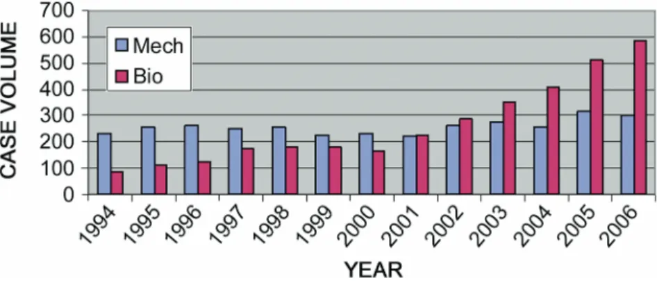
Fig 1. Mechanical aortic valve replacement (blue bars) versus biological aortic valve re-placement (purple bars) for endocarditis (STS National Cardiac Database 2007).
Fig 2. Mitral valve repair (blue bars) versus mitral valve replacement (purple bars) for endocarditis (STS National Cardiac Database 2007).
valve endocarditis. (Class I, Level of evi-dence B)
2. Mechanical or stented tissue valves can be useful in native tricuspid valve endocarditis, if the valve cannot be repaired. (Class IIa, Level of evidence C)
When surgery is indicated for persistent sepsis and severe tricuspid regurgitation, tricuspid valve repair is the treatment of choice. Replacement involves implant-ing prosthetic material in the settimplant-ing of ongoimplant-ing infection, with the risk of reinfection and the need for anticoagu-lation therapy if a mechanical valve is implanted [27]. Thrombus and pannus formation is more frequent, while structural valve degeneration is less extensive in the tricuspid position. These issues are enhanced in the intravenous drug use population in whom right-sided endocarditis is more frequent. This population is younger than the left-sided endocarditis population and more likely not to comply with the anticoagulation regimen.
The feasibility of tricuspid valve repair is based on the extent of the infection and the degree of destruction of the subvalvular apparatus. When two or three leaflets are entirely involved or more than half of the marginal chords of the anterior leaflet are involved, the repair is compromised [38].
When the tricuspid valve cannot be repaired, the choice of prosthesis should follow the same algorithm used for patients without IE. Some authors, however, prefer bioprosthetic valves over mechanical valves owing to concerns about thromboembolic complications [27]. A particular challenge is represented by intravenous drug use patients. In the presence of intravenous drug use, more tissue valves are implanted because of anticipated noncompliance with anticoagulation therapy. Thus, the rate of reoperation for this group is higher. However, the only predictor for poor long-term survival was age.
Tricuspid valve excision without prosthetic valve replace-ment is another option. Valvulectomy may be the appro-priate choice for intractable, extensive endocarditis due to drug addiction. The second-stage operation can be per-formed after controlling drug dependence but only in the absence of pulmonary hypertension and left-sided failure.
V) Multiple Valve Endocarditis
1. In the presence of multiple valve endocarditis involving the aortic valve, the decision to choose a homograft for the aortic valve should follow the same algorithm outlined for isolated aortic valve endocarditis. (Class I, Level of evidence C) 2. In the presence of concomitant aortic or mitral or
tricuspid valve endocarditis, in the aortic, mitral, and tricuspid positions, either a stented tissue or mechanical valve can be implanted. The choice of valve should follow the same algorithm outlined independently for aortic, mitral, and tricuspid valve endocarditis. (Class I, Level of evidence B) 3. When surgery of the mitral and tricuspid valves is
indicated for multiple valve endocarditis, it can be
beneficial to perform mitral and tricuspid valve repair whenever feasible. (Class IIa, Level of evi-dence B)
Ten percent to 25% of all patients with infective endo-carditis require multiple valvular procedures, which usu-ally involve the mitral and aortic valves [39, 40]. Use of either tissue or mechanical valves for double valve en-docarditis yielded the same results in terms of survival and freedom from reinfection [9, 10, 13, 39, 40]. Increas-ingly, groups are using mitral valve repair whenever possible [7, 35, 38].
Some authors advise the use of allografts for aortic valve replacement, citing an increased resistance to rein-fection in the first 6 weeks after surgery, especially in patients with aortic root abscess [16, 21, 39]. Aortic valve endocarditis complicated by annular abscess formation and extension into the mitral annulus is usually present when there is involvement of the aortic-mitral junction or the subannular interventricular septum.
References
1. Bonow RO, Carabello BA, Chatterjee K, et al. ACC/AHA 2006 guidelines for the management of patients with valvu-lar heart disease: a report of the American College of Cardiology/American Heart Association Task Force on Prac-tice Guidelines (writing committee to Revise the 1998 guide-lines for the management of patients with valvular heart disease). J Am Coll Cardiol 2006;48:e1–148.
2. Li JS, Sexton DJ, Mick N, et al. Proposed modifications to the Duke criteria for the diagnosis of infective endocarditis. Clin Infect Dis 2000;30:633– 8.
3. American College of Cardiology Foundation and American Heart Association. Methodology manual and policies from the ACCF/AHA Task Force on Practice Guidelines. Ameri-can College of Cardiology Foundation and AmeriAmeri-can Heart Association, Inc, June 2010.
4. Baddour LM, Wilson WR, Baye AS, et al. Infective endocar-ditis: diagnosis, antimicrobial therapy, and management of complications: a statement for healthcare professionals from the Committee on Rheumatic Fever, Endocarditis, and Ka-wasaki Disease, Council on Cardiovascular Disease in the Young, and the Councils on Clinical Cardiology, Stroke, and Cardiovascular Surgery and Anesthesia, American Heart Association. Circulation 2005;111:394 – 434.
5. Angstwurm K, Borges A, Halle E, Schielke E, Einhaupl K, Weber J. Timing the valve replacement in infective endocar-ditis involving the brain. J Neurol 2004;251:1220 – 6. 6. Gammie JS, O’Brien SM, Griffith BP, Peterson ED. Surgical
treatment of mitral valve endocarditis in North America. Ann Thorac Surg 2005;80:2199 –204.
7. Cooper HA, Thompson EC, Laureno R, et al. Subclinical brain embolization in left-sided infective endocarditis: re-sults from the Evaluation by MRI of the Brains Of Patients With Left-Sided Intracardiac Solid Masses (EMBOLISM) pilot study. Circulation 2009;120:585–91.
8. Horstkotte D, Follath F, Gutschik E, et al. Guidelines on prevention, diagnosis and treatment of infective endocardi-tis executive summary: the task force on infective endocar-ditis of the European Society of Cardiology. Eur Heart J 2004;25:267–76.
9. David TE, Gavra G, Feindel CM, Regesta T, Armstrong S, Maganti MD. Surgical treatment of active infective endocar-ditis: a continued challenge. J Thorac Cardiovasc Surg 2007; 133:144 –9.
10. Moon MR, Miller DC, Moore KA, et al. Treatment of endo-carditis with valve replacement: the question of tissue versus mechanical prosthesis. Ann Thorac Surg 2001;71:1164 –71. 11. Delay D, Pellerin M, Carrier M, et al. Immediate and
long-term results of valve replacement for native and pros-thetic valve endocarditis. Ann Thorac Surg 2000;70:1219 –23. 12. Kaiser SP, Melby SJ, Zierer A, et al. Long-term outcomes in valve replacement surgery for infective endocarditis. Ann Thorac Surg 2007;83:30 –5.
13. Tugtekin SM, Alexiou K, Wilbring M, et al. Native infective endocarditis: which determinants of outcome remain after surgical treatment? Clin Res Cardiol 2006;95:72–9.
14. Aagaard J, Andersen PV. Acute endocarditis treated with radical debridement and implantation of mechanical or stented bioprosthetic devices. Ann Thorac Surg 2001;71: 100 – 4.
15. Alexiou C, Langley SM, Stafford H, Haw MP, Livesey SA, Monro JL. Surgical treatment of infective mitral valve endo-carditis: predictors of early and late outcome. J Heart Valve Dis 2000;9:327–34.
16. Grinda JM, Mainardi JL, D’Attellis N, et al. Cryopreserved aortic viable homograft for active aortic endocarditis. Ann Thorac Surg 2005;79:767–71.
17. Yankah AC, Klose H, Petzina R, Musci M, Siniawski H, Hetzer R. Surgical management of acute aortic root endo-carditis with viable homograft: 13-year experience. Eur J Cardiothorac Surg 2002;21:260 –7.
18. Gulbins H, Kilian E, Roth S, Uhlig A, Kreuzer E, Reichart B. Is there an advantage in using homografts in patients with acute infective endocarditis of the aortic valve? J Heart Valve Dis 2002;11:492–7.
19. Baumgartner FJ, Omari BO, Robertson JM, et al. Annular abscesses in surgical endocarditis: anatomic, clinical, and operative features. Ann Thorac Surg 2000;70:442–7. 20. Murashita T, Sugiki H, Kamikubo Y, Yasuda K. Surgical
results for active endocarditis with prosthetic valve replace-ment: impact of culture-negative endocarditis on early and late outcomes. Eur J Cardiothorac Surg 2004;26:1104 –11. 21. Siniawski H, Grauhan O, Hofmann M, et al. Aortic root
abscess and secondary infective mitral valve disease: results of surgical endocarditis treatment. Eur J Cardiothorac Surg 2005;27:434 – 40.
22. Knosalla C, Weng Y, Yankah AC, et al. Surgical treatment of active infective aortic valve endocarditis with associated periannular abscess—11 year results. Eur Heart J 2000;21: 490 –7.
23. El-Hamamsy I, Clark L, Stevens LM, et al. Late outcomes following Freestyle versus homograft aortic root replace-ment: results from a prospective randomized trial. J Am Coll Cardiol 2010;55:368 –76.
24. Guerra JM, Tornos MP, Permanyer-Miralda G, Almirante B, Murtra M, Soler-Soler J. Long-term results of mechanical prostheses for treatment of active infective endocarditis. Heart 2001;86:63– 8.
25. Lytle BW, Sabik JF, Blackstone EH, Svensson LG, Pettersson GB, Cosgrove DM. Reoperative cryopreserved root and ascending aorta replacement for acute aortic prosthetic valve endocarditis. Ann Thorac Surg 2002;74(Suppl):1754 –9. 26. Leyh RG, Knobloch K, Hagl C, et al. Replacement of the
aortic root for acute prosthetic valve endocarditis: prosthetic composite versus aortic allograft root replacement. J Thorac Cardiovasc Surg 2004;127:1416 –20.
27. Carozza A, Renzulli A, De Feo M, et al. Tricuspid repair for infective endocarditis: clinical and echocardiographic re-sults. Tex Heart Inst J 2001;28:96 –101.
28. Muller LC, Chevtchik O, Bonatti JO, Muller S, Fille M, Laufer G. Treatment of destructive aortic valve endocarditis with the Freestyle Aortic Root Bioprosthesis. Ann Thorac Surg 2003;75:453– 6.
29. Musci M, Siniawski H, Knosalla C, et al. Early and mid-term results of the Shelhigh stentless bioprosthesis in patients with active infective endocarditis. Clin Res Cardiol 2006;95: 247–53.
30. de Kerchove L, Vanoverschelde JL, Poncelet A, et al. Recon-structive surgery in active mitral valve endocarditis: feasibil-ity, safety and durability. Eur J Cardiothorac Surg 2007;31: 592–9.
31. Doukas G, Oc M, Alexiou C, Sosnowski AW, Samani NJ, Spyt TJ. Mitral valve repair for active culture positive infec-tive endocarditis. Heart 2006;92:361–3.
32. Feringa HH, Bax JJ, Klein P, et al. Outcome after mitral valve repair for acute and healed infective endocarditis. Eur J Car-diothorac Surg 2006;29:367–73.
33. Iung B, Rousseau-Paziaud J, Cormier B, et al. Contemporary results of mitral valve repair for infective endocarditis. J Am Coll Cardiol 2004;43:386 –92.
34. Ruttmann E, Legit C, Poelzl G, et al. Mitral valve repair provides improved outcome over replacement in active infective endocarditis. J Thorac Cardiovasc Surg 2005;130: 765–71.
35. Zegdi R, Debieche M, Latremouille C, et al. Long-term results of mitral valve repair in active endocarditis. Circula-tion 2005;111:2532– 6.
36. Mihaljevic T, Paul S, Leacche M, et al. Tailored surgical therapy for acute native mitral valve endocarditis. J Heart Valve Dis 2004;13:210 – 6.
37. Feringa HH, Shaw LJ, Poldermans D, et al. Mitral valve repair and replacement in endocarditis: a systematic review of literature. Ann Thorac Surg 2007;83:564 –70.
38. Couetil JP, Argyriadis PG, Shafy A, et al. Partial replacement of the tricuspid valve by mitral homografts in acute endo-carditis. Ann Thorac Surg 2002;73:1808 –12.
39. Gillinov AM, Diaz R, Blackstone EH, et al. Double valve endocarditis. Ann Thorac Surg 2001;71:1874 –9.
40. Mihaljevic T, Byrne JG, Cohn LH, Aranki SF. Long-term results of multivalve surgery for infective multivalve endo-carditis. Eur J Cardiothorac Surg 2001;20:842– 6.
Appendix 1
Class
I. Evidence and/or general agreement that a given proce-dure or treatment is useful and effective
II. Conflicting evidence and/or a divergence of opinion about the usefulness/efficacy of a procedure or treatment a) Weight of evidence/opinion is in favor of usefulness/
efficacy
b) Usefulness/efficacy is less well established by evidence/ opinion
III. Evidence and/or general agreement that the procedure/ treatment is not useful/effective, and in some cases may be harmful
Level of Evidence
A. Data derived from multiple randomized clinical trials or meta-analyses
B. Data derived from a single randomized trial or from nonrandomized studies
C. Consensus opinion of experts, case studies, or standard of care