Prognosis of Hepatocellular Carcinoma: Comparison of
7 Staging Systems in an American Cohort
Jorge A. Marrero, Robert J. Fontana, Ashley Barrat, Frederick Askari, Hari S. Conjeevaram,
Grace L. Su, and Anna S. Lok
Currently there is no consensus which staging system is best in predicting the survival of patients with hepatocellular carcinoma (HCC). The aims of this study were to identify independent predictors of survival and to compare 7 available prognostic staging systems in patients with HCC. A total of 239 consecutive patients with cirrhosis and HCC seen between January 1, 2000, and December 31, 2003, were included. Demographic, laboratory, and tumor characteristics and performance status were determined at diagnosis and before ther-apy. Predictors of survival were identified using the Kaplan–Meir test and the Cox model. Sixty-two percent of patients had hepatitis C, 56% had more than 1 tumor nodule, 24% had portal vein thrombosis, and 29% did not receive any cancer treatment. At the time of censorship, 153 (63%) patients had died. The 1- and 3-year survival of the entire cohort was 58% and 29%, respectively. The independent predictors of survival were performance status (P<.0001), MELD score greater than 10 (Pⴝ.001), portal vein thrombosis (Pⴝ.0001), and tumor diameter greater than 4 cm (Pⴝ.001). Treatment of HCC was related to overall survival. The Barcelona Clinic Liver Cancer (BCLC) staging system had the best indepen-dent predictive power for survival when compared with the other 6 prognostic systems. In conclusion, performance status, tumor extent, liver function, and treatment were indepen-dent predictors of survival mostly in patients with cirrhosis and HCC. The BCLC staging system includes aspects of all of these elements and provided the best prognostic stratifica-tion for our cohort of patients with HCC.(HEPATOLOGY2005;41:707–716.)
H
epatocellular carcinoma (HCC) is the fifth most common tumor worldwide. In the United States, the incidence of HCC has been rising,1and it is the tumor with the largest increase in incidence over the last 12 years.2Furthermore, the overall survival of
patients with HCC has not improved over the last 20 years, with the incidence rate almost equal to the death rate.3It is projected that the increase in incidence of HCC
will continue over the next 20 years in the United States.4
Therefore, it is important to understand the factors that predict survival of patients with HCC.
Clinical staging of cancers provides a guide to assess prognosis and to direct therapeutic interventions. Well-defined, widely accepted prognostic staging systems are available for many solid tumors, including cancer of the colon5 and prostate.6 These staging systems have been
invaluable in designing tumor surveillance programs and in comparing the efficacy of new therapies. Four key fac-tors that may affect the prognosis of patients with HCC have been identified7: (1) tumor stage at diagnosis; (2)
overall health of the patient; (3) hepatic synthetic func-tion; and (4) efficacy of treatment. Several prognostic staging systems have been proposed for HCC (Table 1),8-14and recently there has been much debate regarding
which prognostic staging system is the best. The lack of a consensus on an HCC staging system is in part related to the heterogeneity in diagnostic criteria of HCC when histological confirmation is not available.7,15 Nonhisto-Abbreviations: HCC, hepatocellular carcinoma; BCLC, Barcelona Clinic Liver
Cancer; CT, computed tomography; MRI, magnetic resonance imaging; AFP, al-pha-fetoprotein; UNOS, United Network of Organ Sharing; ALT, aminotransfer-ase; AST, aspartate aminotransferaminotransfer-ase; CTP, Child-Turcotte-Pugh; MELD, model for end-stage liver disease; TNM, tumor node metastasis; CLIP, Cancer of Liver Italian Program; JIS, Japanese integrated system; GRETCH, Groupe d’Etude de Traitement du Carcinoma Hepatocellulaire; CUPI, Chinese university prognostic index; LR, likelihood ratio.
From the Division of Gastroenterology, Department of Internal Medicine, Uni-versity of Michigan, Ann Arbor, MI.
Received October 6, 2004; accepted January 12, 2005.
Supported by National Institutes of Health Grant CA864000 (Great Lakes New England Clinical Epidemiology Center of the Early Detection Research Network) (J. A. M.) and Grant DK064909 (J. A. M.).
Address reprint requests to: Jorge A. Marrero, M.D., M.S., Division of Gastro-enterology, University of Michigan, 3912 Taubman Center, Ann Arbor, MI 48109-0362. E-mail: jmarrero@umich.edu; fax: 734-936-7392.
Copyright © 2005 by the American Association for the Study of Liver Diseases. Published online in Wiley InterScience (www.interscience.wiley.com). DOI 10.1002/hep.20636
Potential conflict of interest: Nothing to report.
logical criteria for diagnosis of HCC were proposed at a European Association for the Study of the Liver confer-ence,7 but these criteria are not adhered to universally.
There is also a lack of standardization regarding the tests needed to determine tumor burden and extent of spread of HCC, which impede accurate staging. The absence of a consensus on a HCC staging system may hinder progress in critical areas of HCC research, such as evalu-ation of biomarkers for early detection of HCC and de-velopment of new therapeutic modalities.
The aims of this study were to identify independent predictors of survival at the time of HCC diagnosis in a single center and to compare the ability of 7 existing HCC staging systems in predicting survival in a cohort of pa-tients with HCC.
Patients and Methods
Patients. Consecutive patients with HCC seen in the Liver Clinics at the University of Michigan Medical Cen-ter were enrolled into a database that was approved by the Institutional Review Board. Data were extracted from the records of patients seen between January 1, 2000, and December 31, 2003. Follow-up was censored on May 31, 2004. Diagnosis of HCC was based on histology in 192 patients and on nonhistological criteria in 52 patients.7
The nonhistological criteria were two imaging studies— computed tomography (CT) or magnetic resonance im-aging (MRI)—showing an arterial enhancing mass greater than 2 cm (n⫽21), or one imaging study (CT or MRI) showing an arterial enhancing mass greater than 2 cm and an alpha-fetoprotein (AFP) greater than 400 ng/mL (n ⫽ 31). A treatment algorithm was followed (Fig. 1) in which all patients were first assessed for resec-tion; if deemed ineligible, liver transplantation was con-sidered if the patient met United Network of Organ Sharing (UNOS) criteria. Patients who were not
candi-dates for surgical therapy received radiofrequency abla-tion if they had no more than 3 tumor nodules and the maximum diameter of each nodule was less than 5 cm. Patients with diffuse or more extensive tumors were con-sidered for intra-arterial chemoembolization if they had preserved liver function and portal vein was patent. Pa-tients who did not qualify for intra-arterial chemoembo-lization were considered for investigational protocols using radiation, systemic chemotherapy, and/or investi-gational therapies after a multispecialty group evaluation. For all patients, demographic information, etiology of liver disease as previously defined,16biochemical data,
he-matological data, assessment of hepatic function based on Child-Turcotte-Pugh(CTP) and model for end-stage liver disease (MELD) score, and performance status were recorded. All data, including staging of the tumors, were determined at the time of HCC diagnosis and before therapy. Presence of underlying cirrhosis was assessed his-tologically (n⫽188) or via clinical and radiological evi-dence of portal hypertension (n ⫽ 56). Available
Table 1. Variables Included in Seven Staging Systems for HCC
Staging
System Hepatic Function Alpha-fetoprotein Performance Status Tumor Staging
Okuda Ascites, albumin, and bilirubin No No Tumor greater or less than 50% of cross-sectional area of liver
TNM No No No Number of nodules, tumor size, presence of portal vein thrombosis, and presence of metastasis
CLIP CTP ⬍400 orⱖ400
ng/mL
No Number of nodules, tumor greater or less than 50% area of liver, and portal vein thrombosis BCLC CTP No Yes Tumor size, number of nodules, and portal vein
thrombosis CUPI Bilirubin, ascites, alkaline phosphatase ⬍500 orⱖ500
ng/mL
Presence of symptoms TNM
JIS CTP No No TNM
GRETCH Bilirubin, alkaline phosphatase ⬍35 orⱖ35g/L Yes Portal vein thrombosis
Fig. 1. Treatment algorithm of patients with hepatocellular carcinoma at the University of Michigan. HTN, hypertension; RFA, radiofrequency ablation; PVT, portal vein thrombosis; TACE, transarterial chemoemboli-zation; RT, radiation therapy.
abdominal CT or MRI scans at the time of diagnosis were reviewed by 2 radiologists; the number and location of nodules, maximum diameter of the largest nodule, and any evidence of portal vein thrombosis were recorded. Extrahepatic metastasis was evaluated via chest CT (n⫽ 209), bone scan (n⫽232), and/or chest X ray (n⫽35). The date of death was determined by the Social Secu-rity Death Index if more than 3 months had elapsed since the last follow-up visit and death did not occur in our hospital or was not reported by the family. The predom-inant cause of death was extracted from the medical record. Death was attributed to tumor progression in pa-tients with a more than 25% increase in size of any tumor nodule or an increase in the number of nodules based on imaging at the time of death compared with the imaging at diagnosis. Death was attributed to hepatic failure in patients who had a more than twofold increase in biliru-bin, international normalized ratio, or development of ascites, variceal hemorrhage, or hepatorenal syndrome at the time of death. If patients had evidence of hepatic failure and tumor progression, the cause of death was considered to be a combination of both.
Statistical Considerations. Continuous data were expressed as the mean ⫾ SD. A univariate analysis to identify predictors of survival at the time of HCC diag-nosis (baseline) was performed using the Kaplan–Meier method of survival function.17The baseline variables
eval-uated were:
1. demographics: age, sex, ethnicity (non-Hispanic white or not);
2. etiology of liver disease (hepatitis C or not); 3. laboratory values: platelet, international
normal-ized ratio, bilirubin, alanine aminotransferase (ALT), aspartate aminotransferase (AST), albu-min, alkaline phosphatase, creatinine, AFP; 4. hepatic function as determined by CTP
classifica-tion and MELD score;
5. tumor characteristics: number of nodules, maxi-mum tumor diameter, portal vein thrombosis, ex-trahepatic metastasis; and
6. Eastern Cooperative Oncology Group perfor-mance status.
For continuous variables, median values were used to determine the cutoff. Variables other than tumor staging with an alpha less than 0.10 were included in a forward Cox proportional regression model18to identify
indepen-dent predictors of survival.
We next set out to determine which staging systems were the best at predicting survival in our cohort of pa-tients with HCC. Tumor staging was performed in 209 patients who had chest CT. This included the following systems: UNOS-modified tumor node metastasis
(TNM), Barcelona Clinic Liver Cancer (BCLC), Cancer of Liver Italian Program (CLIP), Japanese Integrated Sys-tem (JIS), Groupe d’Etude de TraiSys-tement du Carcinoma Hepatocellulaire (GRETCH), Chinese University Prog-nostic Index (CUPI), and the Okuda staging system. The performance of a prognostic system has been shown to be related to homogeneity (small differences in survival among patients in the same stage within each system), discriminatory ability (greater differences in survival among patients in different stages within each system), and monotonicity of gradients (the survival of patients in earlier stages is longer than the survival of patients in more advanced stages within the same system).19To determine
whether each of the staging systems could predict survival, we used the Kaplan–Meier method as the initial analysis. The Cox regression model was then used to calculate the likelihood ratio (LR)2to determine homogeneity.20In
the LR test, we used the ordinary prognostic score rather than using dummy variables.19 The linear trend2was
then used to measure the discriminatory ability of each of the staging systems.21Both the LR2and linear trend2
were also used to measure the monotonicity of gradients of survival, and the degrees of freedom was 1 so that two prognostic systems with different number of stages could be compared. In addition, the results of the Cox regres-sion were expressed using the Akaike information crite-rion, which shows how the explanatory variable (staging systems) affect the dependent variable (survival of HCC)—the lower the Akaike information criterion, the more explanatory it is and the more informative the model is.22Lastly, the independent contribution of each
staging system to overall prediction of survival in the Cox model was evaluated by comparing the LR test in the full model (all systems included) and in a reduced model when one staging system was removed.23 All statistical
analyses were performed using SAS version 8.1 (Cary, NC), and all graphs were created using MedCalc 7.4 (Mariakerke, Belgium).
Results
Patient Characteristics. A total of 244 HCC
pa-tients were seen during the time period. Table 2 shows the demographic, clinical, and tumor information for all pa-tients. The majority of the patients were men (73%) and non-Hispanic white (74%); the mean age was 57 years. Almost all (n⫽239, 98%) met criteria of having cirrho-sis, the most common cause being hepatitis C (62%). One hundred five (43%) patients were CTP class A. One hun-dred thirty-seven (56%) patients had more than 1 tumor nodule, 60 (24%) had portal vein thrombosis, and 13 (5%) had evidence of extrahepatic metastases.
Ninety-three (38%) patients had an AFP level of 20 ng/mL or less. Ten (4%) patients had surgical resection, 51 (21%) underwent liver transplantation, 46 (19%) had radiofre-quency ablation, 23 (9%) had chemoembolization, and 43 (18%) had other therapies. Seventy-one (29%) did not
receive cancer treatment because of advanced tumor stages (51%), hepatic decompensation (21%), and pa-tient refusal (28%). The following analysis is based on the 239 patients with cirrhosis.
Survival. At the time the data were censored, 153 (63%) patients had died. The overall median survival of the entire cohort was 16.4 months (95% CI 12.9-19.8 mo) (Fig. 2A) and the 1- and 3-year probability of survival was 58% and 29%, respectively. The causes of death were tumor progression (n⫽65, 42%), hepatic failure (n⫽ 38, 24%), combined tumor progression/hepatic failure
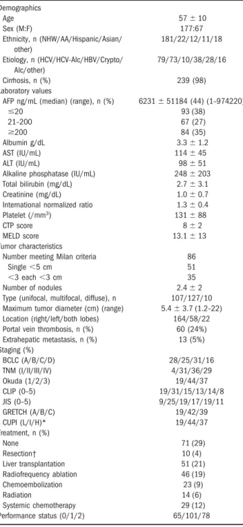
Table 2. Demographic, Clinical, and Tumor Staging Information of 244 Patients With HCC
Demographics Age 57⫾10 Sex (M:F) 177:67 Ethnicity, n (NHW/AA/Hispanic/Asian/ other) 181/22/12/11/18 Etiology, n (HCV/HCV-Alc/HBV/Crypto/ Alc/other) 79/73/10/38/28/16 Cirrhosis, n (%) 239 (98) Laboratory values
AFP ng/mL (median) (range), n (%) 6231⫾51184 (44) (1–974220)
ⱕ20 93 (38) 21–200 67 (27) ⱖ200 84 (35) Albumin g/dL 3.3⫾1.2 AST (IU/mL) 114⫾45 ALT (IU/mL) 98⫾51
Alkaline phosphatase (IU/mL) 248⫾203 Total bilirubin (mg/dL) 2.7⫾3.1 Creatinine (mg/dL) 1.0⫾0.7 International normalized ratio 1.3⫾0.4 Platelet (/mm3) 131⫾88
CTP score 8⫾2
MELD score 13.1⫾13
Tumor characteristics
Number meeting Milan criteria 86
Single⬍5 cm 51
⬍3 each⬍3 cm 35
Number of nodules 2.4⫾2 Type (unifocal, multifocal, diffuse), n 107/127/10 Maximum tumor diameter (cm) (range) 5.4⫾3.7 (1.2–22) Location (right/left/both lobes) 164/58/22 Portal vein thrombosis, n (%) 60 (24%) Extrahepatic metastasis, n (%) 13 (5%) Staging (%) BCLC (A/B/C/D) 28/25/31/16 TNM (I/II/III/IV) 4/31/36/29 Okuda (1/2/3) 19/44/37 CLIP (0–5) 19/31/15/13/14/8 JIS (0–5) 9/25/19/17/19/11 GRETCH (A/B/C) 19/42/39 CUPI (L/I/H)* 19/44/37 Treatment, n (%) None 71 (29) Resection† 10 (4) Liver transplantation 51 (21) Radiofrequency ablation 46 (19) Chemoembolization 23 (9) Radiation 14 (6) Systemic chemotherapy 29 (12) Performance status (0/1/2) 65/101/78
NOTE. Values are the mean⫾SD unless otherwise noted.
Abbreviations: M, male; F, female; NHW, non-Hispanic white; AA, African American; HCV, hepatitis C virus; HCV-Alc, hepatitis C virus⫹alcohol; HBV, hepatitis B virus; Alc, alcohol.
*L, low; I, intermediate; H, high-risk. †Includes 5 patients without cirrhosis.
Fig. 2. Probability of survival (A) in all patients in the entire cohort and (B) according to treatment. Resection/orthotopic liver transplant (. . .. . .), radiofrequency ablation (–—), other treatments such as che-moembolization, radiation and systemic chemotherapy (- - - -), did not undergo treatment (- - - -). RFA, radiofrequency ablation.
(n⫽19, 13%), infections (n⫽19, 13%), and unknown (n⫽12, 8%).
Baseline Predictors of Survival. Univariate analysis showed that AFP, alkaline phosphatase, international normalized ratio, creatinine, MELD score, CTP class,
number of nodules, maximum tumor diameter, portal vein thrombosis, extrahepatic metastasis, and perfor-mance status were significant baseline predictors of sur-vival in patients with HCC (Table 3). Patients who received treatment for HCC had significantly better
sur-Table 3. Univariate Analysis of Baseline Predictors of Survival in 239 Patients With Cirrhosis and HCC
Variables
Number of Patients
Median Survival
(mo) PValue Variables
Number of Patients Median Survival (mo) PValue Age ⬍57 115 16.4 .482 ⱖ57 124 15.2 Sex Male 113 17.1 .933 Female 66 14 Ethnicity Non-Hispanic white 176 16.4 .911 Others 63 22.1 Etiology HCV 151 17.4 .211 Non-HCV 88 18.5 Ascites Present 20 14.7 .318 Absent 219 17.9 AFP ⬍44 119 29.8 ⬍.0001 ⱖ44 120 10.6 AST ⬍110 122 19.4 .357 ⱖ110 117 17.5 ALT ⬍95 125 17.6 .548 ⱖ95 114 18.9 Alkaline phosphatase ⬍220 109 22.5 .010 ⱖ220 130 12.4 International normalized ratio
⬍1.2 153 19.2 .06 ⱖ1.2 86 11.6 Bilirubin ⬍1.5 110 17.6 .283 ⱖ1.5 129 13.3 Creatinine ⬍1.0 148 17.8 .031 ⱖ1.0 91 11.3 Albumin ⬍3.3 118 16.7 .751 ⱖ3.3 121 17.3 Platelet ⬍118 128 16.8 .43 ⱖ118 111 17.9 MELD ⬍10 110 18.5 .020 ⱖ10 129 11.3 CTP class A 100 18.5 .030 B 98 16.5 C 41 10.5 Detected by surveillance Yes 129 17.2 .875 No 110 14.3
NOTE. A dash (—) indicates that the median survival could not be calculated because the last cumulative survival was greater than 50%. Abbreviation: HCV, hepatitis C virus.
*Staging information was available in 209 patients. †L, low; I, intermediate; H, high-risk.
Number of tumor nodules
⬍2 165 18.5 .010
ⱖ2 74 10.1
Maximum tumor diameter (cm)
⬍4 122 33.4 ⬍.0001
ⱖ4 117 7.5
Portal vein thrombosis
Yes 60 5.5 ⬍.0001 No 179 25.4 Extrahepatic metastasis* Yes 13 7.7 .004 No 196 17.5 Performance status 0 63 29.1 ⬍.0001 1 198 16.4 2 78 6 BCLC A 64 — ⬍.0001 B 60 17.1 C 76 9.9 D 39 5.1 TNM I 6 — .0003 II 75 22.5 III 87 16.4 IV 71 5.9 CLIP 0 43 53.1 .001 1 74 16.2 2 36 12.4 3 32 10.8 4 34 3.4 5 20 1.7 Okuda 1 42 18.4 .001 2 107 15.3 3 90 5.4 JIS 0 18 39.8 .0004 1 60 15.8 2 46 17.2 3 42 10.6 4 46 3.3 5 27 1.8 GRETCH A 41 32.3 .003 B 102 17.3 C 96 6.2 CUPI† L 41 20.5 .001 I 108 17.3 H 90 7.8
vival compared with those who did not receive treatment (log rankP⬍ .0001), but there were significant differ-ences between these two groups. Patients who did not receive treatment had significantly more advanced tumors compared with those treated: maximum tumor diameter, 6.9⫾3.6 cm versus 5.1⫾3.4 cm (P ⫽.0001), portal vein thrombosis 46% versus 20% (P ⫽ .0002), and poorer performance status (% Eastern Cooperative On-cology Group 0/1/2: 18/41/41 vs. 31/43/25) (P⫽.01). However, there was no difference with regard to hepatic synthetic function as measured by MELD (P⫽.745) or CTP class (P⫽.132). After controlling for differences in baseline factors and MELD (to also control for hepatic function), a significantly better survival persisted among the patients who received treatment (those treated had a median survival of 13.2 mo vs. 2.8 mo in those untreated;
P⬍.0001). Figure 2B shows the survival according to treatment adjusted for tumor size, portal vein thrombosis, performance status, and MELD score; patients who un-derwent liver transplantation had the best survival. Treat-ment was not included in the multivariate analysis because it is not a variable obtained at diagnosis.
Cox regression analysis identified performance status (P⬍.0001), MELD score (P⫽.001), maximum tumor diameter (P ⫽.001), and portal vein thrombosis (P ⫽
.001) as independent baseline predictors of survival for the entire cohort of HCC patients (Table 4). Performance status of 0 and 1 were protective with hazard ratios of 0.07 (95% CI 0.02-0.16) and 0.46 (95% CI 0.31-0.69), re-spectively.
Staging Systems and Survival. When the seven
prognostic staging systems were analyzed separately using Kaplan–Meier survival analysis (n⫽244), each staging system showed a significant difference in the probability of survival across the different stages (Fig. 3). Figure 3
Table 4. Independent Predictors of Survival
Variables Hazard Ratio (95% CI) PValue All patients (n⫽244) Performance status 0 0.07 (0.02–0.16) ⬍.0001 1 0.46 (0.31–0.69) ⬍.0001 MELD⬎10 1.9 (1.3–2.8) .001 Portal vein thrombosis 2.2 (1.4–3.3) .001 Tumor diameter⬎4 cm 2.4 (1.5–3.9) .001 Nontransplant (n⫽193)* Performance status 0 0.15 (0.03–0.32) .03 1 0.59 (0.38–0.81) .001 MELD⬎10 2.0 (1.4–3.4) .008 Portal vein thrombosis 2.5 (1.3–4.2) ⬍.0001 Tumor diameter⬎4 cm 2.3 (1.4–2.8) .02
*Patients who underwent liver transplantation were not included in the analysis.
Fig. 3. Probability of survival according to (A) UNOS TNM, (B) BCLC, (C) Okuda, (D) CUPI, (E) JIS, (F) CLIP, and (G) GRETCH. UNOS TNM, United Network of Organ Sharing tumor node metastasis; BCLC, Barcelona Clinic Liver Cancer; CUPI, Chinese university prognostic index; JIS, Japanese integrated system; CLIP, Cancer of Liver Italian Program; GRETCH, Groupe d’Etude de Traitement du Carcinoma Hepatocellulaire.
shows that the TNM (stages II and III), JIS (stages 1, 2, and 3), CLIP (stages 1, 2, and 3), and GRETCH (stages B and C) systems had poor stratification of survival at the intermediate stages, while the BCLC, Okuda, and CUPI systems had a better stratification of survival across all stages. The BCLC system had the highest homogeneity (LR 2 76.8), indicating small differences in survival
among patients in the same stages (Table 5). The BCLC classification also had the highest discriminatory score (liner trend228.7) compared with other systems. The
BCLC classification had the best monotonicity of gradi-ent based on the LR2and linear trend2. The Akaike
information criterion was the lowest for the BCLC tem, indicating that the model containing the BCLC sys-tem was the most informative when explaining the survival of HCC patients (see Table 5). Further evidence that the BCLC system provided the best prediction of sur-vival in our cohort was its contribution to the Cox model. The BCLC was the only staging system that had a significant impact on the Cox survival model when it was removed from the model containing all other staging systems (⫺Log likeli-hood⫽903.1; LR242.7;P⬍.0001). Therefore, it was the
only prognostic staging system that had independent predic-tive value on survival in our cohort.
Prediction of Survival in “Non-Transplant” Pa-tients. Liver transplantation can improve survival in
pa-tients with HCC by removing the tumor as well as the underlying cirrhosis. To eliminate the beneficial effects con-ferred by removal of a liver with cirrhosis, predictors of sur-vival were reanalyzed after patients who underwent transplantation were removed from the analysis. The median survival of the 188 patients who did not undergo liver trans-plantation was 11.3 months (95% CI 9.6-15.4) with a mean of 18.8 months; the median survival for the 51 patients who underwent transplantation was more than 50 months, and the mean was 42.8 months, respectively (95% CI, 30-48.9;
P⬍.0001). The 1- and 3-year probability of survival was 48% and 19% for all patients who did not undergo trans-plantation , respectively, and 90% and 74% for those pa-tients who did undergo transplantion, respectively. Cox regression analysis identified the same independent predic-tors of survival in the patients who did not undergo trans-plantion as the entire cohort, but the hazard ratios were slightly different (see Table 4).
Kaplan–Meier analysis of the patients who did not un-dergo transplantation (n⫽193) showed that each staging system— except the JIS— demonstrated significant differ-ences in survival across the different tumor stages (data not shown). The LR2and the linear trend2for the BCLC
system were the highest among the 7 tumor prognostic stag-ing systems for the patients who did not undergo transplan-tation (38.7 and 22.8, respectively) (see Table 5), indicating better homogeneity and discriminatory ability compared with the other systems. The Akaike information criterion was the lowest for the BCLC system, indicating that this system is a more informative model of survival compared with the other systems. The BCLC system had the highest and only significant contribution to the Cox model (⫺Log likelihood⫽468; LR2⫽23.9;P⫽.001) compared to the
other systems in patients who did not undergo tranplanta-tion.
Discussion
Recently there has been much debate regarding which of the existing tumor staging systems has the best prognostic value for HCC. Design of a tumor staging system relies on the identification of individual variables that can predict survival of patients with HCC. In this study, we used data from a large (n⫽239), well-characterized cohort of pa-tients with HCC balanced between early (35% TNM stage I/II) and advanced disease (24% portal vein throm-bosis and 56% multifocal/diffuse tumors), and a substan-tial number of untreated patients (29%) to allow us to study prognostic factors. The extent of tumor (tumor size and portal vein involvement), hepatic function (MELD score), and overall well-being of the patient (performance status)] were independent baseline predictors in our
en-Table 5. Comparison of Prognostic Stratification of Seven HCC Staging Systems Model Discriminatory Ability Linear Trend2 Homogeneity LR2Test Akaike Information Criterion All patients (n⫽244) BCLC 28.7 76.8 943.7 GRETCH 16.3 59.2 970.4 Okuda 11.2 52.9 974.4 CLIP 9.4 51.9 981.5 JIS 8.4 49.7 994.0 TNM 7.2 54.3 978.5 CUPI 9.8 52.3 990.8 With transplantation (n⫽51) BCLC 12.7 23.8 407.1 GRETCH 6.7 10.3 422.5 CUPI 2.9 3.2 427.7 Okuda 2.8 5.3 431.7 CLIP 1.9 2.1 427.3 JIS 0.6 1.8 433.8 TNM 1.1 2.3 425.4 Without transplantation (n⫽193) BCLC 22.8 38.7 534.2 GRETCH 16.2 31.4 549.2 CLIP 11.9 21.3 558.1 TNM 11.1 20.8 560.9 JIS 10.2 16.7 569.4 Okuda 9.8 17.5 566.3 CUPI 8.7 14.3 569.9
tire cohort as well as the subset of patients who did not undergo transplantation. In addition, we also showed that HCC treatment was related to higher overall probability of survival. Therefore, the four key factors affecting HCC prognosis were important in our cohort of patients.
Performance status had been shown to be an indepen-dent predictor of survival in a study on the natural history of untreated HCC and in other solid tumors.24,25Almost
all our patients had underlying cirrhosis, so it is not sur-prising that survival was related to hepatic function. We found that MELD score was a better predictor of survival compared with CTP classification and individual labora-tory tests of hepatic function. Recent studies also found that MELD is a better predictor of survival than CTP classification in patients waiting for a liver transplanta-tion.26 Portal vein thrombosis had been found to be a
poor prognostic variable in multiple studies.27
Micro-scopic and macroMicro-scopic portal vein involvement is one of the major modes of spread of HCC, leading to recurrence after resection28and transplantation.29In addition, portal
venous thrombosis can lead to complications of portal hypertension such as ascites, variceal hemorrhage, and worsening hepatic function in HCC patients.30 Tumor
burden had also been shown to be an important prognos-tic indicator, but the cutoff used in previous studies has varied from more than 5 cm diameter of the largest nod-ule to a tumor involving more than 50% of the liver.31,32
As expected, patients who were eligible for some form of treatment had better survival than those who were too moribund for any treatment. Nevertheless, treatment in general significantly improved survival even after perfor-mance status, MELD score, portal vein thrombosis, and tumor size were controlled for.
Using Kaplan–Meier analysis, we showed that all seven tumor staging systems currently in use for HCC revealed a progressive decrease in survival from the earliest to the most advanced stage. However, the BCLC system was the best at discriminating survival of patients in different stages and had the greatest homogeneity of survival among patients within the same stage. In addition, the BCLC system provided the largest contribution to the Cox model, indicating that it has the best prognostic power for survival compared with the other systems. The superiority of the BCLC system over other tumor staging systems persisted when separate analyses were performed for patients who did not undergo liver transplantation, indicating that it provided better stratification of HCC patients at both intermediate and advanced stages. We believe that the BCLC system had the best prognostica-tion in our cohort because it included the independent predictors of survival we identified: performance status, measure of hepatic function, and tumor stage (size and
portal thrombosis). Although the BCLC system does not include treatment as a variable, it has the advantage of stratifying patients into treatment groups. The superiority of the BCLC system was also demonstrated in a recent study of 187 Italian patients with surgically treated HCC.33
Two staging systems, CUPI and GRETCH (see Table 1), also include performance status, measures of hepatic function, and tumor staging. However, hepatic function was based on bilirubin, presence of ascites, and elevated alkaline phosphatase; the latter has not been shown to be a sensitive marker of liver function. The CUPI system was derived from a cohort of Chinese patients, most of whom had chronic hepatitis B, while the GRETCH system was based on a multicenter French study of patients with al-coholic liver disease. Both CUPI and GRETCH included AFP, which had no prognostic value in our cohort be-cause more than one third of our patients (38%) had an AFP level of less than 20 ng/mL, and only 32% had an AFP level of 500 ng/mL or more. We believe that the Okuda, JIS, and UNOS TNM systems were not predic-tive of survival in our cohort because they included only extent of tumor and a limited assessment of hepatic func-tion.
The CLIP system has been externally validated in Ca-nadian,34Italian,35and Japanese cohorts.19CLIP was
re-cently endorsed by a consensus conference on HCC staging because it was the only staging system externally validated.36 However, one potential limitation of these
validation studies is that the other prognostic systems were not studied. Studies that have evaluated more than three systems have shown an advantage of BCLC33 or
equality of the BCLC, GRETCH, and CLIP systems.37
In our cohort, CLIP was able to discriminate survival of patients with stage 0 from those with stages 4, 5, and 6 (log rankP ⫽ .0001 in Fig. 3). However, it could not differentiate patients with stages 1, 2, and 3, which com-prised 59% of our cohort (poor discriminatory ability). The suboptimal performance of CLIP in our cohort may be related to the inclusion of AFP in the CLIP system. Another limitation of the CLIP system is that treatment decisions often involve overlapping stages. In our cohort, 46% of patients in stage 0, 29% in stage 1, and 42% in stage 2 underwent liver transplantation.
There are several limitations in our study. This is a single-center study, and the results may not be generaliz-able. There may be referral bias, because patients who are moribund may not be referred to a tertiary center. On the other hand, being a tertiary referral center with protocols for investigational treatment, patients with advanced tu-mors are often referred to us as a last resort. Although our treatment algorithm is based on published literature and
recommendations from consensus conference, different centers may have different practice. It is also possible that our results may not apply to patients with HCC in other countries because of differences in demographics, under-lying cause of liver disease, and proportion of patients with cirrhosis. However, the strengths of our study are the complete data in a large number of patients; a full spec-trum of patients with early, intermediate, and advanced tumors at diagnosis; and uniformity with regard to the diagnostic and treatment algorithms. In addition, the ep-idemiological characteristics of our cohort are consistent with that reported in other studies of American patients with HCC.38,39
In conclusion, our study shows that measures of hepatic function (MELD score), performance status, tumor charac-teristics (size and presence of portal vein thrombosis), and the effect of treatment are predictors of survival in cirrhotic pa-tients with HCC. We show that among the seven prognostic staging systems available for HCC, the BCLC system pro-vided the best independent prediction of survival. The supe-rior performance of BCLC may be related to the fact that it includes the same characteristics that had been identified as independent predictive variables in our cohort. Our results should be confirmed in a larger multicenter cohort to study the effect of multiple etiologies, ethnicity, and the effect of various treatments on overall survival. A consensus in prog-nostic staging for HCC is urgently needed to assure progress in the development of biomarkers for early detection and novel therapies.
References
1. El-Serag HB. Hepatocellular carcinoma and hepatitis C in the United States. HEPATOLOGY2002;36:S74-S83.
2. Howe HL, Wingo PA, Thun MJ, Ries LA, Rosenberg HM, Feigal EG, et al. Annual report to the nation on the status of cancer (1973 through 1998), featuring cancers with recent increasing trends. J Natl Cancer Inst 2001;93:824-842.
3. El-Serag HB, Mason AC, Key C. Trends in survival of patients with hep-atocellular carcinoma between 1977 and 1996 in the United States. HEPA
-TOLOGY2001;33:62-65.
4. Tanaka Y, Hanada K, Mizokami M, Yeo AE, Shih JW, Gojobori T, et al. A comparison of the molecular clock of hepatitis C virus in the United States and Japan predicts that hepatocellular carcinoma incidence in the United States will increase over the next two decades. Proc Natl Acad Sci U S A 2002;99:15584-15589.
5. Compton CC, Fielding LP, Burgart LJ, Conley B, Cooper HS, Hamilton SR, et al. Prognostic factors in colorectal cancer. College of American Pathologists Consensus Statement 1999. Arch Pathol Lab Med 2000;124:979-994. 6. Gleason DF, Veterans Administration Cooperative Urological Research
Group. Histologic grading and staging of prostatic carcinoma. In: Tannen-baum M, ed. Urologic Pathology: The Prostate. Philadelphia: Lea & Fe-biger, 1977:171-187.
7. Bruix J, Sherman M, Llovet JM, Beaugrand M, Lencioni R, Burroughs AK, et al. Clinical management of hepatocellular carcinoma. Conclusions of the Barcelona-2000 EASL Conference. J Hepatol 2001;35:421-430. 8. Llovet JM, Bru C, Bruix J. Prognosis of hepatocellular carcinoma: the
BCLC staging classification. Semin Liver Dis 1999;19:329-338.
9. The Cancer of the Liver Italian Program (CLIP) Investigators. Prospec-tive validation of the CLIP score: a new prognostic system for patients with cirrhosis and hepatocellular carcinoma. HEPATOLOGY2000;31:
840-845.
10. Leung TW, Tang AM, Zee B, Lau WY, Lai PB, Leung KL, et al. Con-struction of the Chinese University Prognostic Index for hepatocellular carcinoma and comparison with the TNM staging system, the Okuda staging system, and the Cancer of the Liver Italian Program staging system: a study based on 926 patients. Cancer 2002;94:1760-1769.
11. Kudo M, Chung H, Osaki Y. Prognostic staging system for hepatocellular carcinoma (CLIP score): its value and limitations, and a proposal for a new staging system, the Japan Integrated Staging score (JIS score). J Gastroen-terol 2003;38:207-215.
12. Okuda K, Ohtsuki T, Obata H, Tomimatsu M, Okazaki N, Hasegawa H, et al. Natural history of hepatocellular carcinoma and prognosis in relation to treatment. Study of 850 patients. Cancer 1985;56:918-928. 13. Chevret S, Trinchet JC, Mathieu D, Rached AA, Beaugrand M, Chastang
C, for the Groupe d’Etude et de Traitement du Carcinome He´patocellu-laire. A new prognostic classification for predicting survival in patients with hepatocellular carcinoma. J Hepatol 1999;31:133-141.
14. UNOS/OPTN policy 3.6.4.4. Available at http://www.optn.org. Accessed May 20, 2004.
15. Wildi S, Pestalozzi BC, McCormack L, Clavien PA. Critical evaluation of the different staging systems for hepatocellular carcinoma. Br J Surg 2004; 91:400-408.
16. Marrero JA, Su GL, Wei W, Emick D, Conjeevaram HS, Fontana RJ, et al. Des-gamma carboxyprothrombin can differentiate hepatocellular carci-noma from non-malignant chronic liver disease in American patients. HEPATOLOGY2003;37:1114-1121.
17. Kaplan E, Meier P. Nonparametric estimation from incomplete observa-tions. J Am Stat Assoc 1958;53:457-481.
18. Cox DR. Regression models and life tables. J R Stat Soc 1974;34:187-220.
19. Ueno S, Tanabe G, Sako K, Hiwaki T, Hokotate H, Fukukura Y, et al. Discrimination value of the new western prognostic system (CLIP score) for hepatocellular carcinoma in 662 Japanese patients. HEPATOLOGY2001; 34:529-534.
20. Hosmer DW, Hosmer T, Le Cessie S, Lemeshow S. A comparison of good-ness-of-fit tests for the logistic regression model. Stat Med 1997;16:965-980. 21. Feinsten AR. Clinical biostatistics XVI. The process of prognostic
stratifi-cation. Clin Pharmacol Ther 1972;13:609-624.
22. Foster MR. Key concepts in model selection: performance and generaliz-ability. J Math Psychol 2000;44:205-231.
23. Parzen M, Lipsitz SR. A global goodness-of-fit statistic for Cox regression models. Biometrics 1999;55:580-584.
24. Llovet JM, Bustamante J, Castells A, Vilana R, Ayuso MC, Sala M, et al. Natural history of untreated nonsurgical hepatocellular carcinoma: ratio-nale for the design and evaluation of therapeutic trials. HEPATOLOGY1999; 29:62-67.
25. Buccheri G, Ferrigno D, Tamburini M. Karnofsky and ECOG perfor-mance status scoring in lung cancer: a prospective, longitudinal study of 536 patients from a single institution. Eur J Cancer 1996;32:1135-1141.
26. Wiesner R, Edwards E, Freeman R, Harper A, Kim R, Kamath P, et al. Model for end-stage liver disease (MELD) and allocation of donor livers. Gastroenterology 2003;124:91-96.
27. Fong Y, Sun RL, Jarnagin W, Blumgart LH. An analysis of 412 cases of hepatocellular carcinoma at a Western center. Ann Surg 1999;229:790-799.
28. Poon RT, Fan ST, Lo CM, Liu CL, Wong J. Intrahepatic recurrence after curative resection of hepatocellular carcinoma: long-term results of treat-ment and prognostic factors. Ann Surg 1999;229:216-222.
29. Shetty K, Timmins K, Brensinger C, Furth EE, Rattan S, Sun W, et al. Liver transplantation for hepatocellular carcinoma validation of present selection criteria in predicting outcome. Liver Transpl 2004;10:911-918.
30. Jiang ZB, Shan H, Shen XY, Huang MS, Li ZR, Zhu KS, et al. Transjugu-lar intrahepatic portosystemic shunt for palliative treatment of portal hy-pertension secondary to portal vein tumor thrombosis. World J Gastroenterol 2004;10:1881-1884.
31. Schoniger-Hekele M, Muller C, Kutilek M, Oesterreicher C, Ferenci P, Gangl A. Hepatocellular carcinoma in central Europe: prognostic features and survival. Gut 2001;48:103-109.
32. Calvet X, Bruix J, Gines P, Bru C, Sole M, Vilana R, et al. Prognostic factors of hepatocellular carcinoma in the west: a multivariate analysis in 206 patients. HEPATOLOGY1990;12:753-760.
33. Cillo U, Bassanellos M, Vitale A, Grigoletto FA, Burra P, Fagiuoli S, et al. The critical issue of hepatocellular carcinoma prognostic classification: which is the best tool available? J Hepatol 2004;40:124-131.
34. Levy I, Sherman M. Staging of hepatocellular carcinoma: assessment of the CLIP, Okuda and Child-Pugh staging systems in a cohort of 257 patients in Toronto. Gut 2002;50:881-885.
35. Farinati F, Rinaldi M, Gianni S, Naccarato R. How should patients with hepatocellular carcinoma be staged? Validation of a new prognostic system. Cancer 2000;89:2266-2273.
36. Henderson JM, Sherman M, Tavill A, Abecassis M, Chejfec G, Gramlich T. AHPBA/AJCC consensus conference on staging of hepatocellular car-cinoma: consensus statement. HPB Surg 2003;5:243-250.
37. Giannini E, Risso D, Botta F, Romagnoli P, Malfatti F, Fumagalli A, et al. Prognosis of hepatocellular carcinoma in anti-HCV positive cirrhotic pa-tients: a single-centre comparison amongst four different staging systems. J Intern Med 2004;255:399-408.
38. El-Serag HB. Epidemiology of hepatocellular carcinoma. Clin Liver Dis 2001;5:87-107.
39. Sarbah SA, Gramlich T, Younoszai A, Osmack P, Goormastic M, Grosso L, et al. Risk factors for hepatocellular carcinoma in patients with cirrhosis. Dig Dis Sci 2004;49:850-853.