Pubertal Development and Fertility in Survivors of Childhood Acute Myeloid
Leukemia Treated With Chemotherapy Only: A NOPHO-AML Study
Lene Molgaard-Hansen,
MD,
1Anne-Sofie Skou,
MD,
1Anders Juul,
MD,
2Heidi Glosli,
MD,
3Kirsi Jahnukainen,
MD,
4Marianne Jarfelt,
MD,
5Guðmundur K. J
onmundsson,
MD,
6Johan Malmros,
MD,
7,8Karsten Nysom,
MD,
9Henrik Hasle,
MD,
1* and On behalf of the Nordic Society of Pediatric Hematology and Oncology (NOPHO)
INTRODUCTION
Remarkable progress has been made in the treatment of children with acute myeloid leukemia (AML) over the past decades, and today more than 60% of AML patients become long-term survivors [1]. With increasing long-term survival rates, fertility and pregnancy outcomes have become important issues for AML survivors. Disturbances of pubertal development and impaired fertility have been described in childhood cancer survivors treated with alkylating agent chemotherapy, irradiation, and following hematopoietic stem cell transplantation (HSCT) [2–6]. Previous studies of AML survivors showed that 13–25% had gonadal dysfunction [5–9], however, the endocrinological late effects have mainly been reported in patients treated with HSCT. The previous studies included only few AML survivors treated without HSCT and had considerable treatment heterogeneity limiting the statistical power of reported findings.
The chemotherapy for patients with AML is intensive, based predominantly on anthracyclines and cytarabine, and little is known about the pubertal development and fertility in long-term AML survivors treated with chemotherapy only [10]. The objectives of the present study were to evaluate the pubertal development and fertility in AML survivors and compare it with that of their siblings.
PATIENTS AND METHODS
Eligibility
The first Nordic Society of Pediatric Hematology and Oncology (NOPHO)-AML Study opened in July 1984 including all children diagnosed with AML in the Nordic countries (Denmark, Finland, Iceland, Norway, and Sweden). The enrollment is population-based for patients younger than 15 years of age, and according to local practice for those 15–18-year olds. Patients diagnosed from July 1, 1984 to December 31, 2003 were identified in the database. All
patients completing the treatment according to the NOPHO-AML 84, 88, or 93 protocols and alive by June 30, 2007 were included in the study. We excluded patients with myeloid leukemia of Down syndrome, Fanconi anemia, Kostmann syndrome, preceding myelodysplastic syndrome, therapy-related AML, patients receiv-ing allogeneic in first complete remission (n¼102, 22%) or autologous HSCT (n¼48, 10%), and patients who experienced a relapse or had a secondary malignancy. One patient with non-mosaic Turner syndrome (45,X) as well as her sibling were excluded from the present results, due to the inherent gonadal problems associated with Turner syndrome. A total of 137 patients Background.More than 60% of children with acute myeloid
leukemia (AML) become long-term survivors. Most are cured using chemotherapy without hematopoietic stem cell transplantation (HSCT). We report on pubertal development and compare self-reported parenthood among AML survivors and their siblings.
Procedure.We included 137 children treated for AML according to the Nordic Society of Pediatric Hematology and Oncology (NOPHO)-AML-84, -88, and -93 trials, who were alive by June 2007. Patients with relapse or treated with HSCT were excluded. AML survivors participated in a physical and biochemical examination (n¼102) and completed a questionnaire (n¼101). One of their siblings completed an identical questionnaire (n¼84).Results.At a median follow-up of 11 years (range 5–25) after diagnosis of AML the survivors (median age 16 years, range 5–36) were either prepubertal
or had entered puberty normally. Serum levels of FSH, LH, testosterone, estradiol, sex hormone binding globulin (SHBG), inhibin A and B, and testicular volumes were within normal ranges. Anti-Mu¨llerian hormone (AMH) levels were decreased in 5 of 40 postpubertal females. Mean reported age at menarche was 13.1 (range 11–17) years. Among survivors 15 years of age or older 31% of females reported pregnancies and 9% of males reported pregnancies in their partners, rates comparable with the frequency reported by their siblings. Conclusions. Most AML survivors treated with chemotherapy had normal pubertal development and fertility, however, AMH levels were decreased in 13% of postpubertal females. Longer follow-up is necessary to evaluate possible risk of premature ovarian failure. Pediatr Blood Cancer 2013;60:1988– 1995. #2013 Wiley Periodicals, Inc.
Key words: acute myeloid leukemia; children; fertility; late effects; premature ovarian failure; puberty
1Department of Pediatrics, Aarhus University Hospital Skejby, Aarhus, Denmark;2Department of Growth and Reproduction, Faculty of Health and Medical Sciences, University of Copenhagen, Copenhagen, Denmark; 3Department of Pediatrics, University Hospital Oslo, Rikshospitalet, Oslo, Norway;4Children’s Hospital, Helsinki Univer-sity Central Hospital, Helsinki, Finland; 5Department of Pediatric Oncology, The Queen Silvia Children’s Hospital, Gothenburg, Sweden; 6Department of Pediatrics, Landspitalinn University Hospital, Reykja-vik, Iceland; 7Pediatric Oncology Unit, Astrid Lindgren Children’s Hospital, Stockholm, Sweden;8Department of Women’s and Children’s Health, Karolinska Institutet, Stockholm, Sweden; 9Department of Pediatrics and Adolescent Medicine, University Hospital Rigshospi-talet, Copenhagen, Denmark
Grant sponsor: Danish Cancer Society; Grant sponsor: The Danish Childhood Cancer Foundation; Grant sponsor: The Karen Elise Jensen Foundation; Grant sponsor: The Swedish Childhood Cancer Foundation
Conflict of interest: Nothing to declare.
Correspondence to: Henrik Hasle, Department of Pediatrics, Aarhus University Hospital Skejby, DK-8200 Aarhus, Denmark.
E-mail: hasle@dadlnet.dk
Received 9 November 2012; Accepted 8 July 2013 C 2013 Wiley Periodicals, Inc.
DOI 10.1002/pbc.24715
Published online 23 August 2013 in Wiley Online Library (wileyonlinelibrary.com).
fulfilled the inclusion criteria. The patients were diagnosed in 21 hospitals in Denmark (n¼33), Finland (n¼27), Iceland (n¼2), Norway (n¼33), and Sweden (n¼42).
One sibling of each survivor was invited as control for the questionnaire part of the study whenever available. If the survivor had several siblings, the one closest in age was chosen.
Follow-Up Procedures
Eligible AML survivors were invited to participate in the NOPHO-AML Late Effect Study which has been described in more details previously [11].
Questionnaire
Participants in the study completed a questionnaire with information about pregnancy (either their own for females, or those of female partners for male AML survivors), duration and outcome of pregnancy, and for live births, the birth weight, and any health problems in the children. One sibling of each AML survivor was asked to complete an identical questionnaire. The siblings had no clinical evaluation or blood samples performed.
Clinical Examination
AML survivors had a clinical examination performed at the treating department. The pubertal development was assessed according to the Tanner criteria (stages B1-B5, G1-G5, and PH1-PH5). In males testicular volume was estimated by using a Prader orchidometer. If the testes were not equal in size, the largest was chosen to determine testicular volume.
Biochemical Evaluation
Blood samples were drawn from an antecubital vein between 8 a. m. and 12 a.m. The sample was clotted, centrifuged, and serum was shipped by courier for storage within 24 hours from sampling at 80˚C until hormone analyses were performed. All samples were analyzed after maximum 3 years of storage at the same laboratory using the same assays. Serum follicle stimulating hormone (FSH) and luteinizing hormone (LH) were measured by time-resolved immunofluorometric assays (Delfia; PerkinElmer, Boston, MA) with detection limits of 0.06 and 0.05 IU/L for FSH and LH, respectively. Intra- and interassay coefficients of variation (CV) were less than 5% in both gonadotropin assays. Testosterone was measured by RIA (DPC, Coat-A-Count, Los Angeles, CA) with detection limit of 0.23 nmol/L, and intra- and interassay CVs of less than 17%. Estradiol was measured by RIA (Pantex, Santa Monica, CA). The detection limit was 18 pmol/L, the intra- and interassay CV were less than 8% and 13%, respectively. Sex hormone binding globulin (SHBG) was determined by a time-resolved immunofluo-rescence assay (Delfia, Wallac Oy, Turku, Finland) with a detection limit of 0.23 nmol/L. Intra- and interassay CV were 5.8% and 6.4%, respectively. Inhibin A and B levels were measured in double-antibody immunoenzymetric assays (Beckman Coulter Ltd., High Wycombe, UK and Serotec, Kidlington, UK). The inhibin A and B assays had detection limits of 12 and 20 pg/mL, respectively, and the intra- and interassay CV were less than 16% in both assays. Anti-Mu¨llerian hormone (AMH) levels were determined using the Immunotech Coulter (Immunotech, Beckman Coulter Ltd.) enzyme immunometric assay with detection limit 2 pmol/L. Intra-assay CV
were less than 7.8%, 5.4%, and 6.4% at 13, 123, and 231 pmol/L, respectively. Interassay CV were less than 11.6%, 10.9% and 9.1% at 19, 99, and 209 pmol/L.
We used previously published age-related reference values from the same laboratory for FSH, LH, testosterone, estradiol, SHBG, inhibin A and B, and AMH for healthy females [12–14] and males [15–17]. AMH standard deviation scores (SDS) were calculated using previously published reference values [14]. Hormone analyses were performed in 102 (74%) of 137 AML survivors (participants). Nineteen (34%) of 56 female participants reported taking hormonal contraceptives at the time of blood sampling. None of the males received testosterone substitution at the time of blood sampling.
NOPHO-AML-84/88/93 Treatment
The NOPHO-AML-84, -88, and -93 protocols included cytarabine (cumulative dose 49.6–61.3 g/m2), anthracycline (cu-mulative dose of daunorubicin equivalents 225–450 mg/m2), 6-thioguanine (cumulative dose 800–2,400 mg/m2), and etoposide (0–1,600 mg/m2). Intrathecal methotrexate was given with each course (6 or 7 doses). None of the patients received alkylating agents. Details about the treatment elements and clinical outcome have been reported previously [18].
Statistics
The eligible survivors were classified asrespondentsor non-respondentsdepending on whether they completed the question-naire or not. AML survivors were classified asparticipantsor non-participantsdepending on whether they had the blood sampling and hormone analysis performed or not. Subgroups of patients were compared by Chi-square or Fisher’s exact tests. Age is presented as median with range.
Ethics
The study was approved by the national ethics committees according to national regulations. Written informed consent was obtained from the AML survivors and/or parents/guardians. For siblings, a returned questionnaire was considered as written informed consent.
RESULTS
Clinical examination and blood sampling including hormone analyses were performed in 102 (74%) and the questionnaire was completed by 101 (74%) of the 137 AML survivors (Fig. 1). Three survivors participated in blood sampling and clinical examination but did not complete the questionnaire. Two survivors completed the questionnaire but had no hormonal analyses performed. Of the 137 eligible patients 35 (26%) did not participate in the clinical examination for the following reasons: 21 declined participation, 8 never responded, 5 had moved abroad, and 1 was lost to follow-up. Eight of the 101 AML survivors completing the questionnaire had no siblings. The questionnaire was completed by 84 (90%) of the 93 eligible siblings.
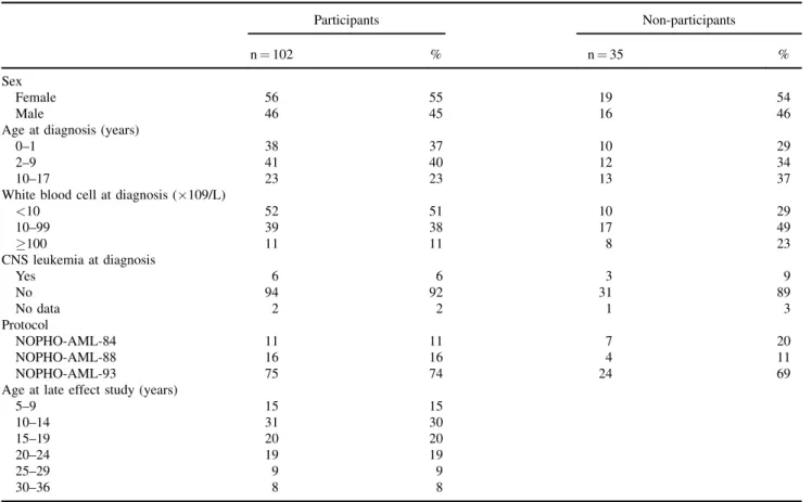
No difference was found concerning sex, age at diagnosis, disease- or treatment-related characteristics when comparing the participants and the non-participants (Table I). The AML survivors did not differ from their siblings in sex and age (Table II) or with
regard to education, employment, or general health status [11]. The median follow-up was 10.7 (range 4.4–25.0) years after diagnosis.
Female AML Survivors
Reported menarche, medication, pregnancies, and off-spring. Sixteen survivors in the age range 5.7–12.8 years were premenarchal at blood sampling. Forty of 56 female respondents reported having had menarche, and they all still had periods. Mean reported age at menarche was 13.1 years [11–17] (n¼39). Menarche occurred within the normal age range (10–16 years) for all postmenarchal survivors, except one girl who had her menarche by the age of 17 years. Except hormonal contraceptives no responders received hormone substitution.
Among 32 female AML survivors>15 years of age (median age 23.7 years, range 15.1–36.6), one had tried for>1 year to become pregnant without success due to impaired spermatogenesis in her partner. Twenty pregnancies were reported in 10 survivors; 13 (65%) live births, no stillbirths, 6 (30%) spontaneous abortions, and 1 (5%) induced abortion. The live-borns were all at term and their birth weights were within the normal range. No offspring had congenital anomalies except two siblings with fragile X syndrome and one child has hemophilia A.
No siblings experienced unwanted childlessness for>1 year. Eight pregnancies were reported in three sisters of AML survivors with four (50%) live births, no stillbirths, three (38%) spontaneous abortions, and one (13%) induced abortion. Ten of 32 (31%) female AML survivors>15 years of age had been pregnant compared with 3 (13%) of their 24 siblings or siblings’ partners (15 sisters, 9 brothers) (P¼0.1).
Clinical assessment. Two female participants were younger than 8 years at blood sampling, and both were prepubertal (B1, P1). Another 10 survivors between ages 8.4 and 11.0 years were also prepubertal. The rest of the participants were either in puberty with Tanner stages appropriate for age or had progressed through puberty normally.
Hormonal levels. The median age at blood sampling was 18.5 (range 5.7–36.6) years. Reproductive hormones in female AML survivors are illustrated according to age and use of hormonal contraceptives in relation to the normal ranges (Fig. 2). A 21-year-old female had raised LH-level whereas the other hormones were within normal range. Two survivors had raised levels of SHBG. The median AMH SDS was0.18 (range2.76 to 1.83) and 5 of 40 postpubertal females had a value below the 2.5 percentile. Serum levels of the other hormones were within normal ranges for all females.
Male AML Survivors
Reported medication, pregnancies of partners, and off-spring. One respondent with delayed puberty at the age of 16 years had been treated with testosterone. Among 22 male AML survivors>15 years of age (median age 19.0 years, range 15.1– 33.0), no one had involuntary childlessness for >1 year. Two pregnancies were reported in the partners of two male survivors. Both resulted in live births at term of healthy babies with normal birth weight. One sibling had involuntary childlessness for>1 year. Twelve pregnancies were reported in six siblings or their partners (4 sisters, 2 brothers) with eight live births, and four spontaneous abortions. Among male respondents>15 years of age 2 of 22 (9%) reported pregnancies in their partners. Among their siblings 2 partners of 8 brothers and 4 of 11 sisters had been pregnant (P¼0.17).
Clinical assessment. Five male participants were younger than 9 years at the time of blood sampling, and they were all prepubertal (G1, testis volume<4 ml). Another six survivors aged 9.3–12.6 years were also prepubertal. The rest of the participants were either in puberty with Tanner stage appropriate for age or had progressed through puberty normally. The 10 AML survivors>20 years at the time of assessment all had testicular volume>15 ml (Fig. 3).
Hormonal levels. The median age at blood sampling was 14.9 (range 5.2–33.0) years. Reproductive hormones in male AML survivors are illustrated according to age and in relation to the normal ranges (Fig. 3). An 18-year-old male had raised levels of both FSH and LH, and a slightly reduced testosterone level but normal inhibin B and testes volume (20 ml). A 17-year-old survivor had a raised FSH level and a reduced inhibin B level but normal testes volume (15 ml). Serum levels of the other hormones were within normal ranges for all subjects.
DISCUSSION
The survival rate of pediatric AML patients has improved dramatically during the past two decades, with former patients now achieving young adulthood and beginning to make decisions regarding partnership and reproduction. In our previous report 19% of AML survivors >10 years of age and 26% of the parents had concerns about the effects of chemotherapy on fertility [11].
Fig. 1. Flowchart of the patients from the NOPHO-AML-84, -88, -93
trials included in the AML late effect study. Vital status was assessed on June 30, 2007. One participant with Turner syndrome was excluded from this study of puberty and fertility. CR1, first complete remission; HSCT, hematopoietic stem cell transplantation; TS, Turner syndrome.
Our results on the pubertal development and fertility in a large unselected series of long-term AML survivors treated with chemotherapy only are reassuring. Both female and male AML survivors were either prepubertal, were in puberty or had progressed through puberty normally. Menarche occurred within the normal age range in 98%. Among survivors >15 years of age, 31% of females and 9% of males reported a frequency of pregnancies which
was comparable with the proportion reported by their siblings. Both female and male AML survivors had reproductive hormones within the normal range. However, the blood samples were taken randomly during menstrual cycle, although there was little variation according to cycle days it may limit the evaluation of the results.
Pubertal development and fertility has been evaluated in four smaller studies including 12–43 AML survivors treated with
TABLE I. Characteristics of AML Survivors Who Participated in the Clinical Examination and Hormone Analyses Versus Those Who Did Not Participate
Participants Non-participants
n¼102 % n¼35 %
Sex
Female 56 55 19 54
Male 46 45 16 46
Age at diagnosis (years)
0–1 38 37 10 29
2–9 41 40 12 34
10–17 23 23 13 37
White blood cell at diagnosis (109/L)
<10 52 51 10 29 10–99 39 38 17 49 100 11 11 8 23 CNS leukemia at diagnosis Yes 6 6 3 9 No 94 92 31 89 No data 2 2 1 3 Protocol NOPHO-AML-84 11 11 7 20 NOPHO-AML-88 16 16 4 11 NOPHO-AML-93 75 74 24 69
Age at late effect study (years)
5–9 15 15 10–14 31 30 15–19 20 20 20–24 19 19 25–29 9 9 30–36 8 8
TABLE II. Sex and Age of the AML Survivors and Their Siblings Completing the Questionnaire Study
AML survivors Siblings
n¼101 % n¼84 %
Sex
Female 56 55 51 61
Male 45 45 33 39
Age at late effect study (years)
0–4 0 0 4 5 5–9 16 16 10 12 10–14 31 31 26 31 15–19 19 19 20 24 20–24 18 18 6 7 25–29 9 9 12 14 30–34 6 6 5 6 35–42 2 0 1 1 Median (range) 15.8 (5.2–36.6) 15.4 (2.0–42.2)
Fig. 2. Serum FSH, LH, testosterone, estradiol, SHBG, inhibin A, inhibin B, and AMH levels according to chronological age in female AML survivors (n¼56). Except for AMH, the lines represent meanþ2 SD in healthy females. For AMH, the lines represent median, 2.5 and 97.5 percentiles., no hormonal contraceptives (n¼37);, taking hormonal contraceptives (n¼14); x, during the one weak break of hormonal contraceptive (n¼5); FSH, follicle stimulating hormone; LH, luteinizing hormone; SHBG, sex hormone binding globulin; AMH, anti-Mu¨llerian hormone; SD, standard deviation.
Fig. 3. Serum FSH, LH, testosterone, estradiol, SHBG, inhibin B, AMH levels, and testicular volume according to chronological age in male AML survivors (n¼46). Except for AMH, the lines represent meanþ2 SD in healthy males. For AMH, the lines represent median, 2.5 and 97.5 percentiles. FSH, follicle stimulating hormone; LH, luteinizing hormone; SHBG, sex hormone binding globulin; AMH, anti-Mu¨llerian hormone; SD, standard deviation.
chemotherapy only [5–7,10]. The endocrine evaluations were normal for age based upon self-reported pregnancies, use of hormones, Tanner staging, and gonadotropins and sex hormones in children >9 years of age (n¼12). Ovarian ultrasound was performed in 14 females who had ovarian volume within the normal range [7]. The lack of hypogonadism and infertility in AML survivors in the previous small studies [5–7,10] is further supported by the findings in our study.
Alkylating agents in particular are known to be gonadotoxic [19]. Treatment of childhood AML without HSCT is based upon anthracyclines and cytarabine. The NOPHO-AML protocols include a higher cumulated dose of cytarabine (49 g/m2or higher) than other AML protocols [18]. Little is known about the gonadotoxic effect of cytarabine [19] but the present study indicates that even very high doses of cytarabine have limited gonadotoxic effect. A large study on male fertility after childhood cancer showed that those who received cytarabine were more likely to ever sire a pregnancy than those who were not exposed to cytarabine [2] and exposure to cytarabine did not influence the chance of pregnancy in female survivors [3].
Although females with AML are cured with non-sterilizing regimens, concern remains about shortened reproductive period due to a possibly reduced ovarian follicle pool and premature ovarian failure [20]. Serum AMH level is the most reliable marker to determine the ovarian reserve and predict the onset of meno-pause [21,22]. A decrease in AMH levels seems to precede changes in conventional parameters associated with peri-menopausal status, such as FSH, inhibin B, and estradiol [23]. AML survivors who received total body irradiation as part of the conditioning regimen for HSCT had undetectable AMH levels [24]. We found that 35 of 40 postmenarchal female AML survivors treated with chemothera-py had AMH levels within the normal range suggesting that previous chemotherapy for childhood AML does not have major impact on ovarian reserve. However, 5 of 40 (13%) postpubertal women had low AMH levels, which could suggest a diminished ovarian follicle pool despite FSH levels being normal. Whether or not the fertile period is compromised remains to be seen.
Inhibin B secretion in adult men requires the presence of intact germ cells, and is the best serum marker of spermatogenesis [25,26]. In our study, only 2 of 46 male AML survivors had either an elevated gonadotropin level combined with a reduced testosterone level or a reduced level of inhibin B but both had testes of normal size. Testicular volume is a sensitive indicator of spermatogenesis in long-term survivors of childhood cancer [27], but since semen analysis was not performed in our study sperm production of the survivors remains unknown.
The frequency of chromosomal abnormalities, single gene defects, and congenital malformations is not increased among the offspring of survivors of childhood cancer [28]. This is in accordance with our findings, where no increased rate of spontaneous abortions, congenital malformations, or other diseases in offspring was observed in AML survivors compared with their siblings.
Although our study included all childhood AML survivors from the five Nordic countries over a 20-year period, the participants were rather few and young, with only 35% being 20 years of age or older. The follow-up was too short to exclude late reduced fertility. Some results were based upon self-reporting, and for instance all subjects may not remember age at menarche accurately. Incomplete participation might influence the study results, but we found no significant differences between respondents and non-respondents.
Our study has several strengths: it is the largest study of AML survivors treated with chemotherapy only, treatment was uniform, the participation rate was high, all participants had hormonal analyses performed in the same laboratory, and we were able to collect basic background and clinical data on non-respondents from the NOPHO-AML database.
In conclusion, many of the AML survivors treated with chemotherapy only had preserved fertility, as evidenced by the number of pregnancies which was comparable to their siblings, and reproductive hormones within the normal range. Although some AML survivors may have a lower ovarian reserve, most survivors seem to have normal gonadal reserve. The cohort is still young and longer follow-up is needed to evaluate the possible risk of premature decrease in fertility.
ACKNOWLEDGMENTS
We thank the participating AML survivors and their siblings as well as the principal investigators of the NOPHO-AML-84/88/93 studies and investigators of the NOPHO-AML Late Effect Studyin Denmark: S. Rosthøj, Aalborg; C. Rechnitzer, Copenhagen; N. Carlsen; and P. Wehner, Odensein Finland: L. Hovi, Helsinki; M. Perkkio¨, Kuopio; M. Arola, Tampere; and A. Harila-Saari, Ouluin Norway: H. Raeder and D. M. Wojcik, Bergen; B. Lund, Trondheim; E. Stensvold, Ullevaal in Sweden: M. Behrendtz, Linko¨ping; L. Hjorth, Lund; U. Hjalmars, Umea˚; J. Arvidson; and C. Rinaldo, Uppsala. Major financial support was received from the Danish Cancer Society, the Danish Childhood Cancer Foundation, the Karen Elise Jensen Foundation, and the Swedish Childhood Cancer Foundation.
REFERENCES
1. Kaspers GJ, Creutzig U. Pediatric acute myeloid leukemia: International progress and future directions. Leukemia 2005;19:2025–2029.
2. Green DM, Kawashima T, Stovall M, et al. Fertility of male survivors of childhood cancer: A report from the childhood cancer survivor study. J Clin Oncol 2010;28:332–339.
3. Green DM, Kawashima T, Stovall M, et al. Fertility of female survivors of childhood cancer: A report from the childhood cancer survivor study. J Clin Oncol 2009;27:2677–2685.
4. Knopman JM, Papadopoulos EB, Grifo JA, et al. Surviving childhood and reproductive-age malignancy: Effects on fertility and future parenthood. Lancet Oncol 2010;11:490–498.
5. Leung W, Hudson MM, Strickland DK, et al. Late effects of treatment in survivors of childhood acute myeloid leukemia. J Clin Oncol 2000;18:3273–3279.
6. Leahey AM, Teunissen H, Friedman DL, et al. Late effects of chemotherapy compared to bone marrow transplantation in the treatment of pediatric acute myeloid leukemia and myelodysplasia. Med Pediatr Oncol 1999;32:163–169.
7. Liesner RJ, Leiper AD, Hann IM, et al. Late effects of intensive treatment for acute myeloid leukemia and myelodysplasia in childhood. J Clin Oncol 1994;12:916–924.
8. Michel G, Socie G, Gebhard F, et al. Late effects of allogeneic bone marrow transplantation for children with acute myeloblastic leukemia in first complete remission: The impact of conditioning regimen without total-body irradiation—A report from the Societe Francaise de Greffe de Moelle. J Clin Oncol 1997;15:2238–2246.
9. Klusmann JH, Reinhardt D, Zimmermann M, et al. The role of matched sibling donor allogeneic stem cell transplantation in pediatric high-risk acute myeloid leukemia: Results from the AML-BFM 98 study. Haematologica 2012;97:21–29.
10. Blijdorp K, van Waas M, van der Lely AJ, et al. Endocrine sequelae and metabolic syndrome in adult long-term survivors of childhood acute myeloid leukemia. Leuk Res 2013;37:367–371. 11. Molgaard-Hansen L, Glosli H, Jahnukainen K, et al. Quality of health in survivors of childhood acute
myeloid leukemia treated with chemotherapy only: A NOPHO-AML study. Pediatr Blood Cancer 2011;57:1222–1229.
12. Sehested A, Juul AA, Andersson AM, et al. Serum inhibin a and inhibin b in healthy prepubertal, pubertal, and adolescent girls and adult women: Relation to age, stage of puberty, menstrual cycle, follicle-stimulating hormone, luteinizing hormone, and estradiol levels. J Clin Endocrinol Metab 2000;85: 1634–1640.
13. Chellakooty M, Schmidt IM, Haavisto AM, et al. Inhibin a, inhibin b, follicle-stimulating hormone, luteinizing hormone, estradiol, and sex hormone-binding globulin levels in 473 healthy infant girls. J Clin Endocrinol Metab 2003;88:3515–3520.
14. Hagen CP, Aksglaede L, Sorensen K, et al. Serum levels of anti-mullerian hormone as a marker of ovarian function in 926 healthy females from birth to adulthood and in 172 turner syndrome patients. J Clin Endocrinol Metab 2010;95:5003–5010.
15. Andersson AM, Juul A, Petersen JH, et al. Serum inhibin b in healthy pubertal and adolescent boys: Relation to age, stage of puberty, and follicle-stimulating hormone, luteinizing hormone, testosterone, and estradiol levels. J Clin Endocrinol Metab 1997;82:3976–3981.
16. Sorensen K, Aksglaede L, Petersen JH, et al. Recent changes in pubertal timing in healthy Danish boys: Associations with body mass index. J Clin Endocrinol Metab 2010;95:263–270.
17. Aksglaede L, Sorensen K, Boas M, et al. Changes in anti-mullerian hormone (AMH) throughout the life span: A population-based study of 1027 healthy males from birth (cord blood) to the age of 69 years. J Clin Endocrinol Metab 2010;95:5357–5364.
18. Lie SO, Abrahamsson J, Clausen N, et al. Long-term results in children with AML: NOPHO-AML study group—Report of three consecutive trials. Leukemia 2005;19:2090–2100.
19. Brougham MF, Wallace WH. Subfertility in children and young people treated for solid and haematological malignancies. Br J Haematol 2005;131:143–155.
20. Larsen EC, Muller J, Schmiegelow K, et al. Reduced ovarian function in long-term survivors of radiation-and chemotherapy-treated childhood cancer. J Clin Endocrinol Metab 2003;88:5307–5314. 21. Visser JA, de Jong FH, Laven JS, et al. Anti-mullerian hormone: A new marker for ovarian function.
Reproduction 2006;131:1–9.
22. Kelsey TW, Wright P, Nelson SM, et al. A validated model of serum anti-mullerian hormone from conception to menopause. PLoS ONE 2011;6:e22024.
23. de Vet A, Laven JS, de Jong FH, et al. Antimullerian hormone serum levels: A putative marker for ovarian aging. Fertil Steril 2002;77:357–362.
24. Lie Fong S, Laven JS, Hakvoort-Cammel FG, et al. Assessment of ovarian reserve in adult childhood cancer survivors using anti-mullerian hormone. Hum Reprod 2009;24:982–990.
25. van Casteren NJ, van der Linden GH, Hakvoort-Cammel FG, et al. Effect of childhood cancer treatment on fertility markers in adult male long-term survivors. Pediatr Blood Cancer 2009;52:108–112. 26. Jorgensen N, Liu F, Andersson AM, et al. Serum inhibin-b in fertile men is strongly correlated with low
but not high sperm counts: A coordinated study of 1,797 European and US men. Fertil Steril 2010; 94:2128–2134.
27. Jahnukainen K, Heikkinen R, Henriksson M, et al. Semen quality and fertility in adult long-term survivors of childhood acute lymphoblastic leukemia. Fertil Steril 2011;96:837–842.
28. Winther JF, Boice JD Jr, Mulvihill JJ, et al. Chromosomal abnormalities among offspring of childhood-cancer survivors in Denmark: A population-based study. Am J Hum Genet 2004;74:1282–1285.