R E S E A R C H
Open Access
LINC01503/miR-342-3p facilitates
malignancy in non-small-cell lung cancer
cells via regulating LASP1
Qiming Shen, Yanbin Sun and Shun Xu
*Abstract
Background:Non-small cell lung cancer (NSCLC) is one of the major types of lung cancer, which is a prevalent human disease all over the world. LncRNA LINC01503 is a super-enhancer-driven long non-coding RNA that is dysregulated in several types of human cancer. However, its role in NSCLC remains unknown.
Methods:Thirty NSCLC patients were recruited between April 2012 and April 2016. Luciferase reporter assay, qRT-PCR, Cell Counting Kit-8 (CCK-8), Transwell migration assay, RNA pull-down assay, western blotting, 5-ethynyl-29-deoxyuridine (EdU) assays, and flow cytometry were utilized to characterize the roles and relationships among LINC01503, miR-342-3p, and LASP1 in NSCLC. The transplanted mouse model was built to examine their biological functions in vivo.
Results:We demonstrated that the expression of lncRNA LINC01503 and LIM and SH3 domain protein 1 (LASP1) were upregulated and miR-342-3p was downregulated in NSCLC samples and cell lines. Functional experiments revealed that inhibiting the expression of LINC01503 or over-expression of miR-342-3p inhibited NSCLC growth and metastasis both in vitro and in vivo. In addition, LINC01503 could bind to miR-342-3p and affect the expression of LASP1.
Conclusion:These results provide a comprehensive analysis of the roles of LINC01503 as a competing endogenous RNA (ceRNA) in NSCLC progression.
Keywords:LINC01503, miR-342-3p, Non-small-cell lung cancer, LASP1
Background
Non-small cell lung cancer (NSCLC) is one of the major types of lung cancer, which is a quite prevalent human disease all over the world [1]. Extensive efforts have been made to discover new therapeutic targets for the treat-ment of NSCLC [2,3]. However, as the early symptoms of NSCLC are not obvious, a large portion of NSCLC patients was diagnosed at late stages, with high malig-nancy and low treatment opportunities [1, 4]. The current overall 5-year survival rate of NSCLC patients
still remains unsatisfactory [5]. In this study, we focused to understand the pathogenesis of NSCLC, as well as to investigate new therapeutic targets based on the tumori-genesis of NSCLC.
Long noncoding RNAs (lncRNAs, > 200 nt) are a major subtype of non-coding RNAs that cannot translate to proteins [6,7]. A large number of lncRNAs have been discovered to play vital roles in tumorigenesis, with an unignorable impact in many biological procedures such as cell proliferation, cell cycle, cell death, as well as tumor metastasis [8, 9]. Previous studies have revealed the expression profiles of several lncRNAs that partici-pate closely in the tumorigenesis of NSCLC, including
© The Author(s). 2020Open AccessThis article is licensed under a Creative Commons Attribution 4.0 International License, which permits use, sharing, adaptation, distribution and reproduction in any medium or format, as long as you give appropriate credit to the original author(s) and the source, provide a link to the Creative Commons licence, and indicate if changes were made. The images or other third party material in this article are included in the article's Creative Commons licence, unless indicated otherwise in a credit line to the material. If material is not included in the article's Creative Commons licence and your intended use is not permitted by statutory regulation or exceeds the permitted use, you will need to obtain permission directly from the copyright holder. To view a copy of this licence, visithttp://creativecommons.org/licenses/by/4.0/. The Creative Commons Public Domain Dedication waiver (http://creativecommons.org/publicdomain/zero/1.0/) applies to the data made available in this article, unless otherwise stated in a credit line to the data.
* Correspondence:[email protected]
lncRNA PVT1 [10], lncRNA AFAP1-AS1 [11], MEG3 [12], and so on. LncRNA LINC01503 is a super-enhancer-driven lncRNA that is dysregulated in several types of human cancer. For example, it was reported that lncRNA LINC01503, regulated by TP63, is over-expressed and oncogenic in squamous cell carcinoma [13]. However, its role in NSCLC remains unknown.
As another major subtype of non-coding RNA, micro-RNAs (mimicro-RNAs, ~ 20 nt) exert tumor-suppressive func-tions in many types of human cancer [14,15]. For NSCL C, miRNAs are usually working together with specific lncRNAs and down-stream factors to regulate their pro-gression and metastasis [16]. MiR-342-3p was reported to target RAP2B to suppress proliferation and invasion of non-small cell lung cancer cells [17]. One report revealed that miR-342-3p suppresses cell proliferation, migration, and invasion by targeting forkhead box protein M1 (FOXM1) in human cervical cancer. In our preliminary experiments, Starbase predicted that lncRNA LINC01503 could target miR-342-3p. LIM and SH3 domain protein 1 (LASP1) is a potential negative predictor of NSCLC [18]. It has been proved to be the down-stream factor of miR-203 [19] and miR-29a [20] in the suppression or proliferation of human NSCLC. Therefore, this study was carried out to investigate the associations among lncRNA LINC01503, miR-342-3p, and LASP1 in the develop-ment and progression of NSCLC.
Methods
Patients and samples
A total of 30 NSCLC patients were recruited in the First Hospital of China Medical University, P.R. China be-tween April 2012 and April 2016. Their ages ranged from 41 to 69 years old, with an average age of 53 years old. These patients did not receive any treatment before the surgeries. After sections, NSCLC tissues and matched normal tissue samples were frozen in liquid ni-trogen and stored at −80 °C. This study was approved by the Ethics Committee of the First Hospital of China Medical University, P.R. China. All the patients signed written informed consent.
Cells culture
NSCLC cell lines A549, SPC-A1, H1299, H1650, H1975, and PC-9 and normal cell line of 16HBE were provided by Institute of Biochemistry and Cell Biology, Chinese Academy of Sciences. Cells were cultured in Dulbeccos modified Eagles medium (DMEM) supplemented with 10% fetal bovine serum (FBS; Invitrogen), 100 U/mL penicillin and 100 mg/mL streptomycin at 37 °C in a hu-midified incubator with 5% CO2.
Plasmid construction and transfection
MiR-342-3p-mimics, miR-342-3p-inhibitor, miR-NC and anti-miR-NC were provided by RiboBio, China. Sh-LINC01503 and sh-NC were provided by Genepharma, Shanghai. The sequences were: LINC01503, sh-1 target 5′-TCTGACAAGTGTGTACCTA-3′; sh-LINC01503, 5′-AATTCTCCGAACGTGTCACGT-3′. LASP1 cDNA was cloned into pcDNA3.1 (Invitrogen, USA). LASP1-WT (wild-type) or LSAP1-MUT (mutant) had amplifica-tion using PCR, and transfecamplifica-tion to A549 cells with Li-pofectamine 2000 (Invitrogen, USA).
Luciferase reporter assays
LINC01503 and LASP1 wild-type or mutant binding miR-342-3p were cloned to pMIR Basic vector (OBiO Biology, Beijing) to generate pMIR-REPOR-LINC01503-wt or pMIR-REPOR-LINC01503-mt, respectively. After being cultivated in 24-well plates for 24 h, A549 cells were transfected with miR-342-3p mimics or control (GenePharma, China) and co-transfected with pMIR-REPOR-NC or constructed recombinant luciferase vec-tors by Lipofectamine 3000 (Invitrogen, USA). Luciferase activities were detected and compared with Renilla lucif-erase activity, which is an internal control for normalization using dual-luciferase reporter assays 48 h after transfection.
qRT-PCR
RNAs were extracted by TRIzol (Invitrogen, USA). Re-verse transcription of RNAs to cDNA was performed using Reverse Transcription Kit (Tiangen, China). qRT-PCR was performed using SYBR (Takara, Japan) on an ABI7300 Real-Time PCR System (Applied Biosystems, USA). U6 and GAPDH were used as endogenous con-trol. The expression levels were calculated using the 2-△△Ctmethod [21]. The primers used were:
LINC01503, 5′-GGGACGGAGACAAATGACGG-3′ (for-ward) and 5′-GCAGGCTCCCTGACACGTA-3′(reverse);
U6, 5′-AACGAGACGACGACAGAC-3′ (forward) and 5′-GCAAATTCGTGAAGCGTTCCATA-3′(reverse);
GAPDH, 5′-ATGTTGCAACCGGGAAGGAA-3′ (for-ward) and 5′-AGGAAAAGCATCACCCGGAG-3′(reverse);
miR-342-3p, 5′-GGGTCTCACACAGAAATCGC-3′ (for-ward) and 5′-CAGTGCGTGTCGTGGAGT-3′(reverse);
LASP1, 5′-TGCGGCAAGATCGTGTATCC-3′ (for-ward) and 5′-GCAGTAGGGCTTC TTCTCGTAG-3′ (reverse).
Mouse model
Male BALB/c nude mice (20–22 g) were provided by Animal Center of the Chinese Academy of Science. A total of 2,000,000 cells in transfection with NC or sh-LINC01503 or miR-342-3p-inh were injected to the mice (n= 5). Tumor sizes were detected every week. After 28
d, tumor volumes were calculated according to length x (width2/2). The tumor weights were measured. Animal experiments were conducted according to Guide for the Care and Use of Laboratory Animals of the National In-stitutes of Health and Ethics Committee of the First Hospital of China Medical University.
Cell counting Kit-8 (CCK-8)
In each well of the 96-well plate, 5000 cells were seeded and cultured. LINC01503 or control was transfected to cells. At 1, 2, 3 and 4 d after transfection, 10μl CCK-8 solution was added to the wells. The absorbance of each plate was examined at 450 nm after 1 h of incubation.
Transwell migration assay
A total of 100,000 cells were seeded on 24-well Trans-well plate. The lower chamber (coated with Matrigel) was placed with medium and 20% FBS. After 1 d of in-cubation, cells were fixed by 4% paraformaldehyde and stained with crystal violet. The migration assay was con-ducted without coating the membranes with Matrigel. Invaded cells were recorded and counted by a microscope.
RNA pull-down assay
Biotin-labeled RNAs were transcribed with AmpliScribe T7-Flash Biotin-RNA Tran-scription Kit (Epicentre) and incubated with RNase-free DNase I and purified with RNeasy Mini Kit (Qiagen). The lambda transcript was generated with the control plasmid provided by the Transcription Kit. Biotinylated RNA supplied with RNA structure buffer (10 mM Tris pH 7, 0.1 M KCl and10 mM MgCl2) was heated to 90 °C for 2 min, incubated on ice for 2 min and then shifted to room temperature (RT) for 20 min. Biotinylated RNAs were mixed with purified proteins and incubated for 1 h. Then, the RNAs were in-cubated by Streptavidin Mag Sepharose (GE Healthcare) for 1 h. After washing, the pull-down assay was mea-sured by PCR.
Western blotting
Cells were lysed and separated by SDS-PAGE. Proteins were transferred to PVDF membranes (Millipore, USA), which were blocked by 5% skimmed milk. Then, the membranes were incubated by anti-LASP1 (1:1000; Abcam, UK) and anti-GAPDH (1:1000; Cell Signal Tech-nology, USA) at 4 °C overnight. Next, the membranes were treated by HRP-labeled anti-IgG for 1 h. Results were measured by the Bio-Rad system.
EdU assays
Cells were digested and seeded them into 96 well plates 1 d post-transfection. After 2 d, the EdU proliferation as-says were conducted by EdU Apollo (RiboBio, China).
The proliferated cells (EdU positive) were observed under the microscope.
Flow cytometry
Cell apoptosis was measured by Annexin V (Biosea, China) at 2 d post-transfection and Cell Quest (BD, USA). For cell cycle, 1,000,000 cells were collected, fixed, stained by propidium iodide, and measured by flow cytometry.
Immunohistochemical staining
Samples were formalin fixed and paraffin embedded, and were subjected to immunohistochemical evaluation for Ki-67, according to the method described in previous lit-erature [22].
Statistical analyses
Statistical analyses were conducted by SPSS 19. Data were shown as mean ± SD and comparisons were per-formed by student’s t-test (2 groups) or ANOVA test (> 2 groups). The χ2 test was used to analyze the relation-ship between the expression of LINC01503 and LASP1. Kaplan-Meier and log-rank were utilized to generate and analyze survival curves. P< 0.05 was considered as sig-nificant differences.
Results
The expression of LINC01503 was upregulated in NSCLC patients and NSCLC cells
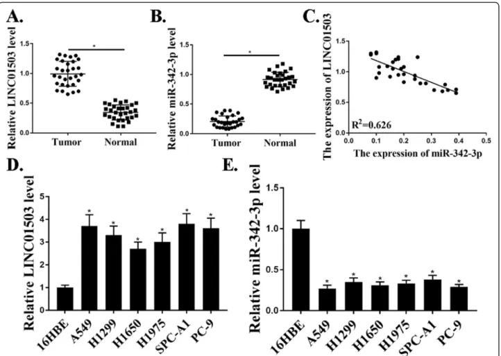
To identify the roles of LINC01503 in NSCLC progres-sion, qRT-PCR analysis was used to investigate the ex-pression of LINC01503 in 30 pairs of NSCLC samples compared to that in adjacent normal samples. Our re-sults revealed that the expression level of LINC01503 in tumor samples were markedly elevated compared with those in the corresponding normal samples (P<0.05) (Fig. 1a). However, the expression levels of miR-342-3p were significantly lower in tumor samples compared with that in normal control samples (P<0.05) (Fig. 1b). Moreover, the expression of miR-342-3p was negatively related to the expression of LINC01503 in NSCLC pa-tients (Fig. 1c). We also examined the expression of LINC01503, miR-342-3p in NSCLC cells. As shown in Fig.1d, e, the expression levels of LINC01503 were also markedly increased and the expression levels of miR-342-3p were decreased in NSCLC cells compared with that in control 16HBE cells (P<0.05). These results dem-onstrated that the expression of LINC01503 was upregu-lated in NSCLC patients and NSCLC cells.
Knockdown of LINC01503 suppressed proliferation, migration, and invasion of NSCLC cells in vitro
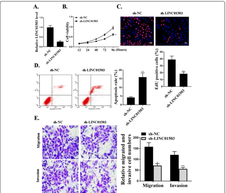
expressing specific shRNA targeting LINC01503 (sh-LINC01503) and negative control shRNA (sh-NC) were used in cells. The reduced expression levels of LINC01503 in these cells transfected with sh-LINC01503 were confirmed by qRT-PCR (Fig. 2a). The effect of LINC01503 on cell proliferation was evaluated by CCK-8 assay and Edu assay, and the data revealed that sh-LINC01503 inhibition signifi-cantly reduced cell proliferation compared with that in the control groups (P<0.05) (Fig. 2b and c). Trans-well assay further revealed a notable reduction in the number of migrated (P<0.05) and invaded cells (P< 0.01) in LINC01503 shRNA transfection group com-pared to the negative control shRNA transfection group (Fig. 2e). As shown in Fig. 2d, sh-LINC01503 transfection significantly increased cell apoptosis com-pared with that in sh-NC transfection group (P<0.01). It is obvious that knockdown of LINC01503 could suppress the proliferation, migration, and invasion of NSCLC cells in vitro.
MiR-342-3p mediated proliferation, migration, and invasion of NSCLC cells in vitro
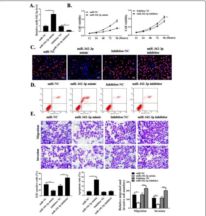
To understand the functional roles of miR-342-3p in NSCLC progression, knockdown and over-expression ex-periments were performed. MiR-342-3p mimic or miR-342-3p inhibitor and negative control NC were used in cells. The expression of miR-342-3p in these cells was de-tected by qRT-PCR (Fig.3a). The effect of miR-342-3p on cell proliferation was evaluated by CCK-8 assay and EdU assay, and it revealed that transfection of miR-342-3p mimic significantly reduced cell proliferation compared with that in the control groups (P<0.01) (Fig. 3b and c). As shown in Fig.3d, miR-342-3p mimic transfection sig-nificantly increased cell apoptosis compared with that in the control groups (P<0.01). Transwell assay was further utilized to assess the influence of miR-342-3p on the mi-gration and invasion. The data revealed a notable reduc-tion in the number of migrated and invaded cells in miR-342-3p mimic transfection group compared with that in the negative control groups (P<0.001) (Fig. 3e). In Fig. 1Up-regulation of LINC01503 in NSCLC patients and NSCLC cells.a.Relative expression of LncRNA-LINC001503 in NSCLC samples and adjacent normal samples (n= 30).b. Relative expression of miR-342-3p in NSCLC samples and adjacent normal samples (n= 30).c. The correlation between LINC01503 and miR-342-3p levels was measured.d. The expression levels of LINC01503 in NSCLC cells.e. The expression levels of miR342-3p in NSCLC cells.N= 3, *p< 0.05
summary, miR-342-3p could mediate the proliferation, migration, and invasion of NSCLC cells in vitro.
MiR-342-3p was a target of LINC01503
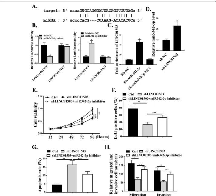
We next explored the underlying mechanism of how LINC01503 regulates NSCLC progression. The starBase online tool was used to explore the association of LINC01503 and microRNAs. We noticed that miR-342-3p, a tumor suppressor microRNA, was predicted to interact with LINC01503 (Fig.4a). As shown in Fig. 4b, miR-342-3p markedly inhibited the luciferase activities in cells in transfected with the LINC01503-WT (P<0.05), but did not affect the luciferase activities transfected
with LINC01503-MUT (P>0.05). The results from RNA pull-down assay showed a markedly elevated fold enrich-ment of LINC01503 using Bio-miR-342-3p, compared with the assays using NC-bio or Bio-miR-342-3p MUT (P<0.05) (Fig. 4c). Results also showed that the expres-sion levels of miR-342-3p were significantly increased after transfection of sh-LINC01503 (P<0.01) (Fig.4d).
CCK-8 assay and EdU assay, and the data revealed that sh-LINC01503 transfection reduced cell proliferation compared with that in the control group cells (P<0.01), and this effect was attenuated by co-transfection with miR-342-3p inhibitor (P<0.001) (Fig.4e and f). Transwell
assay was further utilized to assess the influence of LINC01503 on the migration and invasion. The data re-vealed a notable reduction in the number of migrated and invaded cells in transfection with sh-LINC01503 compared with that in the negative control shRNA group, Fig. 3MiR-342-3p mediates proliferation, migration, and invasion of NSCLC cells in vitro.a. The expression levels of miR-342-3p in cells
transfected with miR-NC, miR-342-3p-mimic, inhibitor NC, or miR-342-3p inhibitor.b. CCK8 assay for cell viabilities in cells transfected with miR-NC, miR-342-3p-mimic, inhibitor miR-NC, or miR-342-3p inhibitor.c. Edu staining in cells transfected with miR-NC, miR-342-3p-mimic, inhibitor NC, or miR-342-3p inhibitor. For the colors, blue indicates Hoechst, and red indicates Edu.d. Cytoflow to examine cell apoptosis in cells transfected with miR-NC, miR-342-3p-mimic, inhibitor NC, or miR-342-3p inhibitor.e. Transwell assays for cell invasive and migrative abilities in cells transfected with miR-NC, miR-342-3p-mimic, inhibitor NC, or miR-342-3p inhibitor.N= 3, *p< 0.001,**p< 0.01, ***p< 0.05
and this effect was attenuated by co-transfection with miR-342-3p inhibitor (P<0.01, P<0.001) (Fig.4h). As shown in Fig.4g, sh-LINC01503 promoted cell apoptosis and this ef-fect was attenuated by co-transef-fection with miR-342-3p in-hibitor (P<0.001). These results indicated that miR-342-3p was a target of LINC01503.
LASP1 was a target of miR-342-3p
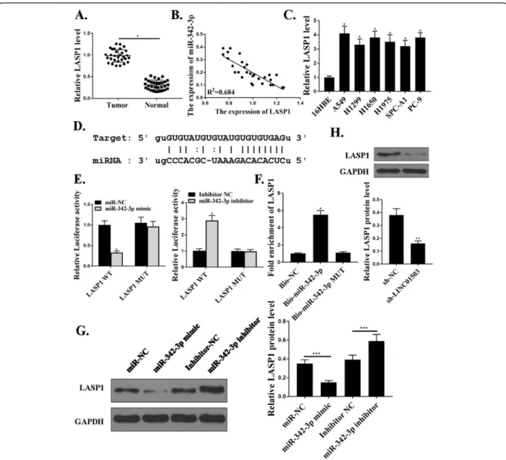
The expression of LASP1(LIM and SH3 domain protein 1) was upregulated in tumor samples (Fig. 5a). Moover, the expression of miR-342-3p was negatively re-lated with that of LASP1 in NSCLC patients (Fig. 5b). We also examined the expression of LASP1 in NSCLC
342-3p and LINC01503 regulated the expression of LASP1 (Fig. 5g and h). As a result, we established that LASP1 was a target of miR-342-3p.
MiR-342-3p/LASP1 axis mediated proliferation, migration, and invasion of NSCLC cells in vitro
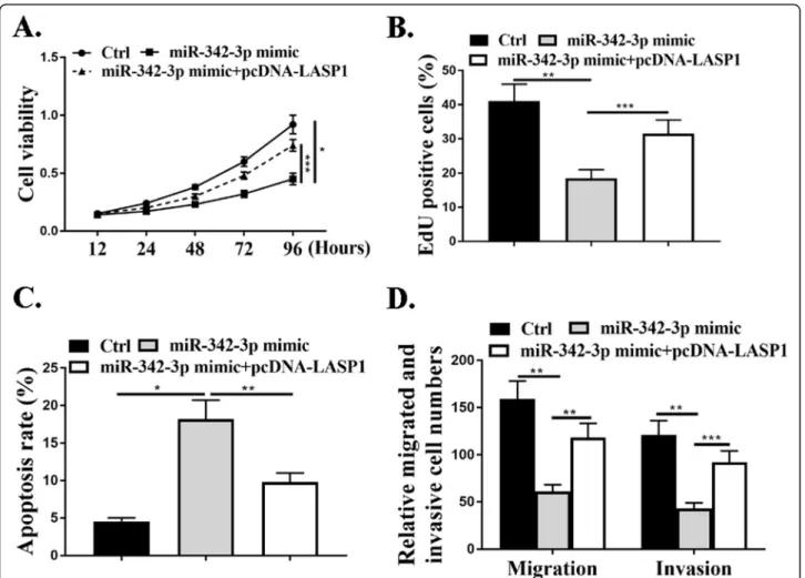
The effect of miR-342-3p on cell proliferation was evaluated by CCK-8 assay and EdU assay and the data revealed that miR-342-3p mimic reduced cell proliferation compared with that in the control cells (P<0.05), and this effect was attenu-ated by co-transfection with pcDNA-LASP1 (P<0.001) (Fig.6a and b). Transwell assay results revealed a notable re-duction in the number of migrated and invaded cells in
transfection with miR-342-3p mimic relative to the negative control group (P<0.01) and this effect was attenuated by co-transfection with pcDNA-LASP1(P<0.01,P<0.001) (Fig.6d). As shown in Fig.6c, miR-342-3p mimic promoted cell apop-tosis and this effect was attenuated by co-transfection with pcDNA-LASP1 (P<0.01). The above data revealed that miR-342-3p/LASP1 axis could mediate the proliferation, migra-tion, and invasion of NSCLC cells in vitro.
LINC01503/miR-342-3p promotes NSCLC cell tumor growth in vivo
We next injected shLINC01503 knockdown cells, shLINC01503 and miR-342-3p reducing cells, or control Fig. 5LASP1 is the target ofmiR-342-3p.a. Relative expression of LASP1 in NSCLC samples and adjacent normal samples (n= 30).b. The
correlation between miR-342-3p and LASP1 levels was measured.c. The expression levels of miR342-3p in NSCLC cells. H. The expression levels of LASP1 in NSCLC cells.d. The putative target sequence for miR-342-3p on the 3′-UTR of LASP1.eThe luciferase activity was detected by luciferase reporter assay.f. RNA pull-down assay.g. andh. The LASP1 protein expression.N= 3, *p< 0.001, **p< 0.01. ***p< 0.05
cells into nude mice to investigate whether LINC01503/ miR-342-3p would influence NSCLC cells tumorigenesis in vivo. The data revealed that tumors grown from cells with the knockdown of LINC01503 were significantly smaller compared with tumors grown from control cells and this effect was attenuated by a miR-342-3p inhibitor (P<0.01, P<0.001) (Fig. 7a-c). Moreover, tumor samples collected from the LINC01503 knockdown group had fewer Ki67-positive cells, whereas the miR-342-3p in-hibitor had more Ki67-positive cells in contrast to the LINC01503 group (Fig. 7d). In addition, shLINC01503 reduced the expression levels of LASP1 and co-transfection with miR-342-3p inhibitor promoted the ex-pression of LASP1 (Fig.7e). In summary, we proved that LINC01503/miR-342-3p promoted NSCLC cell tumor growth in vivo.
Discussions
LINC01503 was reported to promote colorectal cancer cell proliferation and invasion by regulating the miR-4492/FOXK1 signaling [23]. In colorectal cancer tissues,
they found that LINC01503 was upregulated and miR-4492 was significantly down-regulated [23]. In our ex-periments, qRT-PCR analysis revealed that the expres-sion of LINC01503 in tumor samples was markedly elevated in contrast to those in the corresponding nor-mal samples. However, the expression levels of miR-342-3p were lower in tumor samples. The expression of miR-342-3p as negatively corelated with that of LINC01503 in NSCLC patients. The expression levels of LINC01503 were also markedly increased and the ex-pression levels of miR-342-3p were reduced in NSCLC cells compared with that in 16HBE cells. Similar to pre-vious published literature, we found that the expression of LINC01503 was up-regulated in NSCLC patients and NSCLC cells, and the expression of miR-342-3p was suppressed.
EdU assay revealed that sh-LINC01503 inhibition re-duced cell proliferation compared with that of the con-trol cells. Migration and invasion assays revealed a notable reduction in the number of migrated and in-vaded cells in transfection with LINC01503 shRNA rela-tive to the negarela-tive control shRNA group. In consistence with previous studies, we found that LINC01503 pro-moted proliferation, migration, and invasion of NSCLC cells in vitro.
MiR-342-3p, a well-studied miRNA that has been proved to be a tumor suppressor for the proliferation and invasion of NSCLC [17, 25]. In addition, miR-342-3p was also demonstrated to bind with several different lncRNAs to exert biological functions [26,27]. From our results, miR-342-3p was predicted to interact with LINC01503 directly. MiR-342-3p markedly inhibited the luciferase activities in cells transfected with the LINC01503-WT but did not affect LINC01503-MUT. RNA pull-down assays pointed out a markedly higher expression levels of LINC01503 in contrast to the assays using NC-bio or hsa-mir-miR-342-3p probes.
Sh-LINC01503 reduced the expression levels of miR-342-3p. Moreover, CCK-8 assays and EdU assays revealed that sh-LINC01503 inhibition reduced cell proliferation compared with the control group in cells and this effect was attenuated by co-transfection with miR-342-3p in-hibitor. Migration and invasion assays revealed a notable reduce in the number of migrated and invaded cells in transfection with LINC01503 shRNA relative to the negative control shRNA group and this effect was atten-uated by co-transfection with miR-342-3p inhibitor. For the first time, we revealed that miR-342-3p was a target of LINC01503 in NSCLC samples and cells.
LASP-1, a cyclic adenosine monophosphate (cAMP) and cGMP-dependent signaling protein, was recently found to play important roles in NSCLC progression. Studies have demonstrated that LASP-1 was notably up-regulated and promoted tumor proliferation, invasion and metastasis in multiple malignant tumors, including NSCLC. LASP-1 was reported to be regulated by miR-203 and facilitate tumor proliferation and aggressiveness in human NSCLC [19]. LASP-1 was also reported to Fig. 7LINC01503/miR-342-3p promotes NSCLC cell tumor growth in vivo.a.Tumor volumes were measured every 1 week, and growth curves are shown.b. Representative images of mice bearing tumors.c. Tumor weight.d. Representative images for Ki67 immunostaining of tumor samples from different groups.e. The LASP1 protein expression,N= 5. *p< 0.001, **p< 0.01, ***p< 0.05
promote the malignant phenotype of NSCLC via indu-cing phosphorylation of the FAK-AKT pathway [28]. From our experiments, LASP1 had a higher expression levels in tumor samples, which was consistent with pre-vious publications [19]. The expression of miR-342-3p was negatively corelated to that of LASP1 in NSCLC pa-tients. LASP1 was also upregulated in NSCLC cells com-pared with that in 16HBE cells. Moreover, miR-342-3p can directly binding to LASP1. MiR-342-3p mimic re-duced cell proliferation compared with the control group in cells and this effect was attenuated by co-transfection with pcDNA-LASP1. PcDNA-LASP1 can also reverse the migration and invasion abilities of miR-342-3p mimic, suggesting that LASP1 and miR-miR-342-3p play opposite roles in NSCLC progression. As far as we know, we are the first to report that the miR-342-3p/ LASP1 axis mediated proliferation, migration, and inva-sion of NSCLC cells in vitro. Besides, we also confirmed the above resutls by in vivo assays.
Conclusions
LINC01503 as a competing endogenous RNA (ceRNA) regulates NSCLC proliferation and migration via miR-342-3p-LASP1 axis in vitro and in vivo.
Abbreviations
NSCLC:Non-small cell lung cancer; lncRNAs: Long non-coding RNAs; LASP1: LIM and SH3 domain protein 1; ceRNA: Competing endogenous RNA; miRNAs: MicroRNAs; FOXM1: Forkhead box protein M1; EdU: 5-ethynyl-29-deoxyuridine; CCK-8: Cell Counting Kit-8; cAMP: Cyclic adenosine monophosphate
Acknowledgements Not applicable.
Authors’contributions
QMS: experiment studies, clinical studies, Data collection and Manuscript writing; YBS: interpreted the patient data, Data collection and Manuscript writing; SX: Project development, data analysis, manuscript editing, and literature research. All contributing authors have read and agreed to the final version of the manuscript. The author(s) read and approved the final manuscript.
Funding Not applicable.
Availability of data and materials
The datasets used and/or analyzed during the current study are available from the corresponding author on reasonable request.
Ethics approval
All procedures performed in studies involving humans were reviewed and permitted by The First Hospital of China Medical University. Written informed consent was obtained from all individual participants included in the study. Animal experiments were conducted according to Guide for the Care and Use of Laboratory Animals of the National Institutes of Health and Ethics Committee of The First Hospital of China Medical University approved this study.
Consent for publication Not applicable.
Competing interests
The authors declare that they have no competing interests.
Received: 21 February 2020 Accepted: 22 July 2020
References
1. Zarogoulidis K, Zarogoulidis P, Darwiche K, Boutsikou E, Machairiotis N, Tsakiridis K, et al. Treatment of non-small cell lung cancer (NSCLC). J Thor Dis. 2013;5(Suppl 4):S389.
2. Scagliotti G, Hanna N, Fossella F, Sugarman K, Blatter J, Peterson P, et al. The differential efficacy of pemetrexed according to NSCLC histology: a review of two phase III studies. Oncologist. 2009;14(3):253–63.
3. Chang A. Chemotherapy, chemoresistance and the changing treatment landscape for NSCLC. Lung Cancer. 2011;71(1):3–10.
4. Cappuzzo F, Gregorc V, Rossi E, Cancellieri A, Magrini E, Paties CT, et al. Gefitinib in pretreated non–small-cell lung cancer (NSCLC): analysis of efficacy and correlation with HER2 and epidermal growth factor receptor expression in locally advanced or metastatic NSCLC. J Clin Oncol. 2003; 21(14):2658–63.
5. Hof H, Herfarth KK, Münter M, Hoess A, Motsch J, Wannenmacher M, et al. Stereotactic single-dose radiotherapy of stage I non–small-cell lung cancer (NSCLC). Int J Rad Oncol Biol Phys. 2003;56(2):335–41.
6. Zhang C, Yin D, Sun S, Han L. The use of lncRNA analysis for stratification management of prognostic risk in patients with NSCLC. Eur Rev Med Pharmacol Sci. 2017;21(1):115–9.
7. He R. Hu Zhang F, Shen N. LncRNA FEZF1-AS1 enhances epithelial-mesenchymal transition (EMT) through suppressing E-cadherin and regulating WNT pathway in non-small cell lung cancer (NSCLC). Biomed Pharmacother. 2017;95:331–8.
8. Tang Q, Ni Z, Cheng Z, Xu J, Yu H, Yin P. Three circulating long non-coding RNAs act as biomarkers for predicting NSCLC. Cell Physiol Biochem. 2015; 37(3):1002–9.
9. Zhang Y, Chen WJ, Gan TQ, Zhang XL, Xie ZC, Ye ZH, et al. Clinical significance and effect of lncRNA HOXA11-AS in NSCLC: a study based on bioinformatics, in vitro and in vivo verification. Sci Rep. 2017;7(1):5567. 10. Yang Y-R, Zang S-Z, Zhong C-L, Li Y-X, Zhao S-S, Feng X-J. Increased
expression of the lncRNA PVT1 promotes tumorigenesis in non-small cell lung cancer. Int J Clin Exp Pathol. 2014;7(10):6929.
11. Deng J, Liang Y, Liu C, He S, Wang S. The up-regulation of long non-coding RNA AFAP1-AS1 is associated with the poor prognosis of NSCLC patients. Biomed Pharmacother. 2015;75:8–11.
12. Lu KH, Li W, Liu XH, Sun M, Zhang ML, Wu WQ, et al. Long non-coding RNA MEG3 inhibits NSCLC cells proliferation and induces apoptosis by affecting p53 expression. BMC Cancer. 2013;13(1):461.
13. Xie J-J, Jiang Y-Y, Jiang Y, Li C-Q, Lim M-C, An O, et al. Super-enhancer-driven long non-coding RNA LINC01503, regulated by TP63, is over-expressed and oncogenic in squamous cell carcinoma. Gastroenterology. 2018;154(8):2137–51 e1.
14. Yu T, Liu L, Li J, Yan M, Lin H, Liu Y, et al. MiRNA-10a is upregulated in NSCL C and may promote cancer by targeting PTEN. Oncotarget. 2015;6(30): 30239.
15. Jiang Z, Yin J, Fu W, Mo Y, Pan Y, Dai L, et al. MiRNA 17 family regulates cisplatin-resistant and metastasis by targeting TGFbetaR2 in NSCLC. PLoS One. 2014;9(4):e94639.
16. Enfield KS, Pikor LA, Martinez VD, Lam WL. Mechanistic roles of noncoding RNAs in lung cancer biology and their clinical implications. Genet Res Int. 2012;2012.
17. Xie X, Liu H, Wang M, Ding F, Xiao H, Hu F, et al. miR-342-3p targets RAP2B to suppress proliferation and invasion of non-small cell lung cancer cells. Tumor Biol. 2015;36(7):5031–8.
18. Orth MF, Cazes A, Butt E, Grunewald TG. An update on the LIM and SH3 domain protein 1 (LASP1): a versatile structural, signaling, and biomarker protein. Oncotarget. 2015;6(1):26.
19. Zheng J, Wang F, Lu S, Wang X. LASP-1, regulated by miR-203, promotes tumor proliferation and aggressiveness in human non-small cell lung cancer. Exp Mol Pathol. 2016;100(1):116–24.
20. Hu Z, Cui Y, Zhou Y, Zhou K, Qiao X, Li C, et al. MicroRNA-29a plays a suppressive role in non-small cell lung cancer cells via targeting LASP1. OncoTargets Ther. 2016;9:6999.
22. Byers R, Ward T, Womack C. Dive. C. Effect of prolonged formalin fixation on immunohistochemical staining for the proliferation marker Ki67.
Histopathology. 2011;59:1261–79.
23. Lu SR, Li Q, Lu JL, Liu C, Xu X, Li JZ. Long non-coding RNA LINC01503 promotes colorectal cancer cell proliferation and invasion by regulating miR-4492/FOXK1 signaling. Exp Ther Med. 2018;16(6):4879–85.
24. Qu Y, Qu X, Chen G, Feng Y, Teng X, Liu W, et al. LINC01503 promotes cell proliferation, invasion and EMT process in cholangio-carcinoma. Eur Rev Med Pharmacol Sci. 2019;23(15):6445–52.
25. Xue X, Fei X, Hou W, Zhang Y, Liu L, Hu R. miR-342-3p suppresses cell proliferation and migration by targeting AGR2 in non-small cell lung cancer. Cancer Lett. 2018;412:170–8.
26. Wang S-H, Ma F, Tang ZH, Wu XC, Cai Q, Zhang MD, et al. Long non-coding RNA H19 regulates FOXM1 expression by competitively binding
endogenous miR-342-3p in gallbladder cancer. J Exp Clin Cancer Res. 2016; 35(1):160.
27. Cheng D, Fan J, Ma Y, Zhou Y, Qin K, Shi M, et al. LncRNA SNHG7 promotes pancreatic cancer proliferation through ID4 by sponging miR-342-3p. Cell Bioscience. 2019;9(1):28.
28. Zhang X, Liu Y, Fan C, Wang L, Li A, Zhou H, et al. Lasp1 promotes malignant phenotype of non-small-cell lung cancer via inducing phosphorylation of FAK-AKT pathway. Oncotarget. 2017;8(43):75102.
Publisher’s Note
Springer Nature remains neutral with regard to jurisdictional claims in published maps and institutional affiliations.