ANTI-BACTERIAL
ACTIVITY STUDIES ON
TEXTILES
MODIFIED WITH SILVER METAL,
COPPER, ZINC AND
MAGNESIUM
OXIDES NANOPARTICLES
Nehad Hamdi
[a], Doaa M. El-Mekkawi
[b]*, Mohamed M. Selim
[b], S. A.
Hassan
[c]and Amir Ezzat
[c]Keywords: inorganic antibacterial, antibacterial textile, metal and metal oxide nanoparticle.
The anti-bacterial characteristics of different nano-structured metal and metal oxides modified cotton fabrics were investigated. In this study, silver metal, copper, zinc and magnesium oxides have been supported on bleached cotton fabrics. Reduction, wet method, sol gel and precipitation methods were used in the preparation of the antibacterial nanoparticles (NPs). The preparation of the antibacterial-loaded cotton was carried out in-situ and ex-situ by pad dry methods. Formation of the supported nanoparticles was confirmed using x-ray diffraction (XRD), scanning electron microscopy studies (SEM) and energy dispersive x-ray (EDX) analyses. Antibacterial studies on the supported nanoparticles were done on gram positive (Bacillus and S. aureus) and gram negative (E-coli) bacteria by agar diffusion method. The loaded antibacterial nanoparticles are effective against the bacteria under investigation. Under the given experimental conditions, the maximum inactivation performances of each loaded inorganic agent were investigated. The loaded fabrics show the following antibacterial performance order against Bacillus subtilis is Ag=CuO>ZnO>MgO. However, the activity order is CuO>Ag>ZnO=MgO against S.aureus
and E. coli. The inactivation performances depend on the type, purity and the amount of antibacterial nanoparticles on the textile surfaces. * Corresponding Authors
Fax: (+202) 33370931
E-Mail: [email protected]
[a] Textile Lab., Chemistry Administration, 12 Ramses st., Cairo, Egypt.
[b] Physical Chemistry Department, National Research Centre, 33 EL Bohouth st., Dokki, P. O. 12622, Giza- Egypt. [c] Faculty of Science, Ain Shams University, Cairo, Egypt.
Introduction
Inorganic materials such as metal and metal oxides have extensively considered as promising antibacterial agents.1-5 Several kinds of metals and metal oxides posses obvious killing abilities to most microbes even at very low concentrations.6 Different metal, oxide or salt compounds, mostly based on silver, copper, zinc and magnesium kill microbes by binding to intracellular proteins and inactivating them. These materials can react with cell enzymes leading to either generating or catalyzing reactive oxygen species. Reactive oxygen can then induce an oxidative stress, damaging cellular proteins, lipids and DNA.7 Recently, much attention has been paid on the loading of metal and metal oxides nanoparticles (NPs) onto cotton substrate due to their potential applications.8-12 The modified cotton fabrics have exhibited excellent antimicrobial activities against bacteria.7 There is a growing awareness of the use of antibacterial fabrics in the form of medical clothes, protective garments, and bed spreads to minimize the chance of the nosocomial infections.
In the last few years, environmental and technical efforts have been made to develop new efficient antimicrobial textiles. For example, silver is now used in a large number of commercial antimicrobial products, either as an isolated form, for fiber finishing or incorporation, or even already in fiber or fabric form,13 Copper particles also show antifungal and antiviral properties.14 Impregnation of cellulose textiles with copper salt, Cu-alginate or CuO nanoparticles
including release properties has been studied.13-19 Different techniques to insert/deposit copper in cellulose fibers have been also proposed. Zinc oxide has also been explored as a powerful antibacterial agent.7-13 The relation between the antibacterial activity versus the ZnO particles size was studied.17 It was observed that the antibacterial activity is inversely proportional to the nanoparticles’ size. Arouk et al. have developed a ZnO nanoparticle-chitosan composite to coat cotton fabrics in order to conjugate the antimicrobial power of ZnO and chitosan and even to overcome the lack of strong chemical bonding with textile substrates presented by chitosan.20 Magnesium oxide nanoparticles has been also reported as an acceptable mild inorganic antibacterial agent due to its abundance and low cost.21 It also can work in simple antibacterial conditions and safe materials to human beings.22
The aim of the present research is to prepare inorganic antibacterial-loaded cotton for antibacterial studies. The prepared nanoparticles act as antibacterial agent, while cotton act as a substrate which can be used to in many biomedical applications. Silver metal and metal oxides (CuO, ZnO and MgO) supported cotton have been prepared and characterized for the antibacterial applications. The antibacterial modified cotton fabrics were characterized by XRD, SEM and EDX. The killing abilities of the modified cotton fabrics were tested against Gram-negative
Escherichia coli (E. coli) and Gram-positive Staphylococcus aureus (S. aureus) and Bacillus subtilis (B. subtilis) bacteria.
Experimental
Materials
Switzerland), sodium dodecyl sulfate (SDS, ≥ 98 %, Aldrich, U.S.A), agar culture medium (Oxoid, UK), CuSO4.5H2O (≥ 99 %, Oxford, India), acrylic binder (purity > 99 %, Pachin – Obour Paints & Chemical Industries, Egypt), Zn(NO3)2.3H2O and Mg(NO3)2 (> 98 %, Merck ,Germany), soluble starch (≥ 98 %, WINLAB, UK) and NaOH (purity > 98 %, El Nasr for Intermediate Chemicals, Egypt) were used as received.
Preparation of cotton loaded with Agmetal nanoparticles Silver metal nanoparticles were prepared by the reduction of silver ions using trisodium citrates (TSC) as reducing agent.23 AgNO3 solution was heated to boiling at 70 oC and 10 mL of TSC solution (36×10−3 mol L-1) were added drop by drop. During the process, the solution was mixed vigorously and heated until a pale yellow color appeared. Reduction of silver ions (Ag+) to silver nanoparticles was evident in the colloid synthesis process, as the colorless solution became pale yellow with the formation of the silver nanoparticles. Next, the solution was removed from the heating and stirred until cooled to room temperature. Cotton fabric samples were then immersed in the prepared silver nanocolloids heated at 50 oC and stirred rapidly for 30 min. The cotton samples subsequently were padded. The padding process ensured the solution was coated evenly, and the excess chemicals were removed. The padded samples were air dried and finally cured at 120 oC for 3 min in a pre-heated curing oven. The preparation steps were repeated with different initial concentrations of AgNO3 (0072, 0.018 and 0.0288 mol /l).
Preparation of CuO loaded textile
Copper oxide nanoparticles were prepared by sonochemical method as indicated previously.24 Cotton woven fibers were first washed in a water bath containing 5 % of sodium dodecyl sulfate at 40 °C for 1 h. After rinsing with distilled water, the fibers were dried in vacuum at 60 °C for 24 h. Dry cotton (5×5 cm) was first soaked into 100 mL of aqueous solution of CuSO4.5H2O (4.8 × 10⁻⁴ mol L-1) solution in a sonicated flask and irradiated for 10 min with Ultrasonic generator. Then 0.06 g of NaOH was added to the mixture while stirring. The mixture was then re-sonicated at 35 to 40 °C for 1 h, the strong blue color was gradually converted into brown after 15 min. The bath temperature was kept at a constant temperature around 40 °C. The product was then washed thoroughly several times with distilled water to remove any excess hydroxide and dried in vacuum at 60 °C overnight. The preparation experiment was repeated using different initial concentrations of copper sulphate (0.0012, 0.00192, 0.00216 and 0.0024 mol L-1).
Preparation of textile loaded with ZnO NPs
The zinc oxide nanoparticles were prepared by wet chemical method.25 Zinc nitrate and sodium hydroxide were used as precursors while soluble starch was added as stabilizing agent. Soluble starch (0.1 %) was dissolved in 500 ml of distilled water then zinc nitrate was added. Then the solution was kept under constant stirring using magnetic stirrer till completely dissolve the zinc nitrate. After
complete dissolution of zinc nitrate, 0.2 M of sodium hydroxide solution (20 mL was used in our study) was added under constant stirring, drop by drop touching the walls of the vessel. The reaction was allowed to proceed for 2 h after complete addition of sodium hydroxide. After the completion of reaction, the solution was allowed to settle for overnight and the supernatant solution was then discarded carefully. The remaining solution was centrifuged at 35,000 rpm for 10 min and the supernatant was discarded. The obtained nanoparticles were washed three times using distilled water. Washing was carried out to remove the byproducts and the excessive starch that were bound with the nanoparticles. After washing, the nanoparticles were dried at 80 oC for overnight. During drying, complete conversion of zinc hydroxide into zinc oxide takes place.
ZnO nanoparticles were applied on cotton using immersion method. The cotton fabric cut to the size of 5×5 cm was immersed in the solution containing ZnO (1 %) and acrylic binder (1 %) for 5 min and then it was padded. After padding, the fabric was air-dried and then cured for 3 min at 140 °C. The fabric was then immersed for 5 min in sodium dodecyl sulfate solution (2 g L-1) to remove unbound nanoparticles. Then the fabric was rinsed at least 10 times to completely take out all the soap solution. The fabric was washed then air-dried. The loading procedure was repeated with different solutions containing different percentages of ZnO suspension (1, 2, 2.5, 4.5, 5 and 8 % w/v).25
Preparation of textile loaded with MgO
Magnesium oxide nanoparticles were prepared by wet chemical method.26 Magnesium nitrate and sodium hydroxide were added to soluble starch as stabilizing agent. Starch act as a stabilizing agent and also prevents the agglomeration of nanoparticles. Starch (0.1 % w/v) solution was dissolved in 100 mL distilled water then magnesium nitrate was added to the above solution. Then the solution was kept under constant stirring using magnetic stirrer for complete dissolution of contents. After complete dissolution, 4 g (0.2 M) sodium hydroxide solution (25 ml) was added in drops along the sides of the container under constant stirring for 2 h and allowed to settle for 24 h. The supernatant liquid was then discarded carefully and the remaining solution was centrifuged (35,000 rpm at 25 °C) for 10 min. Centrifugate was washed three times using distilled water to remove the byproducts and the excessive starch that bound with the nanoparticles. The nanoparticles of magnesium hydroxide were placed in furnace at 300 °C. During this process, conversion of magnesium hydroxide into magnesium oxide takes place. The following reaction explains the formation of magnesium oxide nanoparticles.
Mg(NO3)2.6H2O+2NaOH → Mg(OH)2+2NaNO3 (1)
Mg(OH)2→MgO +H2O (2)
dodecyl sulfate solution (2 g L-1) to remove unbound nanoparticles. Then the fabric was rinsed at least 10 times to completely take out all the soap solution. The fabric thus washed was air-dried. The loading procedure was repeated with different solutions containing different percentages of MgO suspension (3, 4 and 5 % w/v).
Characterization of the prepared samples X-ray diffraction measurements
To determine the crystal phase composition of the synthesized samples, X-ray diffraction (XRD) measurements were taken using a Bruker D8 advance instrument with CuKα1 target with secondary monochromator 40 kV, 40 mA. The reference data for the interpretation of the X-ray diffraction patterns were obtained from the International Centre for Diffraction Data Joint Committee on Powder Diffraction Standards ICDD-JCPDS card files. The crystallite size (D) determined by the Scherrer’s formula: 27
(3)
where
λ is the wavelength of X- ray radiation (CuKα1= 0.15406 nm),
K is a constant taken as 0.89,
is the line width at half maximum height (FWHM) of the peak, and
θ is the diffracting angle.
Scanning electron microscope
In SEM measurements, an electron beam was passed through the specimens followed by scattering them back as electrons and secondary electrons. Back scattered secondary electrons were used to form the image on the computer monitor. The acceleration of the electron beam was 10 kV. This was carried out on Quanta FEG250 Instrument.
Energy dispersive X-ray
The spectra of NP coated cotton fabrics were obtained by EDX measurements. A field emission scanning electron microscope (QUANTA FEG 250) coupled with an energy dispersive X-ray spectrometer (EDX) unit was employed to evaluate the elemental composition of NP/cotton sample. Semi-quantitative analyses in the inspection field were conducted using ZFA software where the energy of the emitted electrons for each element was counted in units of weight percent.
Antibacterial activity
The antibacterial activity was carried out by the agar diffusion method using the suspension of bacteria spread on nutrient agar. The swab was dipped into the broth culture of
the organism and was gently squeezed against the tube inside to remove excess fluid. The swab used to streak agar plate or a nutrient agar plate for a lawn of growth. This is best accomplished by streaking the plate in one direction, then streaking at right angles to the first streaking, and finally streaking diagonally. End by using the swab to streak the outside diameter of the agar. The inoculated plates were incubated at appropriate temperature for 24 h. The supported fabric placed on the surface of the agar using a dispenser that dispenses multiple pieces at the correct distance apart, on the surface of the agar using flame sterilized forceps. The antibacterial activity was evaluated by measuring the zone of inhibition against the test organisms. Zone of inhibition is the area in which the bacterial growth is stopped due to bacteriostatic effect of the compound and it measures the inhibitory effect of compound towards a particular microorganism. Finally diameters of zones of inhibition (mm) were measured and illustrated.
Results and Discussion
XRD analyses
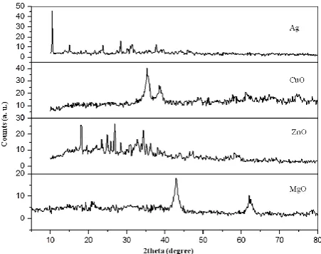
The XRD measurements of the prepared inorganic antibacterial agents were carried out. The estimated average crystals sizes and the purity of the samples were estimated. Figure 1 shows the x-ray diffractograms of Ag, CuO, ZnO and MgO, respectively. As shown in Figure 1, the diffractogram of Ag shows good agreement with the characteristic peaks of pure Ag.
The characteristic peaks of pure Ag appeared in the prepared sample at 2 =10.54°, 15.02 °, 23.80 °, 28.43 °, 31.44 ° and 37.74 ° degrees. This data is consistent with the standard XRD data in Joint Committee on Powder Diffraction Standards (JCPDS) card file no. 74-1828. The calculated average crystal size of Ag using Scherrer relation is 64.8 nm. On the other hand, the characteristic peaks of pure CuO appeared at 2 =35.48 °, 38.64 °, 48.64 °, 61.33 °, 66.97 ° and 79.56 ° degrees. The data matches the recorded peaks in JCPDS card no.48-1548. The calculated average crystal size of CuO is 5.8 nm. Further, the diffractograms of the prepared MgO NPs are in good agreement with the JCPDS data (89-7746).
Figure 1. X-ray diffractograms of the prepared Ag, CuO, ZnO and MgO nanoparticles.
K
D
cos
This indicates the formation of pure nanocrystalline MgO without any foreign material. The estimated average crystal size of MgO is 10.5 nm. However, XRD data of the prepared ZnO indicated the presence of foreign phases. Data indicated the formation of only 11.5 % zinc oxide phase. Characteristic peaks of zinc hydroxide and nitrate appeared and the estimated percentage of each phase was 33.5 and 55.0 %, respectively.
SEM and EDX analyses
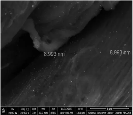
The SEM micrographs of the supported inorganic agents on cotton fabric were measured. For example, the supported Ag nanoparticles are illustrated in Figure 2. As shown in Figure 2, the Ag nanoparticles appear to be homogeneously distributed on the cotton surface. On the other hand, EDX spectroscopy reveals the good binding of Ag to cotton surfaces. As shown in Figure 3, the characteristic signals of Ag atom were observed in the EDX spectra of Ag/cotton sheets. In addition, EDX data indicated that the percentage amount of Ag on cotton surfaces was 28.48 %.
Figure 2. SEM image of Ag nanoparticles loaded on cotton.
Figure 3. EDX spectra of Ag nanoparticles loaded on bleached cotton.
Antibacterial studies
Antibacterial activities of the loaded samples have been tested against gram positive (B. subtilis and S. aureus) and gram negative (E. coli) bacteria by agar diffusion method (Figure 4).
Figure 4. Zones of inhibition of zinc (left) and copper (right) oxides nanoparticles against a gram positive bacteria (B. subtilis).
Overall, results revealed different killing abilities of bacteria. It has been found that the inactivation performances depend on the type and the amount of antibacterial nanoparticles on the textile surfaces as well as the type of bacteria. First, the influence of the amount of the loaded antibacterial nanoparticles was investigated (Figure 5). The amount of loaded NPs was initially increased as previously illustrated in the experimental section. The amount of loaded MgO or ZnO were varied by initially increase the amount of suspended oxides during the loading processes. However, loading of Ag and CuO were carried in-situ. Therfore, the influence of the amount of loaded Ag or CuO were controlled by varying the initial amounts of the added AgNO3 or CuSO4, respectively. Generally, according to each loading procedure, the increase in the amount of the loaded NPs was proceeded till the maximum inhibition zones were acheived (i. e. no further increase in the inhibition zone upon increasing the amount of loaded NPs). This may be attributed to the maximum loading capacity of the cotton fabric toward the loaded NPs.
Contrary, in case of CuO, loading of CuO on cotton was acheived through the in-situ sonochemical conversion of copper sulphate into CuO in the presence of NaOH. Change of the characteristic blue color of copper hydroxide into the brown CuO was ceased when the initial concentration copper sulphate exceeds 0.0024 mol L-1. Therefore, this concentration was taken as the maximum initial loading concentration.
However, loaded MgO shows no killing abilities toward the three kinds of bacteria. It worthy to mention that preliminarily antibacterial activity tests of both MgO and ZnO in its powder form was tested against the three bacteria. The preparation of the oxide powders were carried out as previously discussed but in the absence of the cotton fabric. The unsupported ZnO powder showed lower activity against
S. aureus and E. coli than that toward B. subtilis. However, MgO powder shows only very low antibacterial activities against all bacteria.
a)
b)
c)
Figure 5. The effect of the initial concentration of inorganic nanoparticles on the antibacterial activity of the loaded samples against B. subtilis, E.coli and S.aureus. (a) Ag, (b) CuO and (c) ZnO.
Obviously, loading of the inorganic antibacterial agents into textile fabrics leads to decrease in the amount of active agents and consequently decrease in their killing abilities (see for example the estimated amount of loaded Ag on cotton using EDX technique).
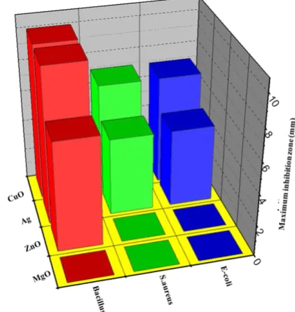
The maximum inactivation performances of each loaded inorganic agent against the three bacteria are illustrated in figure 6. Under the given experimental conditions, the loaded fabrics show the following antibacterial performance order against Bacillus: Ag=CuO>ZnO>MgO. However, the activity order is CuO>Ag>ZnO=MgO against S.aureus and
E.coli.
Figure 6 . The effect of the type of loaded inorganic agent on the antibacterial activity against B. subtilis, B. subtilis and B. subtilis.
The maximum inactivation performances of each loaded inorganic agent against the three bacteria are illustrated in figure 6. Under the given experimental conditions, the loaded fabrics show the following antibacterial performance order against B. subtilis: Ag=CuO>ZnO>MgO. However, the activity order is CuO>Ag>ZnO=MgO against S.aureus
and E.coli.
Overall, the results revealed that not all of the active antibacterial agents in its powder form can be loaded into fabrics for the biomedical application purposes. It is not recommended to load fabrics with NPs that posses low antibacterial activities (such as MgO) as an adequate perfomances is not expected. One important issue that should be taken in consideration is the purity of the inorganic agents. The low purity of the prepared ZnO NPs negatively impacts the killing ability of the loaded samples.
Conclusion
confirmed by XRD analyses. SEM measurements indicate the homogeneous distribution of NPs on the cotton surface. However, EDX spectroscopy reveals the good binding of NPs to cotton surfaces. As the initial concentrations of the loaded NPs increase, the killing abilities of bacteria increase. The zero antibacterial activity of ZnO toward S. aureus and
E. coli is probably due to its low purity. Whereas, zero killing ability of loaded MgO is due to its very low antibacterial activity in its unsupported form. Under the given experimental conditions, the loaded fabrics show the following antibacterial performance order against B. subtilis: Ag=CuO>ZnO>MgO. However, the activity order is CuO>Ag>ZnO=MgO against S. aureus and E. coli. On the light of this work, it can concluded that the inactivation performances depend on the type, purity and amount of antibacterial nanoparticles on the textile surfaces.
Acknowledgement
The authors are thankful to Chemistry Administration, for providing them necessary facilities and support to carry out this work and Dr. Shimaa Ashour Zaid, Microbiology Department, Chemistry Administration for her aid in carrying out the antimicrobial tests.
References
1Sahooli, M., Sabbaghi, S., Saboori, R., Matter. Lett., 2012, 81,
169–17.https:// doi: 10.1016/j.matlet.2012.04.148
2Khawla, K., Ghassan, S., Farah. A., Arab. J. Sci. Eng., 2016, 41,
301–310.doi:10.1007/s1336
3Ahamed, M., Siddiqui, M. A., Akhtar, M. J., Ahmad, I., Pant, A.
B., Alhadlaq, H. A., Biochem. Biophys. Res. Comm. 2010, 396, 578–583. doi:10.1016/j.bbrc.2010.04.156
4Mortimer, M., Kasemets, K., Kahru, A., Toxicology, 2010, 269,
182–189.doi: 10.1016/j.tox.2009.07.007
5Rashu K, Harpeet K., Gurav S., Mu, N., Deepak, P., J. Ind.., Eng.
Chem., 2015, 31, 173–184. doi: 10.1016/j.jiec.2015.06.021. 6Jones, N., Ray, B., Ranjit, K.T., Manna, A.C., FEMS Microbiol.
Lett., 2008, 279(1), 71–76. doi:10.1111/j.1574-6968.2007.01012.x
7Lura K. A., Delina, Y. L., Pedro, J. J., Water Res.,2006, 40(19),
3527–3532. doi: 10.1016/j.watres.2006.08.004.
8Zahran, M. K., Hanan. B. A., El Rafie, M. H., Carbohyd. Polym., 2014, 108, 145-152. doi: 10.1016/j.carbpol.2014.03.005 9Hossam, E. E., Avinash, P. M., Barbora, S., Heinz, D., Petra, M.,
Bernhard, R., Thomas B., Surf. Coat. Tech., 2014, 254, 344– 351. doi:10.1016/j.surfcoat.2014.06.036
10Eunmi, K., and Kyung, H. H., Dyes Pigments., 2014, 103,
222-227. doi: 10.1016/j.dyepig.2013.09.015
11Rajendran, R., Balakumar, C., Hasabo, A., Ahammed, M.,
Jayakumar, S., Vaideki, K., Rajesh, E., Int. J. Eng. Sci., 2010, 2, 202-208. doi: 10.4314/ijest.v2i1.59113
12Anita, S., Ramachandran, T., Rajendran, R., Koushik, C.V.,
Mahalakshli, M., Text. Res. J., 2011, 81, 1091–1099. doi: 10.1177/0040517510397577
13Seil J. T., and Webster, T. J., Int. J Nanomed., 2012, 7, 2767–
2781. doi: 10.2147%2FIJN.S24805
14Zarrindokht E. K., Pegah C., Afr. J. Microbiol. Res., 2011, 5(12),
1368–1373. doi: 10.5897/AJMR10.159
15Xie, Y., He, Y., Irwin, P. L., Jin, T., Shi, X., Appl. Environ.
Microb., 2011, 77, 2325–233. doi:10.1128/AEM.02149-10 16Krishna, R. R., Ranjit, T. K., Adhar, M., Langmuir., 2011, 27(7),
4020–4028. doi: 10.1021/la104825u
17Pal, S., Tak, Y. K., Song, J. M., Appl. Environ. Microb., 2007,
73(6), 1712–1720. doi: 10.1128%2FAEM.02218-06
18Lingling, Z., Yunhang, J., Vulang, D., Malclm, P., David, Y., J.
Nanopart. Res., 2007, 9(3), 479–489.
doi:10.1002/9781118150887.ch13
19Peng, X., Palma, S., Fisher, N. S., Wong, S. S., Aquat. Toxicol., 2011, 102(3), 186–196. doi: 10.1016/j.aquatox.2011.01.014 20Hebeish, A., Sharaf, S., Farouk, A., Int. J. Biol. Macromol., 2013,
60, 10–17. doi: 10.1016/j.ijbiomac.2013.04.078
21Di, D. R., He, Z. Z., Sun, Z. Q., Liu, J., Nanomedicine UK.,
2012, 8, 1233–1241. doi: 10.1016/j.nano.2012.02.010 22Banele, V., Phumlan, T., Poslet, M. S., Jane, C.N., Lucky,
Richard, M., J. Biomater. Nanobiotechnol., 2013, 4, 365-373.
doi: 10.4236/jbnb.2013.44046
23Srimala, P., Bharat, B., Rathnayake, B., Gamini, R., Sanath, R.,
Chaturanga, B., Colloids. Surf. A., 2013, 436, 975– 989.
doi:10.1016/j.colsurfa.2013.08.038
24El-Nahhal, M. I., Shehata, Z. M., Fawzi, K. S., Mohamed, S.,
Isabelle G., and Florence, B., Int. Nano. Lett.,2012, 2, 62-67.
doi:10.1186/2228-5326
25Rajendran, R., Balakumar, C., Hasabo, A., Jayakumar, S.,
Vaideki K., and Rajesh, Int. J. Eng. Sci.,2010, 2(1), 202- 208.
doi: 10.4314/ijest.v2i1.59113
27Sundrarajan, M., Suresh, J., Rajiv, R. G., Dig. J. Nanomater.
Bios., 2012, 7, 983 – 989.
28Patterson, A. L., Phys. Rev., 1939, 56, 978–982. doi:
10.1103/PhysRev.56.978