SENSING ASCORBIC ACID WITH DNA/PRUSSIAN BLUE
CARBON PASTE ELECTRODE
Yugandhar Parepalli,
[a]T. Ravi Sankar,
[b]P. T. S. R. K. Prasada Rao,
[c]Manju
Shree Nair,
[a]Sudhakar Reddy Pamanji
[d]and Murthy Chavali
[a]*Keywords: DNA, Ascorbic Acid (AA), vitamin-c tablets, electrochemical, sensor, carbon paste electrode.
A simple sensor for the detection of ascorbic acid (AA) has been developed. The electrochemical behaviours were studied after successfully constructing the electrode through DNA electro-deposition on carbon paste electrode using Prussian blue (PB). The electrodes were characterized by Raman spectroscopy that confirmed electro-deposition of PB, functioning as a redox mediator. The detection method was based on DNA damage induced by hydroxyl radical generated by Fenton reaction in which ascorbic acid effectively scavenges the hydroxyl radical. The amount of DNA damage is proportional to the amount of ascorbic acid concentration which was analyzed by cyclic voltametry (CV). Obtained results show that DNA-based biosensor can be utilized for estimating ascorbic acid in vitamin-c tablets. The detection range of this biosensor was in the range of 1.14 µM to 12.54 µM. Interference effects and stability of the biosensor were also investigated, results show that the biosensor has high sensitivity and selectivity towards ascorbic acid concentrations.
* Corresponding Author: Tel: +91-863-234-4756 Fax: +91-863-253-4468 E-Mail: [email protected]
[a] Division of Chemistry, Department of Sciences and Humanities, Vignan’s Foundation for Science, Technology and Research University (VFSTRU; Vignan University), Vadlamudi, Guntur 522 213 Andhra Pradesh, India [b] Department of Chemistry, Acharya Nagarjuna University,
Nagarjuna Nagar, Guntur 522 510 Andhra Pradesh India [c] PG Department of Chemistry, P. B. Siddhartha College of
Arts & Science, Moghalrajpuram, Vijayawada 520 010 Andhra Pradesh, India
[d] Department of Zoology, Vikrama Simhapuri University Post-Graduate Centre, Kavali 524 201 Andhra Pradesh, India
Introduction
Oxidative metabolism is extremely critical for proper functioning of cells as it produces free radicals and other reactive oxygen species that can cause oxidative damage.1
Free radicals have unpaired electrons that are neutralized by the antioxidants produced within the body. The formation of a large number of free radicals stimulates the formation of more free radicals, leading to even more damage. Excess free radicals produced cause oxidative stress. Oxidative stress induces damage to lipids, proteins or deoxyribonucleic acid (DNA), obstructing normal cell function. These damages cause tissue damage and diseases like cancer and Alzheimer’s disease.2 Hydroxyl radical
(•OH) is the main free radical that causes DNA damage. The hydroxyl radical if generated next to the DNA attacks deoxyribose sugar and the purine and pyrimidine bases, the resulting intermediates radicals are the immediate precursors for DNA base damage.3 However the •OH formation can
occur in several ways, the most important mechanism in vivo is likely to be the transition metal catalyzed decomposition of superoxide and hydrogen peroxide. Transition metals, iron and copper, play a key role in the production of hydroxyl radicals in vivo.4 Hydrogen peroxide
can react with iron II (or copper I) to generate the •OH, a reaction first described by Fenton.5
In order to prevent the adverse effect of free radicals, body uses antioxidants to reduce the oxidative damage, acting as hydrogen or electron donors and scavenge the free radicals.6
In the recent past, there is an enhanced interest to analyze substances that exert some antioxidant potential. The evaluation of antioxidant property is not an easy task, many methods are based on free radical scavenging activity for the determination of antioxidant capacity.7 Most of these
methods are time consuming and expensive. Electro-chemical methods are alternative easier and cheaper methods than those are employed for the antioxidant analysis.8a
L-Ascorbic acid (AA, vitamin-c) is the major antioxidant found in many plants. AA is an essential nutrient that has been widely used on a large scale as an antioxidant agent in foods, beverages and pharmaceutical applications, due to its participation in several human metabolic reactions.8b AA, a
naturally occurring organic antioxidant, has a significant role in normal neuronal physiology and acts as an important antioxidant, enzyme co-factor, and neuromodulator in the brain.8c Chemically it is
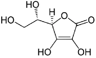
(5R)-[(1S)-1,2-dihydroxyethyl]-3,4-dihydroxyfuran-2(5H)-one (Figure 1). The analytical determination of AA has been reported by many methods, such as enzymatic,8d iodometric titration using
2,6-dichlorophenol-indophenol as indicator,8e spectroscopic,8f
chromatographic,8g fluorimetric8h and electrochemical.8i
Due to their quick response, high sensitivity, low detection limit and simple use, electrochemical methods are currently of much interest for AA determination, mostly by the electrocatalytic oxidation reaction on conventional electrodes. Though AA is an important antioxidant compound, it is difficult to determine it by direct oxidation on conventional electrodes because of interfering species such as dopamine and glucose.8j
In this work, we report the use of cyclic voltammetry (CV) technique to estimate the amount of ascorbic acid using a DNA based biosensor. The biosensor was developed by adsorbing DNA on a carbon paste electrode (CPE) electrodeposited with Prussian Blue (PB). Prussian blue is an efficient redox mediator which is efficiently used in many biosensors.9-11 DNA damage is caused by the Fenton
reaction and the effect of antioxidant on free radical scavenging is studied by using CV technique. The effect of DNA concentration, electrolyte, scan rate, interference and stability on the biosensor was also studied. The results were compared with pharmaceutical tablets containing ascorbic acid.
Experimental
Materials and Methods
All the chemicals were of analytical reagent grade. Disodium hydrogen phosphate was obtained from Nice Chemicals Pvt. Ltd., Kochi. Fructose, glucose, H2O2, H2SO4,
K3[Fe(CN)6] and KCl were obtained from Spectrum, Kochi.
NaCl and FeCl3 were obtained from Loba Chemie Pvt. Ltd.,
Mumbai. Ascorbic acid, paraffin liquid (heavy), NaOH and KH2PO4 were obtained from Merck, India. Graphite
(particle size < 50 µm) was obtained from Merck, Germany. Sucrose, oxalic acid, citric acid and DNA were obtained from HIMEDIA India Ltd., Mumbai. All aqueous solutions were freshly prepared using deionized water (Resistivity, ρ≥18 MΩ cm) from Elga Purelab Option-Q system (ELGA LabWater, UK).
Vitamin-C was purchased from the market in the form of a tablet, containing 500 mg Vitamin-C (C6H8O6). This was
used as a standard sample to compare the results.
Raman Spectra were measured in air using EZRaman-N-785. Electrochemical Workstation CHI 604D (CH Inc., USA was used for CV measurements.
Measurement setup
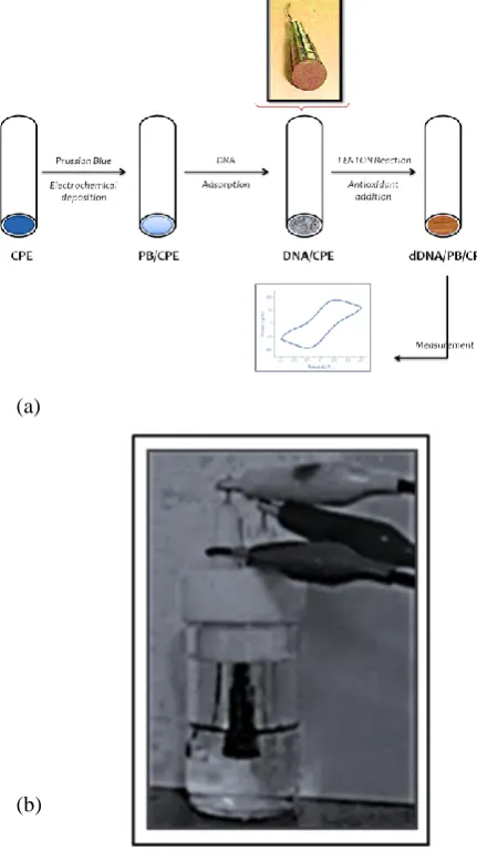
A conventional three electrode system is used with homemade CPE as working electrode, platinum wire counter electrode and Ag/AgCl reference electrode. The CPE is prepared by mixing paraffin liquid and graphite (30:70). The unmodified carbon paste is introduced into pipette tips as shown in figure 2a with measurement setup in figure 2b and the side with maximum area is polished by using white paper. Electrical contacts were taken out from the other side using copper wire.
Electrode preparation
The CPE is prepared by the procedure mentioned above and it is subjected to pretreatment steps12 in order to obtain
reproducible results from different electrodes. The following steps were carried out for pretreatment of the electrodes.
Newly prepared electrode surface was immersed in 0.1 M H2SO4 solution which was held with stirring at +1.30 V for
2 min. Then the electrode surface was immersed in 0.1 M NaOH solution and held with stirring at +1.25 V for 2 min. It was then immersed in 0.1 M H2SO4 solution and held for
2 min while stirring, at +1.30 V. Electrode was then washed in 0.01 M phosphate buffered saline (PBS) of pH 7.4 for 1 min with stirring.
Electrodeposition of PB
The pretreated CPEs were modified by means of electro-deposition and activation of a PB film.11The PB layer was
electro-deposited using cyclic voltammetry by applying 12 cyclic scans within the limits of –0.2 to +0.4 V at scan rate of 0.1 V s-1 in a solution containing 1.5 mM K
3[Fe(CN)6]
and 1.5 mM FeCl3 in 0.1 M KCl and 0.1 M HCl. These
PB/CPEs were cleaned in water and activated by applying another 50 cycles in electrolyte solution (0.1 M KCl and 0.1 M HCl), using the same protocol. Before being used, the PB/CPE was cleaned again in water for several seconds. This CPE electro-deposited with PB is mentioned as PB/CPE.
Assay procedure
Assay experiments were carried out in three steps. DNA immobilization on the PB/CPE (DNA adsorbed electrode is mentioned as DNA/PB/CPE), damage of oligonucleotide by the immersion of DNA/PB/CPE on the Fenton mixture (DNA damaged electrode is mentioned as dDNA/PB/CPE) and analyze the dDNA/PB/CPE. For adsorbing DNA on the PB/CPE, 30 µL of 1 mg/mL DNA (unless stated otherwise) solution is placed on the polished, pretreated PB/CPE.Then it is air-dried overnight. Later, the modified electrode is soaked in ultrapure water for 4 h, in order to remove the unabsorbed DNA from the electrode surface. The electrodes are freshly prepared and for each experiment new electrode is used. Then the DNA damage of different DNA/PB/CPE was checked. This is carried out by dipping the DNA/PB/CPE in a reagent solution containing iron (freshly prepared ferrous sulphate solution, 0.416 mM FeSO4) for 2
min in the presence or absence of antioxidant (1 mg/L, unless stated otherwise). The reaction was started by the addition of hydrogen peroxide (12.5 mM H2O2) into the
non-stirred solution. After a short period of time (1-2 min) the electrode was washed with 0.01 M PBS and then it is immersed in 0.1 M PBS (pH 7.4) and CV is recorded.
Results and Discussion
The studied system involved basically three important steps. It comprises of preparation of DNA-based biosensor, interaction of the biosensor with Fenton solution in absence and presence of antioxidant samples and the evaluation of the event that occurs at electrode surface.
(a)
(b)
Figure 2. (a) Schematic representation of the preparation of DNA/PB/CPE and measurement of DNA damage b) Measurement setup
Characterization of electrodes
Raman Spectroscopy - Carbon materials are studied
extensively with the help of Raman Spectroscopy. The study of carbon materials with Raman Spectroscopy showed that the frequencies and intensity of characteristic peaks changes with change in laser energy.13,14 Figure 3 shows the Raman
spectra of different materials and electrodes fabricated. Graphite powder showed the presence of D, G, D’, G1’, G2’
bands corresponding to 1336, 1542, 1646, 2705, 2741 cm-1. 14-16 The peaks at 1878, 1016, 954 cm-1 are characteristic for
Prussian blue.17 This indicates that PB has been electro
deposited on the CPE. The bands in the region of 600-800 cm-1 are assigned to breathing vibration in the rings in the
bases, the bands in the region 1200-1400 cm-1 are related to
the stretching vibration of the rings of the bases.18 After
DNA adsorption, the PB/CPE electrode showed peaks at
978, 1020, 1118 cm-1 corresponding to DNA backbone,
1285 cm-1 corresponds to cytosine, 1624 and 2783 cm-1
correspond to tyrosine, 1459 cm-1 corresponds to CH 2
deformation in DNA backbone19
Cyclic voltametry Studies - Electrochemical behaviour of the different electrodes is shown in figure 4. Figure 4a illustrates the response of DNA/CPE and CPE. No redox peaks were observed in these electrodes. This suggested the use of Prussian blue as the redox mediator.10 Different
electrolytes were also studied (Figure 4). PBS showed better redox peaks in all the experiments. The PB modified electrodes, DNA/PB/CPE (Figure 4b) shows perfect redox peaks, which is linked to the redox process of Prussian blue
Figure 3. Raman spectra of different materials and electrodes - Graphite, DNA, CPE, Pretreated CPE, PB/CPE, DNA/PB/CPE and dDNA/PB/CPE
Figure 5 (i). CV at different electrolytes (before causing DNA damage) (a) 1 mM K3[Fe(CN)6] in 0.1 M KCl (b) 6 mM K3[Fe(CN)6] in 1 M Na2SO4 (c) 0.1 M H2SO4 (d) PBS (0.1 M, pH 7.4)
Figure 5 (ii). CV at different electrolytes (after causing DNA damage) (a) 1 mM K3[Fe(CN)6] in 0.1 M KCl (b) 6 mM K3[Fe(CN)6] in 1 M Na2SO4 (c) 0.1 M H2SO4 (d) PBS (0.1 M, pH 7.4)
Figure 6. CV of DNA damage a) different scan rate b) different DNA concentrations c) varying ascorbic acid concentration. d) plot of ipa vs conc. of ascorbic acid.
An optimized concentration of FeSO4 and H2O2 is used for
the production of •OH.20 Experiments were conducted to
conclude the DNA damage is caused only by •OH and not by other reagents used, the DNA damage was caused by
•OH and not by the other reagents FeSO
4 and H2O2 used to
produce •OH is given in figure 4c. The CV of DNA/PB/CPE with damaged and undamaged DNA (Figure 4d) explains that there is an increase in current at the electrode after DNA damage induced by Fenton reaction. Before DNA damage the anodic and cathodic peak current (ipa and ipc) values were calculated as -7.99 and 8.59 µA. After DNA damage the values increased to -67.7 and 67.4 µA. With increasing scan rate (Figure 6a) both redox peak currents and peak-to-peak separation increased. The peak-to-peak currents are proportional to the square root of scan rate from 10 to 250 mVs-1 shown in figure 6a (inset), indicating that the redox
process is diffusion limited.21
Influence of ascorbic acid on DNA damage
The amount of DNA adsorbed on the PB/CPE electrode also has an effect on the response of the sensor (figure 6b). As the concentration of DNA increased from 0.2 mg/mL to 1.0 mg/mL the ipa and ipc increases linearly shown in Figure. 6b (inset). This may be due to the fact that as the concentration of DNA increases the number of reactive species available for oxidation also increases.22 The
overcrowding of DNA on the electrode can hinder the electron transfer rate.23 Ascorbic acid is a good
antioxidant.24 Ascorbic acid scavenges the •OH radical and
thereby prevents DNA damage. Figure 6c illustrates the effect of ascorbic acid on the DNA damage induced by •OH. As the concentration of ascorbic acid is increased, there is a decrease in the ipa and ipc. This shows there is a decrease in DNA damage, which may be due to DNA crowding. At above 2.2 mg/mL ascorbic acid concentrations, all the •OH is scavenged indicating no DNA damage. This is the optimal concentration of ascorbic acid for •OH scavenging under these experimental conditions. With further decrease of ascorbic acid concentration below 0.2 mg/L, the ipa and ipc value retails towards a constant value. Figure 6d shows the linear response between ascorbic acid concentration and anodic peak current.
Figure 7. CV of Vitamin-C tablet
Interference Study
The influence of the possible interfering compounds commonly present in pharmaceutical formulations such as glucose, fructose, sucrose, citric acid and oxalic acid were studied. These compounds were substituted for the ascorbic acid and the measurements were carried out at a concentration of 1 mg/L. The compounds glucose, fructose, sucrose and oxalic acid did not cause any interference and the ipa and ipc values were in par with the stationary values obtained with ascorbic acid concentrations less than 0.2 mg/L. The presence of citric acid caused a decrease in ipc indicating its function as an antioxidant.25
Stability of the Biosensor
The stability of the fabricated DNA biosensor was investigated. The DNA/PB/CPE electrode was stored in the refrigerator at 4 °C for 4 weeks and then the experiments were carried out. The results showed that the biosensor retains about 93% of its original response. This result show that the electrode can stably operate after a long period of storage.
Real sample analysis of the biosensor
To demonstrate the practical use of this biosensor samples were measured. Ascorbic acid tablets were used for this purpose. The tablets were weighed, ground, and an accurate weight of the powder assigned to contain 0.5 mg/L ascorbic acid was dissolved in ultrapure water, this solution is used as the antioxidant in the experiments and the CV is measured in 0.1 M PBS. Figure 7 gives the CV response for the real samples (n=3). The ipa values for the measurements were calculated as –79±0.36 µA. This when converted to concentration by using the calibration curve (Figure 6d), the concentration was 0.475±0.009 mg/L, RSD=2.77%. The recovery of the biosensor is 95%. The proposed results show that the biosensor can be used to determine ascorbic acid in pharmaceutical samples. The selectivity and sensitivity of the proposed biosensor appear to be a promising candidate for measurements in real samples.
Conclusions
We have demonstrated, here, a DNA based biosensor for the determination of ascorbic acid in pharmaceutical tablets. The electrodes were prepared by electro deposition of PB on CPE, and DNA is adsorbed on to this electrode, as the DNA/PB/CPE showed higher peak current than the PB/CPE, conforming PB as a perfect redox mediator. The Fenton reagents provided a good environment for the generation of
•OH and thereby inducing DNA damage. The antioxidant
scavenges the •OH thereby preventing DNA damage. The amount of DNA damage is related to the concentration of antioxidant. This is measured by using CV technique. The results showed a linear response in the range of 1.14 µM (0.2 mg/L) to 12.54 µM (2.2 mg/L), R2=0.9825. The sensor
exhibited high sensitivity and selectivity in the determination of ascorbic acid. This biosensor can be used for rapid, selective and precise in the determination of ascorbic acid in pharmaceutical samples. Disposable
electrode with suitable activation has extensively improved the selectivity and sensitivity of analytical systems especially towards biological targets and also clearly made it possible to detect certain analytes that are otherwise challenging. Usage of other nanomaterials via screen printing and synchronization of chemical modification with more sophisticated electrode designs can play a vital role in future applications.
Acknowledgement
Authors thank Department of Science and Technology, Govt. of India for the three R&D projects towards purchase of minor and major instruments required for material characterization, No. SR/FTP/CS-116/2007 dated 21st July
2008 (PI - Prof. Dr. techn. Murthy Chavali), No. SR/FT/CS-134/2010 dated 08th February 2012 (PI - Dr. Joseph Joly)
and No. GITA/DST/TWN/P-002/2009 dated 24th March
2009 (PIs - Prof. Dr. techn. Murthy Chavali & Prof. Dr. Wu Ren-Jang). MSN also thank Mr. R. Iseac, for his assistance in performing CV experiments.
References
1Pihlanto, A., Intl. Dairy J.,2006, 16, 1306.
2Prieto-Simon. B., Cortina, M., Campas, M., Calas-Blanchard, C., Sens. Actuat. B-Chem.,2008, 129, 459.
3Jaruga, P., Dizdaroglu, M., Nucleic Acids Res.,1996, 24, 1389.
4Madhavi, D. L., Deshpande, S. S., Salunkhe, D. K., Food Antioxidants: Technological: Toxicological and Health Perspectives, 1995, Taylor & Francis.
5Fenton, H., J. Chem. Soc. Trans.,1894, 65, 899.
6Halliwell, B., Gutteridge, J., Cross, C., J. Lab. Clin. Med., 1992, 119, 598.
7Sanchez-Moreno, C., Food Sci. Technol. Intl.,2002, 8, 121.
8(a) Buratti, S., Scampicchio, M., Giovanelli, G., Mannino, S., Talanta, 2008, 75, 312; (b) Zhang, L., Wang, Z., Xia, Y., Kai, G., Chen, W., Tang, K., Crit. Rev. Biotechnol., 2007, 27, 173; (c) Rice, M. E., Trends Neurosci.,2000, 23, 209; (d) Zhu, M., Huang, X.-M., Shen, H.-X., Li, R., Anal. Chim. Acta, 1996, 334, 303; (e) Svehla, G., Koltai, L., Erdey, L., Anal. Chim. Acta,1963, 29, 442; (f) Chan, H. S. O., Ng, S. C., Seow, S. H., Synth. Met.,1994, 66, 177; (g) Tai, A., Gohda, E., J. Chromatogr. B,2007, 853, 214; (h) Wu, X., Diao, Y., Sun, C., Yang, J., Wang, Y., Sun, S., Talanta,2003, 59, 95; (i) Ragupathy, D., Gopalan, A.I., Lee, K.-P., Sens. Actuat. B-Chem., 2010, 143, 696; (j) Zhao, Y., Gao, Y., Zhan, D., Liu, H., Zhao, Q., Kou, Y., Shao, Y., Li, M., Zhuang, Q., Zhu, Z., Talanta, 2005, 66, 51.
9Wang, X., Gu, H., Yin, F., Tu, Y., Biosens. Bioelectron.2009, 24, 1527.
10Zhao, H., Yuan, Y., Adeloju, S., Wallace, G., Anal. Chim. Acta,
2002, 472, 113.
11Salazar, P., Martín, M., O’Neill, R., Roche, R., González-Mora, Intl. J. Electrochem. Sci.,2012, 7, 5910.
12Gonzalez-Garcı́a, M. B., Costa-Garcı́a, An., Biosens. Bioelectron.,
2000, 15, 663.
13Dresselhaus, M. S., Dresselhaus, G., Saito, R., Jorio, A., Phys. Rep.,2005, 409, 47.
15Ferrari, A. C., Solid State Commun.,2007, 143, 47.
16Vidano, R., Fischbach, D. B., J. Am. Chem. Soc.,1978, 61, 13.
17Chaplin, T. D., Clark, R. J., and Beech, D. R., J. Raman Spectr.,
2002, 33, 424.
18Zhao, Y. D., Pang, D. W., Hu, S., Wang, Z. L., Cheng, J. K., Qi, Y. P., Dai, H. P., Mao, B. W., Tian, Z. Q., Luo, J., Anal. Chim. Acta,1999, 388, 93.
19Prescott, B., Steinmetz, W., Thomas, G., Biopolymers, 1984, 23, 235.
20Wang, Y., Xiong, H., Zhang, X., and Wang, S., Sens. Actuat. B-Chem.,2012, 161, 274.
21Jiang, Y., Zhang, X., Shan, C., Hua, S., Zhang, Q., Bai, X., Dan, L., Niu. L., Talanta, 2011, 85, 76.
22Ghanbari, K., Bathaie, S., Mousavi, M., Biosens. Bioelectron.,
2008, 23, 1825.
23Ricci, F., Lai, R. Y., Heeger, A. J., Plaxco, K. W., Sumner, J. J., Langmuir, 2007, 23, 6827.
24Carr, A., Frei, B., FASEB J,1999, 13, 1007.
25Van Den Berg, A., Halkes, S., Van Ufford, H. Q., Hoekstra, M., Beukelman, C., J. Wound Care, 2003, 12, 413.
![Figure 5 (i). CV at different electrolytes (before causing DNA damage) (a) 1 mM K3[Fe(CN)6] in 0.1 M KCl (b) 6 mM K3[Fe(CN)6] in 1 M Na2SO4 (c) 0.1 M H2SO4 (d) PBS (0.1 M, pH 7.4)](https://thumb-us.123doks.com/thumbv2/123dok_us/7831056.2089191/4.595.58.272.62.208/figure-different-electrolytes-causing-dna-damage-kcl-pbs.webp)
