AJPRHC Volume 4 Issue 3 95-104
AJPRHC
Research Article
NEW SPECTROPHOTOMETRIC METHODS DEVELOPMENT FOR THE DETERMINATION OF OSELTAMIVIR PHOSPHATE IN CAPSULES BASED ON THE OXIDATION REACTIONS OF THE
OLEFENIC DOUBLE BOND
B. KALYANA RAMU*2, M. SYAM BAB*1, U. VIPLAVA PRASAD1
For Author affiliations see end of the text
This paper is available online at www.jprhc.in
ABSTRACT
Simple, sensitive and selective spectrophotometric methods (M1 and M2) for the assay of oseltamivir phosphate (OP) through the olefenic double bond are proposed. Method M1 is based on the reaction of potassium permanganate to the olefenic double bond in OP and estimating the unreacted permanganate with fast green FCF (FGFCF). Method M2 involves the treatment of the olefenic double bond in OP with a Lemieux reagent (mixture of KMnO4 and NaIO4) and estimating the aldehyde formed with 3-methyl-2-benzothiazolinone hydrazone (MBTH). The color produced in M1 and M2 methods has maximum absorption at 620nm and 654nm respectively. Beer’s law obeyed in the concentration range of 4-20µg/ml and 4-12 µg/ml for method M1 and M2 respectively. The precision and accuracy of the methods are checked by the reported UV reference method. No interference was observed from the usually existing additives in pharmaceutical formulations and the applicability of the methods was examined by analyzing NATFLUE capsules containing OP. The both methods are found to be suitable for the determination of oseltamivir phosphate.
KEY WORDS: Assay, FGFCF, Lemieux reagent, MBTH, Beer’s law
INTRODUCTION
AJPRHC Volume 4 Issue 3 95-104
COOC
2H
5O
N
H
NH
2O
H
3PO
4.
Fig.1: Chemical structure of OP
In literature, OP can be identified by thin layer chromatography, specific optical rotation, infrared spectrophotometry and tests characteristic for ortho phosphates1, Determination, by International Pharmacopeia 2, can be done by high-performance liquid chromatography 1-2 or by titration with perchloric acid 1. Other analytical methods such as UV spectroscopy 3-5, visible spectrophotometric 6-9, colorimetric and LC10, spectrofluorimetric 11, HPLC with UV detection 12-19 and mass spectrometry20-23, Micellar electrokinetic chromatography24, capillary electrophoresis25 voltammetry26 and potentiometry 27 have been reported for the determination of OP in biological fluids and formulations. In the current scenario for the analysis of drugs many oxidants have been applied for oxidation. Very few workers have used oxidants to exploit olefenic double bonds. This paper describes analytical studies on the role of oxidants in the olefenic double bond in oseltamivir phosphate. It is well known that compounds of the R-CH=CH-R1 type undergo oxidation with acid permanganate, directly yielding a mixture of carboxylic acids , while in the presence of sodium metaperiodate(Lemieux reagent) they yield a mixture of aldehydes. Existing analytical methods reveal that relatively little attention has been paid to develop visible spectrophotometric methods. The low λmax value of the colored species in many of the reported methods prompted us to explore the possibility of developing new methods with a higher λmax. The efforts of this accord resulted in two such procedures, based on the oxidation of OP with KMnO4 and a Lemieux reagent and estimating the unreacted permanganate with FGFCF 28(Method M1) or the aldehyde formed with MBTH 29 (Method M2). The results of these methods are statistically validated. These methods can be extended for the routine quality control analysis of pharmaceutical products containing OP.
MATERIALS & METHODS (EXPERIMENTAL)
Apparatus and chemicals
A Milton Roy UV/Visible spectrophotometer model-1201 with 10mm matched quartz cells was used for all spectral measurements. All the chemicals used were of analytical grade and the solutions wee freshly prepared. Aqueous solutions of acid KMnO4 (BDH, 0.0316%, 2.0x10⁻³M for M1 or 0.01% 6.32x10⁻⁴M for M2 in 2.0M H2SO4), FGFCF solution (Chroma, 0.01%, 1.23x10⁻⁴M prepared by dissolving 10mg of fast green FCF in 100ml of 1.0M sulphuric acid. 10ml of this solution was further diluted to 100ml with the same strength of acid), Na2SO4 solution (BDH,4.2%, 1.0M, prepared by dissolving 4.2g of sodium sulphate in 100ml of distilled water), NaIO4 (Qualigens, 0.05%, 2.33x10⁻³M , prepared by dissolving 50mg of sodium meta periodate in 100ml of distilled water and MBTH (Fluka, 0.2%, 8.55x10⁻³M, prepared by dissolving 200mg of MBTH in 100ml of distilled water) were used.
Preparation of standard drug solution: A 1mg/mL solution was prepared by dissolving 100mg of oseltamavir phosphate in 100ml of 20% acetic acid and the stock solution was diluted stepwise with distilled water to obtain working standard solutions of 100µg/mL for the both methods (M1 and M2).
AJPRHC Volume 4 Issue 3 95-104 Determination of wavelength maximum (λmax):
Method M1:
Method M1: 5.0ml of Standard OP solution was transferred into 25ml calibrated tube. To this 0.5ml of KMnO4
(2.063x10⁻³M) solution was added. And the total volume in tube was brought to 10ml with distilled water and set aside for 10 min at laboratory temperature. Then 4.0ml each of the FGFCF (1.236x10⁻⁴M) solution and sodium sulphate (1.0M) solution were added successively After 10 min, the volume was made up to the mark with distilled water. In order to investigate the wavelength maximum, the above colored solution was scanned in the range of 400-660 nm UV-Visible spectrophotometers against a reagent blank. From the absorption spectra (Fig.2), it was concluded that 620nm is the most appropriate wavelength for analyzing OP with suitable sensitivity.
Fig.2: Absorption spectra of OP-KMnO4-FGFCF system
Method M2: 3.0 ml of Standard OP solution was transferred into 25ml calibrated tube. Then 0.5mL of KMnO4
(6.32x10⁻⁴M) and 1.0mL of NaIO4 (2.33x10⁻³M) solutions were added successively and kept in a boiling water bath for 10 min. After that 1.0mL of MBTH (8.56x10⁻³ M) solution was added and heated for another 3 min. The solution was cooled to room temperature and the total volume in tube was made up to the mark with distilled water. In order to investigate the wavelength maximum, the above colored solution was scanned in the range of 400-660 nm UV-Visible spectrophotometers against a reagent blank. From the absorption spectra (Fig.3), it was concluded that 654nm is the most appropriate wavelength for analyzing OP with suitable sensitivity.
AJPRHC Volume 4 Issue 3 95-104 Preparation of calibration curve:
Method M1: Aliquots of Standard OP solution (1.0-5.0mL, 100µg/mL) were transferred into a series of 25mL
calibrated tubes. To each tube 0.5mL of KMnO4 (2.063x10⁻³M) solution was added and the total volume in each tube was brought to10mL with distilled water and set aside for 10 min at laboratory temperature. Then 4.0mL each of the FGFCF solution and sodium sulphate solution were added successively and set aside for 5 min. for complete color development and then diluted to the mark with distilled water. The absorbance was measured at 620nm against a reagent blank prepared simultaneously. The decrease in absorbance corresponding to the drug content was obtained by subtracting the absorbance of the blank from that of test solution. The amount of drug was computed from its calibration graph (Fig.4).
Fig.4: Beer’s law plot of OP-KMnO4-FGFCF system
Method M2:
Aliquots of Standard OP solution (1.0-3.0mL, 100µg/mL) were transferred into a series of 25mL calibrated tubes. Then 0.5mL of KMnO4 (6.32x10⁻⁴M) and 1.0mL of NaIO4 (2.33x10⁻³M) solutions were added successively and kept in a boiling water bath for 10 min. After that 1.0mL of MBTH (8.56x10⁻³ M) solution was added and heated for another 3 min. The solution was cooled to room temperature and the total volume in each tube was made up to the mark with distilled water. The absorbance was measured after 5 minutes before 60minutes at 654nm against the reagent blank prepared similarly. The content of the drug computed from the appropriate calibration graph (Fig.5).
AJPRHC Volume 4 Issue 3 95-104 For pharmaceutical formulations:
Preparation of Sample solution
About 10 capsules were weighed to get the average weight and pulverized and the powder equivalent to 100mg of OP was weighed, dispersed in 25ml of isopropyl alcohol (IPA), sonicated for 30minutes and filtered through whatman filter paper no.41. The filtrate was evaporated and the residue was used for the preparation of working sample solution in the same way as under working standard solutions and analyzed under the procedures for the bulk samples. The UV method reported earlier using 0.1M NaOH (λmax =216nm) as a solvent was chosen as the reference method for ascertaining the accuracy of the proposed methods.
RESULTS AND DISCUSSION
The working conditions for the color developments of methods M1 and M2 were established by varying the parameters on at a time and keeping the others fixed and observing the effect produced on the absorbance of the colored species. The following experiments were conducted for this purposed and the conditions so obtained were incorporated into the recommended procedures. Method M1: 0.4 to 0.6ml of KMnO4 (2.063x10⁻³M) and a waiting
time of 5 to 15 min at room temperature were found to be adequate. A prolonged waiting period or an increase in temperature has no additional advantage. Hence 0.5ml of KMnO4 and a waiting time of 10 min were preferred. To maintain the linear relationship between the un-reacted KMnO4 and FGFCF, the addition of 4.0ml each of 1.23x10 -4
M and 1.0M sodium sulphate were found to be optimum. The consistency in absorbance after the gradual decrease of FGFCF was attained within 5 min and remained stable for further 45 min, and was measured at 620nm.
Method M2: In the first step, 0.4 to 0.6ml mL of 6.32x10⁻⁴M KMnO4 and 0.5to 1.5mL of (2.33x10⁻³M) NaIO4 and
heating on a boiling water bath for 10to20 min. were found to be necessary to get constant and reproducible absorbance values. The values were erratic beyond this range. In the second step, an optimum range of 0.5 to 1.5mL of MBTH (8.56x10⁻³ M) and further heating on a boiling water bath for 2 to5min were found to be adequate to get the maximum absorbance. Thus list volumes of KMnO4 (0.5mL, 6.32x10⁻⁴M), NaIO4(1.0mL, 2.33x10⁻³M) and MBTH(1.0mL, 8.56x10⁻³ M) and heating times of 10 before and 3 min after the addition of MBTH were preferred .The color product was stable for one hour and was measured at 654nm.
Analytical Data
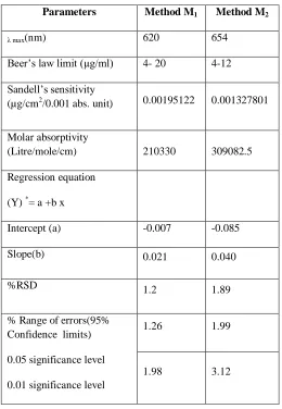
In order to test whether the colored species formed in the methods adhere to Beer’s law, absorbances at appropriate lengths of a set of solutions containing varying amounts of OP and specified amounts of reagents(as given the recommended procedures for each method ) were recorded against the corresponding reagent blank. The optical characteristics such as Beer’s law limit, Sandell‘s sensitivity, molar absorptivity, percent relative standard deviation, (calculated from the six measurements containing 3/4th of the amount of the upper Beer’s law limits), Regression characteristics like standard deviation of slope (Sb), standard deviation of intercept (Sa), standard error of estimation (Se) and % range of error (0.05 and 0.01 confidence limits) were calculated and the results are summarized in Table-1.Nat flu capsules were successfully analyzed by the proposed methods. The values obtained by the proposed and reference methods for formulations were compared statistically by the t-and F-test and found not to differ significantly. As an additional demonstration of accuracy, recovery experiments were performed by adding a fixed amount of the drug to the pre-analyzed formulations at three different concentration levels. MS Excel Software-2007 used for calculations and graphs. These results are summarized in Table-2.
AJPRHC Volume 4 Issue 3 95-104 Table - 1 Optical characteristics, precision and accuracy of the proposed methods
*Y = a + b x, where Y is the absorbance and x is the concentration of OP in µg/ml
Parameters Method M1 Method M2
λ max(nm) 620 654
Beer’s law limit (µg/ml) 4- 20 4-12 Sandell’s sensitivity
(µg/cm2/0.001 abs. unit) 0.00195122 0.001327801
Molar absorptivity
(Litre/mole/cm) 210330 309082.5 Regression equation
(Y) *= a +b x
Intercept (a) -0.007 -0.085
Slope(b) 0.021 0.040
%RSD 1.2 1.89
% Range of errors(95% Confidence limits) 0.05 significance level 0.01 significance level
1.26 1.99
AJPRHC Volume 4 Issue 3 95-104 Table-2 Analysis of OP in pharmaceutical formulations
Method *Formulations Labeled Amount (mg)
Found by Proposed Methods Found by Reference Method ± SD
#% Recovery by Proposed Method ± SD **Amount
found ± SD
t F
M1
Capsule-1 30 29.59±0.32 1.64 3.94 29.80±0.16 98.62±1.07
Capsule-2 75 74.44±0.49 0.54 3.35 74.65±0.91 99.26±0.66 M2 Capsule-1 30 29.79±0.13 0.35 1.55 29.80±0.16 99.29 ± 0.43
Capsule-2 75 74.49 ±0.46 0.41 3.8 74.65±0.91 99.30 ± 0.62 * Capsule- 1 and capsule-2: Natflu capsules of NATCO PHARMA LIMITED, Hyderabad (India)
**Average ± Standard deviation of six determinations, the t- and f-values refer to comparison of the proposed method with UV reference method. Theoretical values at 95% confidence limits t =2.57 and F = 5.05.
# Recovery of 10mg added to the pre analyzed sample (average of three determinations). Reference method (reported UV method) using 0.1M NaOH (λ max=216nm).
Chemistry of the color species:
Method M1: This method was based on the reaction of permanganate to the olefenic double bond of cyclo hexene
moiety in OP (first step) and an estimation of the un-reacted permanganate with FGFCF (second step). The probable sequence of reactions are presented in the scheme (Fig.6)
Method M2: This method involves the treatment of an olefenic double bond with Lemieux reagent (first step) and
AJPRHC Volume 4 Issue 3 95-104
O H
SO3Na
N
N
CH2
C2H5 SO3
-C2H5
CH2
SO3Na
O O
+
+
Drug analyte
KMnO4
+
+
KMnO4
+
Mn(II)unreacted
Step-2
KMnO4
+
Unreacted
+
FGFCF
Unreacted dye
+
Mn(II)Mixture of compounds with rupture of conjugate system (colorless)
(reproducible but not stoichiometric
as several alternate pathways are possible) (colored)
+
C C C C
Step-I
Fig.6: Probable scheme of the reaction for Method M1
Fig.7: Probable Scheme of the reaction for Method M2
AJPRHC Volume 4 Issue 3 95-104 The proposed methods are attractive ones compared to the reported methods since the proposed methods have higher λmax. The contaminants do not interfere in the color development. This was further proven by the resulting percentage recoveries of the proposed methods. Hence the proposed methods are simple selective and reliable and can be used for the assay of OP in bulk samples and pharmaceutical formulations. These methods can be used as alternative methods to the reported ones.
Ackmowledgements
The authors (MS Bab & BK Ramu) are thanks to the University Grants Commission, New Delhi for providing financial assistance under teacher fellow ship and also thanks to University authorities for providing facilities in this work.
REFERENCES
1. AHFS Drug information 2008, p.782.
2. WHO, Oseltamivir phosphate, Final text for addition to The International Pharmacopoeia. Document QAS/06.190/FINA., December 2008.
3. Chourasia A, Acharya S, Sahu S.K., Method development and validation of oselamivir phosphate in bulk drug by UV spectroscopy, International Journal of Pharmaceutical and Biological Research 2011, 2(5), 132-136.
4. Mehar D, Rajesh Y, Rajakumar V, Ravi kumar BVV, Arun kumar S. Development, estimation and validation of oseltamivir phosphate in bulk and in its pharmaceutical formulations by UV spectroscopic method, International Journal of Pharma and Bio Sciences 2010, 1(4), 579-586.
5. Rault C.S., Gharge DS, Dhabale PN, Onjari ID, Hosmani AH, Hosmani Abhijeet H. Development and validation of oseltamivir phosphate in fluvir by UV-spectrophotometer, International Journal of Phar Tech Research 2010, 2(1), 363-366.
6. Shanmuka kumar JV, Prasanthi S, Guravaiah M, Sakaram CH.B. Application of potassium permanganate to the spectrophotometric determination of oseltamivir phosphate in bulk and capsules, Asian Journal of Pharmaceutical Clinical Research 2012, 5(2), 18-22.
7. Ashish Ashok Thatte, Pramila T, Simple extractive colorimetric determination of oseltamivir phosphate by ion-pair complexation method in bulk and capsule dosage form. International Journal of Research in Pharmaceutical and Biomedical Sciences 2011, 2(2), 543-547.
8. Aydogmus Z, Sari F, A new spectrophotometric method for the determination of oseltamivir phosphate in bulk and capsules, OZET, 40, 47-55.
9. Jahn K, Deepthi M, Malipatil S, Spectrophotometric determination of oseltamivir phosphate in bulk drug and pharmaceutical formulations, Res J Pharm Biol Chem Sci 2010, 1(4), 933.
10. Michael D. Green, Henry Nettey and Robert A.Wirtz, Determination of oselamivir quality by colorimetric and liquid chromatographic methods, Emerging Infectious Diseases 2008,14(4),552-556.
11. Aydogmus Z, Simple and sensitive spectrofuorometric method for the determination of oseltamivir phosphate in capsules through derivatization with fluorescamine, Journal of Fluorescence 2009, 19(4), 673-679.
12. Aydogmus Z, Caglar S, Toker S, RP-HPLC methods for determination of oseltamivir phosphate in capsules and spiked plasma, Analytical Letters, 2010, 43(14), 2200-2209.
13. Lindegardh L, Hein TT, Farrar J, Shinghasivanon P, White NJ, Day NPJ, A simple and rapid LC assay for evaluation of potentially counterfeit Tamiflu, J Pharm BiomedAnal.2006,42,430-433.
14. Joseph-Charles J, Geneste C, Laborde -Kummer E, Gheyouche R, Boudis H, Dubost JP, Development and validation of a rapid HPLC method for the determination of oseltamivir phosphate in Tamiflue and generic versions, J Pharm Biomed Anal 2007,44,1008-1013.
AJPRHC Volume 4 Issue 3 95-104 16. Fuke C, Ihama Y, Miyazaki T, Analysis of oseltamivir active metabolite, oseltamivir carboxylate, in
biological materials by HPLC-UV in a case of death following ingestion of Tamiflue Legal Medicine 2008, 10, 83-87.
17. Bahrami G, Mohammadi Bahareh , Amir Kiani, Determination of oseltamivir carboxylic acid in human serum by solid phase extraction and HPLC with UV detection, Journal of Chromotography B, 2008, 864, 38-42.
18. Nagarajan JSK, Muralidharan S, A validated RP-HPLC method for estimation of oseltamivir in pharmaceutical formulation, Der Pharmacia Lettre, 2009, 1(1), 162-168.
19. Eisenberg EJ, Cundy KC, High performance liquid chromatographic determination of GS4071, a potent inhibitor of influenza neuraminidase in plasma by precolumn fluorescence derivatization with naphthalenedialdehyde, J Chrom B Biomed Sci Appl. 1998, 716, 267-273.
20. Wiltshire H, Wiltshire B, Citron A, Clarke T, Serpe C, Gray D, Herron, Development of a high-performance liquid chromatography-mass spectrometric assay for the specific and sensitive quantification of Ro-64-0802, an anti-influenza drug and its prodrug, oseltamivir, in human and animal plasma and urine. J Chrom B Biomed Sci App 2000,745, 373-388.
21. Lindegardh N, Hanpithakpong W, Wattanagoon Y, Singhasivanon P, White NJ, Day NPJ, Development and validation of a liquid chromatographic-tandem mass spectrophotometric method for determination of oseltamivir and its metabolite oseltamivir carboxylate in plasma, saliva and urine, Journal of Chromatography B, 2007,859, 74-83.
22. Heinig K, Bucheli F, Sensitive determination of oseltamivir and oseltamivir carboxylate in plasma, urine, cerebrospinal fluid and brain by liquid chromatography-tandem mass spectrometry, Journal of Chromatography B 2008, 876, 129-136.
23. Chang Q, Chow MS, Zuo Z, Studies on the influence of esterase inhibitor to the pharmacokinetic profiles of oseltamivir and oseltamivir carboxylate in rats using an improved LC/MS/MS method, Biomed Chromatography 2009, 23(8), 852-857.
24. Jabbaribar F, Mortazavi A, Jalali-Milani R, Jouyban A, Analysis of oseltamivir in Tamiflu capsules using Micellar electrokinetic chromatography, Chem Pharm Bull. 2008, 56, 1639-1644.
25. Laborde-Kummer E, Gaudin K, Joseph-charles J, Gheyouche R, Boudis H, Dubost Jean-Pierre, Development and validation of a rapid capillary electrophoresis method for the determination of oseltamivir phosphate in Tamiflu and generic versions, Journal of Pharmaceutical and Biomedical Analysis 2009, 50, 544-546.
26. Milka L. Avramov Ivic, Slobodan D. Petrovic, Dusan Z. Mijin, Katica M. Drljevic-Duric. The qualitative determination of oseltamivir phosphate in tamiflu capsule by cyclic voltammetry, Hem. ind. 2011, 65(1), 87-91.
27. Salem M. Hamza Nashwa MH. Rizk Hamdy AB. Matter. A Novel potentiometric method for determination of oseltamivir phosphate (Tamiflu) and its pharmaceutical applications. International Journal of Pharma. Research &Development 2010, 2(6), 1-11.
28. Gordon HT, Anal. Chem, 1951, 23(12), 1853.
29. Sawicki E, Mauser TR, Stanley TW and Elbert W, Anal Chem 1961, 33, 93. AUTHORS AFFILIATION AND ADDRESS FOR CORRESPONDENCE
1. Department of Organic Chemistry& Analysis of Foods Drugs &water Laboratories, School of Chemistry, Andhra University, Visakhapatnam-530003 Andhra Pradesh (India)