The later larval life of Arenicola marina L.
6
0
0
Full text
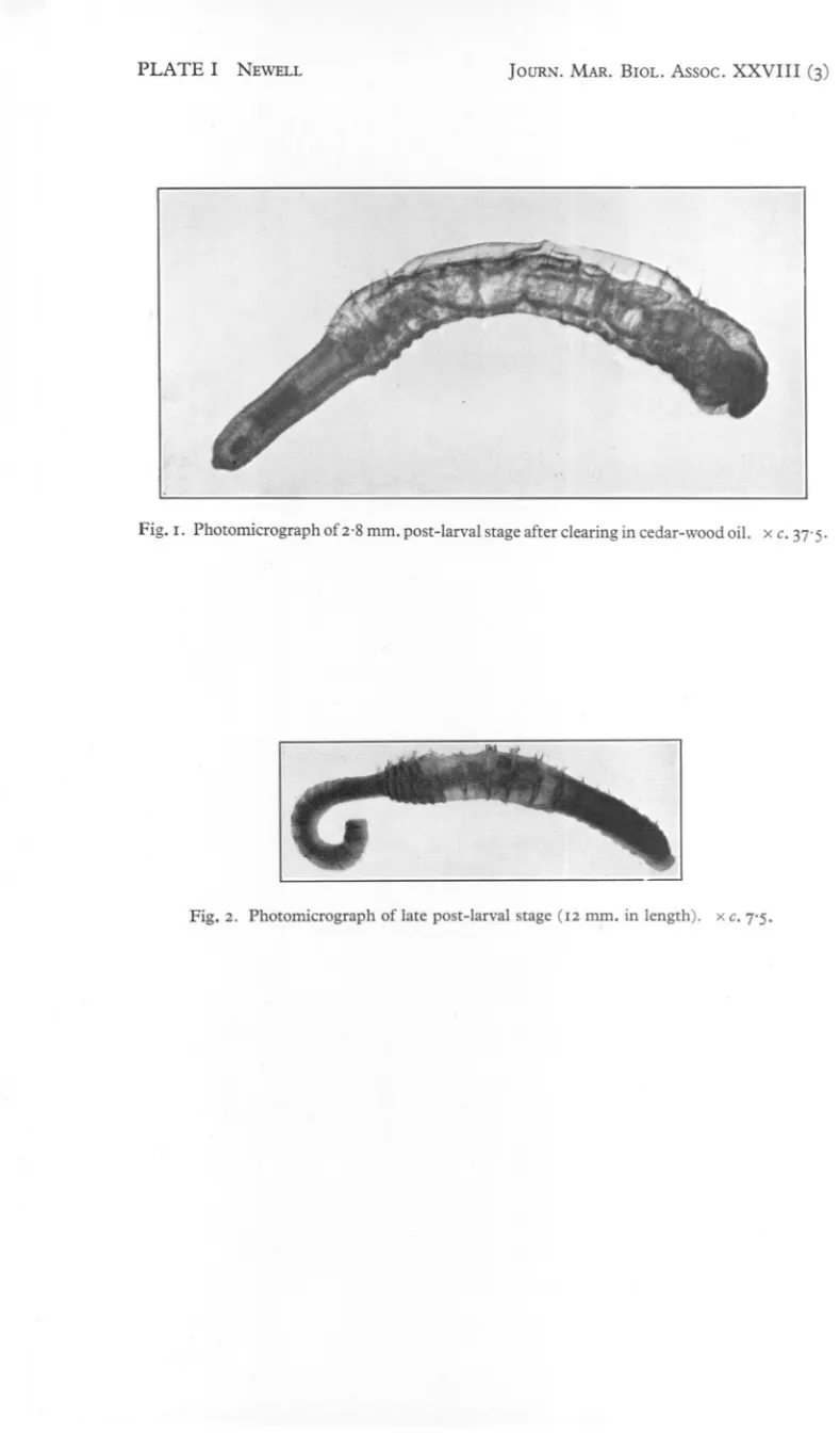
(2) 636. G. E. NEWELL. debris in suspension was caused to drain into the pit. The water in the pit was then poured, a pint or so at a time, through a small plankton net whose contents were then transferred at intervals to a jar of sea water. Usually about 20 gallons or so of sea water were filtered in this way, and the resulting suspension was then examined in Petri dishes for polychaete larvae. These were removed by means of a pipette and reserved for further examination. Trial and error soon showed that it was only in the pebbly Fucus zone that lugworm larvae were to be found and, it may be remarked, that it was only in this zone that the larvae of other polychaetes were at all plentiful. DESCRIPTION OF LARVAE. Larva with threechaetigeroussegments(Text-fig. I) On 17November 1947,and 29November 1948,larvaewiththree chaetigerous segmentswere found. Otherwisethey differedbut little in general appearance from larvae with two chaetigerspreviouslydescribed. Although able to swim by means of their cilia, they were usually enclosed in a layer of dense mucus. The prostomium was the largest division of the body and bore an apical tuft and one pair of eyes. A broad prototroch encircled the greater part of the prostomium and peristomium. The three segments behind the achaetous peristomium each bore a pair. of dorsal chaetae and a minute crotchet. The dorsal chaetae were of two kinds; a capillary chaeta and a shorter one which was broadly winged or spatulate. Both neurotroch and telotroch were still prominent. The total length of the, larva was 0'33 mm. Larva with four chaetigerous segments This was found on 14 November 1948. It measured 0'35 mm. in length and much resembled the three segment larva. Early post-larval stage (Text-fig. 2 and PI. I, fig. I) An early post-larval stage was collected on 14 February 1949. Its total length when alive varied between 2'5 and 3'2 mm. and when fixed was 2.8 mm. with a diameter of about 0'4 mm. in the thoracic region. The tail was about half the length of the thoracic region from which it was sharply distinguishable by being narrower and achaetous. The prostomium, still a prominent lobe, bore six eyes, two of which were larger than the others and probably represented the single pair of eyes persisting from earlier stages. The otocysts in the peristomium were clearly visible in transparency and contained minute particles of sand. Behind the peristomium were twenty segments bearing chaetae, but in the first chaetiger the chaetae, of which there was but one on each side, were difficult to detect. The next nineteen segments bore both notopodial and neuropodia! chaetae. The notopodial chaetae (Text-fig. 3A) were of two kinds: (a) capillary with narrow wings, and (b) shorter ones with broad wings..
(3) LARV..s\L. LIFE. 637. OF ARENICOLA. The neuropodial chaetae (Text-fig. 3B) were minute crotchets with a curved rostral hook and one (possibly two) smaller hooks. The number of crotchets varied from five in the second chaetiger to nine in the twentieth chaetiger.. E E VI 0. '". B A 0'1 mm.. Fig. 3.. Fig. I. ot.. Fig. 2. Text-fig. 1. Larva with three chaetigerous segments, dorsal view. XC. 175. Text-fig. 2. Early post-larval stage 2.8 nun. in length, side view. XC. 43. mth, mouth; prost., prostomium; ot., otolith; seg. 2, segment 2 (first chaetigerous segment). Text-fig. 3. Notopodial chaetae from third chaetigerous segment (A)and crotchets from third chaetigerous segment (B) of 2.8 nun. post-larval stage. xc. 400.. The tail consisted of about fifty segments and ended in a cone-like pygidium still bearing a trace of a telotroch. None of the segments of the body was as yet divided into annuli; neither were there any traces of gills. The gut was fully 41-2.
(4) 638. G. E. NEWELL. functional and the worm seemed to feed by means of its eversible proboscis which engulfed food in much the same way as does that of the adult. Faecal matter was seen in the intestine and the food probably consisted of fine organic debris. The main regions of the gut, as well as the blood vessels, which contained blood with haemoglobin, were already differentiated. Nevertheless, the mode of life differed from that of the adult in that the larva was enclosed in a thick mucous tube, a good deal longer than the total length . of the. worm, instead of inhabiting a burrow excavated in the soil. In fact, the smallest size of worm found living in the adult manner, as shown .. by the castings at the exit of the burrow, was 8 mID.in length. This figure is probably on the low side, owing to shrinkage during preservation and even larger specimens of up to 18 mID. in length, which were collected between 19 and 3° April 1949, were still living in mucous tubes like the earlier postlarval stages. Reference to PI. I, fig. 2, will show that one of these specimens, which was 12 mm. long, has all the main adult features including the thirteen pairs of branched gills, annulation of the segments and papillae on the tail segments. Indeed, except in size, so closely does it resemble the mature worms that detailed description seems unnecessary. Shortly after finding these specimens young worms began to make their appearance in the shoreward edge of the muddy sand flats which are the main habitat of the adults, and by 7 May 1949 their minute castings were seen in great profusion, indicating that seaward migration from the shore took place somewhat later in 1949 than in 1947. DISCUSSION. Thorson (1946) figures a larva (fig. 59, p. 1°9) which he found in a mid-water bottle collector at Ven which he believes should be referred to Arenicola marina. It is true that his larva bears some resemblances to a young lugworm, but although it measured 2.8 mID. in length the two main regions of the body are not demarcated. It also differs from early post-Iarvallugworms collected at Whitstable in that: (I) there is no clear prostomium; (2) in the absence of capillary chaetae, all the chaetae apparently are crotchets; (3) there are twentyone chaetigerous. segments; and (4) despite the general lack of differentiation the segments are already marked off into annuli. When it is remembered that this larva was collected in the bottle between 27 July and 13 August, that is, at a time when Thorson states that the gametes are far from mature in the adult worms, it becomes reasonably certain that the larva was not that of Arenicola marina and may not even have been that of any species of Arenicola. The 2.8 mID. post-larval stage from Whitstable also differs in several important respects from Benham's larva. Among these differences may be listed: (I) the general appearance; (2) the fact that Benham figures considerably fewer segments (about 30) in the tail region; (3) only two capillary chaetae are. ..
(5) PLATE I. NEWELL. Fig. I. Photomicrograph. JOURN. MAR. BIOL. Assoc.. XXVIII. (3). of 2.8 mm. post-larval stage after clearing in cedar-wood oil. x c. 37' 5.. Fig. 2. Photomicrograph. of late post-larval stage (12 mm. in length).. xc. 7'5..
(6) \. LARVAL. LIFE. OF ARENICOLA. 639. figured in the anterior dorsal bundles, whereas, since Benham's larvae is much larger, even more bristles would be expected, not less; (4) the absence of notopodiallobes; (5) apparently the notopodial bristles are all of one king., and (6) the smaller number of crotchets in the neuropodial regions. Ashworth (19°4) also succeeded in obtaining post-larval stages of Arenicola, but only when they were in the plankton. Reference to his descriptions and figures shows that they agree well with the older post-larval stages collected at Whitstable. SUMMARY. Larvae with three and four chaetigerous segments and also an early post-larval stage of Arenicola marina are described with figures. These are all bottomdwelling stages enclosed in mucous tubes and were collected from the pebble and Fucus zone at Whitstable. The finding of these larvae furnishes additional evidence for the view that, except when migrating in the water to new habitats, the lugworm possesses no pelagic larval stage in its life history. REFERENCES ASHWORTH,J. H., 1904. Arenicola. L.M.B.C. Memoir, No. XI. Liverpool. BENHAM,W. B., 1893. The post-larval stage of Arenicola marina. Journ. Mar. BioI. Assoc., Vol. III, pp. 48-53. THORSON,G., 1946. Reproduction and larval development of Danish marine bottom invertebrates, with special reference to the planktonic larvae in the Sound (0resund). Medd. Komm. Danmarks Fisk. Havund., K{l}benhavn,Ser. Plankton, Bd. 4, No. I, 523 pp. NEWELL,G. E., 1948. A contribution to our knowledge of the life history of Arenicola marina L. Journ. Mar. BioI. Assoc., Vol. XXVII,pp. 554-80..
(7)
Figure
Related documents
The nutrition policy sub-system in Zambia is therefore split between an international coali- tion promoting action on child stunting, and a national coalition focused on food
HCC is developing in 85% in cirrhosis hepatis Chronic liver damage Hepatocita regeneration Cirrhosis Genetic changes
amphivasal bundles to the center and few collateral bundles to periphery.The petiole exhibits epidermis with regularlly arranged cutinized cells, without
The synthesized MEPCMs were incorporated into white paint at three different concentrations, and temperature profiling revealed that the paint’s temperature
This narrative inquiry bears ontological, epistemological, and ethi- cal implications for teacher education programs because identity joins emotions and knowledge
In this project we were able to use a Software Defined Radio platform in order to achieve reliable communications between Unmanned Aerial Vehicles (UAVs).. Our goal was to
Using text mining of first-opinion electronic medical records from seven veterinary practices around the UK, Kaplan-Meier and Cox proportional hazard modelling, we were able to
After examining the circuit hierarchy, our tool creates a partition into blocks (modeling macros and standard cells) and analyses its array information to estimate dataflow
