In vitro thyroid hormone metabolism:
Effects of nuclear receptor activation on the metabolic profiles of thyroxine in rat and human hepatocytes
Vicki Michele Richardson
A dissertation submitted to the faculty of the University of North Carolina at Chapel Hill in partial fulfillment of the requirements for the degree of Doctor of Philosophy in the
Curriculum of Toxicology.
Chapel Hill 2013
Approved by:
Michael DeVito, PhD Louise Ball, PhD
ii ©2013
iii
ABSTRACT
VICKI RICHARDSON: In Vitro Thyroid Hormone Metabolism:
Effects of nuclear receptor activation on the metabolic profiles of thyroxine in rat and human hepatocytes
(Under the direction of Michael J. DeVito, PhD)
Thyroid hormones are critical in the normal growth and development of amphibians, birds, fish, and mammals. There are numerous xenobiotics that interfere with thyroid
iv
measurement of T4 metabolites. Here we report that hepatic glucuronidation may be a more important pathway for T4 metabolism in rats whereas T4 deiodination may be a favored pathway in humans. Following nuclear receptor activation, glucuronidation is a primary route of T4 metabolism in rat and humans hepatocytes. Agonists of CAR/PXR are more consistent in the induction of T4 glucuronidation in rat and human hepatocytes. We also show
similarities in the in vivo and in vitro effect on T4 metabolism in response to the
v
ACKNOWLEDGEMENTS
I would like to express my deepest gratitude to my advisor, Dr. Michael DeVito, whose guidance on this project has been invaluable. To my committee members, Drs. Louise Ball, Kim Brouwer, Edward LeCluyse, and Mary Paine, thank you for your time and
thoughtful criticism. I would especially like to thank Dr. Stephen Ferguson for his
willingness to provide his support and expertise on numerous occasions. I must also express my appreciation to Dr. Linda Birnbaum for her encouragement and counsel over the years.
To my wonderful group of friends, thank you for your forgiveness and words of comfort. To David Ross and Dr. David Szabo, thank you for the laughter and friendship. To my dearest friend Monica Richard, thank you for being the voice of reason and my greatest cheerleader.
vi
TABLE OF CONTENTS
LIST OF FIGURES ... ix
LIST OF TABLES ... x
LIST OFABBREVIATIONS ... xiii
CHAPTER 1: INTRODUCTION ... 1
A. Overview... 1
B. Nuclear Receptor ... 2
1. Aryl Hydrocarbon Receptor ... 2
2. Constitutive Androstane Receptor ... 3
3. Pregnane X Receptor ... 4
C. Thyroid Hormones ... 5
1. Synthesis and Feedback ... 5
2. Production and Clearance in Humans and Rats ... 6
3. Serum Binding Proteins ... 7
4. Metabolism ... 8
1. Deiodination ... 8
2. Glucuronidation ... 9
3. Sulfation ... 10
vii
D. Thyroid Hormone Disruption ... 12
1. Xenobiotics and Hepatic Thyroid Hormone Metabolism... 12
2. Animal to Human Extrapolation ... 13
E. Rationale for the Proposed Project ... 15
References ... 24
CHAPTER 2: IN VITRO METABOLISM OF THYROXINE BY RAT AND HUMAN HEPATOCYTES ... 35
A. Introduction ... 35
B. Materials and Methods ... 38
C. Results ... 45
D. Discussion ... 50
References ... 56
CHAPTER 3: EFFECTS OF PROTOTYPICAL NUCLEAR RECEPTOR AGONISTS ON THYROXINE METABOLISM IN RAT AND HUMAN HEPATOCYTES ... 75
A. Introduction ... 75
B. Materials and Methods ... 79
C. Results ... 83
D. Discussion ... 87
References ... 112
CHAPTER 4: EFFECTS OF BDE 47 ON THYROXINE METABOLISM ... 121
viii
B. Materials and Methods ... 125
C. Results ... 131
D. Discussion ... 134
References ... 168
CHAPTER 5: CONCLUSION ... 174
References ... 181
APPENDIX: POSSIBLE MECHANISMS OF THYROID HORMONE DISRUPTION IN MICE BY BDE 47, A MAJOR POLYBROMINATED DIPHENYL ETHER CONGENER ... 184
A. Introduction ... 184
B. Materials and Methods ... 187
C. Results ... 190
D. Discussion ... 193
ix
LIST OF FIGURES
Figure 1.1 Structure of thyroxine (T4) and triiodothyronine (T3) ... 19
Figure 1.2 Deiodinase conversion of thyroid hormones... 20
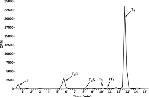
Figure 2.1 Representative chromatogram of T4 and its metabolites in medium ... 68
Figure 2.2 T4 metabolite levels in media during time in culture ... 69
Figure 2.3 Accumulation of [125I]-derived radioactivity in rat and human hepatocytes... 70
Figure 2.4 T4 metabolite levels in media during incubation time ... 71
Figure 2.5 [125I]-T4 clearance in the media of rat and human hepatocytes ...72
Figure 2.6 Metabolite levels in media of hepatocytes exposed to [125I]-T4 on culture day 6 for 24 hours ... 73
Figure 2.7 Comparison of metabolites in the media of SCRH and SCHH following treatment with PCB 153 ... 74
Figure 3.1 Comparison of appearance of metabolites in medium of SCRH following treatment with nuclear receptor agonists ... 99
Figure 3.2 Comparison of appearance of metabolites in medium of SCHH following treatment with nuclear receptor agonists ... 100
Figure 3.3 Accumulation of [125I]-derived radioactivity in rat and human hepatocytes following treatment with nuclear receptor agonists ... 109
Figure 4.1 Total serum T4 concentrations in rats treated with BDE 47 ... 145
Figure 4.2 Comparison of metabolites in the medium of SCRH treated with BDE 47 ... 146
Figure 4.3 Comparison of metabolites in the medium of SCHH treated with BDE 47 ... 148
Figure 4.4 Comparison of in vivo and in vitro responses based on either medium concentrations (in vitro) or estimated blood concentrations (in vivo) ... 150
x
LIST OF TABLES
Table 1.1 Physiological differences in thyroid hormone parameters ... 21
Table 1.2 Inconsistencies in T4-UGT activity and serum total T4 concentrations ... 22
Table 1.3 Key events in thyroid hormone disruption and relevance to humans ... 23
Table 2.1 Donor information for human hepatocytes ... 62
Table 2.2 Liver microsomes and cytosol ... 63
Table 2.3 Comparison of hepatic and intestinal T4G formation rates in microsomes and hepatic T4S formation rates in cytosols from rats and humans ... 64
Table 2.4 Mass balance of T4 and T4 metabolites after exposure of human hepatocytes on culture day 6 to 0.1µM [125I]-T4 ... 65
Table 2.5 Mass balance of T4 and T4 metabolites after exposure of rat hepatocytes on culture day 6 to 0.05µM [125I]-T4 ...66
Table 2.6 Intrinsic clearance of [125I]-T4 following treatment with PCB 153 ... 67
Table 3.1 Donor information for human hepatocytes ... 96
Table 3.2 TaqMan rat gene expression assays ... 97
Table 3.3 TaqMan human gene expression assays ... 98
Table 3.4 Fold change in rat hepatic P450 mRNA expression ... 101
Table 3.5 Fold change in human hepatic P450 mRNA expression ... 102
Table 3.6 Fold change in rat hepatic UGT mRNA expression ... 103
Table 3.7 Fold change in human hepatic UGT mRNA expression ... 104
Table 3.8 Fold change in rat hepatic SULT mRNA expression ... 105
Table 3.9 Fold change in human hepatic SULT mRNA expression ... 106
Table 3.10 Fold change in rat hepatic deiodinase I mRNA expression ... 107
xi
Table 3.12 Fold change in rat hepatic efflux and uptake transporter
mRNA expression ... 110
Table 3.13 Fold change in human hepatic efflux and uptake transporter mRNA expression ... 111
Table 4.1 Donor information for human hepatocytes ... 142
Table 4.2 Taqman rat gene expression assays ... 143
Table 4.3 Taqman human gene expression assays ... 144
Table 4.4 Intrinsic clearance of [125I]-T4 following treatment with BDE 47 ... 151
Table 4.5 Effects of BDE 47 on rat body weight, organ weights, and liver-to-body weight ratio ... 152
Table 4.6 Effects of BDE 47 on P450s in rat liver ... 153
Table 4.7 Effects of BDE 47 on P450s in rat hepatocytes ... 154
Table 4.8 Effects of BDE 47 on P450s in human hepatocytes ... 155
Table 4.9 Effects of BDE 47 on T4-UGT activity and Ugt mRNA expression in rat liver... 156
Table 4.10 Effects of BDE 47 on Ugt mRNA expression in rat hepatocytes ... 157
Table 4.11 Effects of BDE 47 on UGT mRNA expression in human hepatocytes ... 158
Table 4.12 Effects of BDE 47 on T4-SULT activity and SULT mRNA expression in rat liver ... 159
Table 4.13 Effects of BDE 47 on and SULT mRNA expression in rat hepatocytes ... 160
Table 4.14 Effects of BDE 47 on SULT mRNA expression in human hepatocytes ... 161
Table 4.15 Effects of BDE 47 on efflux and uptake transporter mRNA expression in rat liver ... 162
xii
Table 4.17 Effects of BDE 47 on efflux and uptake transporter mRNA
expression in human hepatocytes ... 164
Table 4.18 Effects of BDE 47 on thyroid hormone responsive
genes in rat liver ... 165
Table 4.19 Effects of BDE 47 on thyroid hormone responsive genes in
rat hepatocytes ... 166
Table 4.20 Effects of BDE 47 on thyroid hormone responsive genes in
human hepatocytes ... 167
Table A.1 Body weight, organ weights, and liver-to-body weight ratio ... 199
Table A.2 Fold change in hepatic UDPGT activity and mRNA expression ... 200
Table A.3 Fold change in hepatic cytochrome P450 activity and
mRNA expression ... 201
Table A.4 Fold change in hepatic efflux transporter mRNA expression ... 202
Table A.5 Fold change in hepatic thyroid hormone transporter
xiii
LIST OF ABBREVIATIONS
3MC 3-methylcholanthrene
ABC Adenosine triphosphate binding cassette ANOVA Analysis of variance
ACN Acetonitrile
AhR Aryl hydrocarbon receptor
ALB Albumin
BLQ Below limits of quantitation
β-NF Beta naphthoflavone
BDE-47 2,2′,4,4′-tetrabromodiphenyl ether
BSA Bovine serum albumin
Clint Intrinsic clearance
CAR Constitutive androstane receptor
CITCO 6-(4-chlorophenyl)imidazo [2,1-b] thiazole-5-carbaldehyde O-(3,4 dichlorobenzyl) oxime
CO2 Carbon dioxide
CYP Cytochrome P450
D1 Type 1 deiodinase
D2 Type 2 deiodinase
D3 Type 3 deiodinase
DE-71 Pentabromodiphenyl ether
DMSO Dimethyl sulfoxide
xiv EDTA Ethylenediaminetetraacetic acid
GAPDH Glyceraldehyde 3-phosphate dehydrogenase
HCl Hydrogen chloride
HEPES 4-(2-hydroxyethyl)-1-piperazineethanesulfonic acid HPT Hypothalamus-pituatary-thyroid
ITS+ Insulin, transferrin, and selenium complex], gentamicin, L-glutamine, and HEPES
MCT Monocarboxylate transporters MDR Multidrug resistance proteins
MgC12 Magnesium chloride
mRNA Messenger ribonucleic acid
MRP2 Mutidrug resistance-associated protein 2
MRP3 Mutidrug resistance-associated protein 3
N2 Nitrogen
Na Sodium
NaOH Sodium hydroxide
NHANES National health and nutrition examination survey NTCP Na+/taurocholate-cotransporting polypeptide OAT Organic anion transporter
OATP Organic anion transporting polypeptide
P450 Cytochrome P450
PAPS 3’-Phoshoadenosine-5’-phosphsulfate
PB Phenobarbital
xv PBDE Polybrominated diphenyl ether PCB Polychlorinated biphenyl
PCB 153 2,2′,4,4′,5,5′-hexachlorobiphenyl PCDD Polychlorinated dibenzo-p-dioxins PCDF Polychlorinated dibenzofurans PCN Pregnenolone-16α-carbonitrile PTU Propylthiouracil
PXR Pregnane x receptor
Rif Rifampicin
RT-PCR Real time polymerase chain reaction SCH Sandwich-cultured hepatocytes SCRH Sandwich-cultured rat hepatocytes SCHH Sandwich-cultured human hepatocytes SULT Sulfotransferase
T1/2 Half-Life
T3 3,3’,5-triiodothyronine
T4 Thyroxine
rT3 3,3’,5’-triiodothyronine
T2 3,3’-diiodothyronine
T4S T4-sulfate
T4G T4-glucuronide
xvi
TCDD 2,3,7,8-tetrachlorodibenzo-p-dioxin
TCPOBOP 1,4-bis[2-(3,5-dichloropyridyloxy)]benzene
Tg Thyroglobulin
TH Thyroid hormone
TPO Thyroperoxidase
TRH Thyrotropin-releasing hormone TSH Thyroid stimulating hormone
TTR Transthyretin
UPLC Ultra Performance Liquid Chromatography
UDP Uridine diphosphate
UDPGA Uridine diphosphate -glucuronic acid
UDPGT Uridine-diphosphate glucuronosyl transferase
CHAPTER 1
INTRODUCTION
A. Overview
Thyroid hormones (THs) are essential to development, growth and metabolism in humans with its most prominent effects occurring during fetal development and early childhood. The lack of TH in childhood delays growth and in adults the primary effect is an alteration in metabolism. A broad spectrum of xenobiotics decrease serum THs levels in rodents and these decreases are often associated with the induction of xenobiotics
metabolizing enzymes which result in increases in thyroxine (T4) metabolism and biliary elimination (Barter and Klaassen, 1992; Liu et al., 1995; Kolaja and Klaassen, 1998; Hood et al., 1999; Hood and Klaassen, 2000a; Hood and Klaassen, 2000b; Hood et al., 2003). Xenobiotics that activate the nuclear receptors, such as aryl hydrocarbon receptor (AhR), constitutive androstane receptor (CAR), and pregnane X receptor (PXR), appear to
2
which is associated with an increase in the biliary elimination of T4-glucuronides
(Oppenheimer et al., 1968; McClain et al., 1989; Wong et al., 2005). PB increases [125I] liver accumulation in rats (Kato et al., 2010) and increases biliary elimination of [125I]-T4 and [125I]-T4G suggesting the involvement of hepatic transporters in cellular uptake and biliary excretion (Mitchell et al., 2005; Visser et al., 2011). PB also decreases serum T4
concentrations in humans (Ohnhaus et al., 1981; Eiris-Punal et al., 1999). Mechanistic studies in humans are limited; consequently, the hypothyroid effect of PB in humans is thought to occur through the same mechanism as rats. Induction of hepatic UGTs by xenobiotics appears to be a common mechanism in thyroid hormone disruption in rodents; however, it is uncertain if this occurs in humans. The goals of this research are to examine the species differences in T4 metabolism using human and rat hepatocytes and further explore how these differences are affected by exposure to AhR, CAR, and/or PXR agonists.
B. Nuclear receptors
B.1. Aryl Hydrocarbon Receptor
3
HAHs, which include polychlorinated biphenyls (PCBs), polychlorinated dibenzo-p-dioxins (PCDD), and polychlorinated dibenzofurans (PCDF) (Safe, 1990). There are species
differences in the ligand binding domain of AhR, which may be responsible for species differences in ligand binding and response (Bisson et al., 2009; Pandini et al., 2009). 2,3,7,8-tetrachlorodibenzo-p-dioxin (TCDD) a prototypical ligand for AhR, produces toxic effects some of which include tumor promotion, dermal toxicity, disruption of endocrine homeostasis, wasting, and lethality. In humans, a point mutation in the ligand binding domain was found to lower the ability of AhR to bind TCDD by 10-fold compared to mouse AhR (Ema et al., 1994; Ramadoss and Perdew, 2004). TCDD and 3-methylcholanthrene (3MC) induced AhR-regulated genes, CYP1A1 and CYP1A2, to a greater degree in wild type mice compared to AhR humanized mice, demonstrating mouse AhR has a higher binding affinity for TCDD and 3MC than human AhR (Moriguchi et al., 2003).
B.2. Constitutive Androstane Receptor
4
example, 1,4-bis-[2-(3,5,-dichloropyridyloxy)] benzene (TCPOBOP) is a potent CAR activator in rodents, but lacks activity in humans (Moore et al., 2000). In contrast, 6-(4-chlorophenyl :imidazo [2,1-b] thiazole-5-carbaldehyde O-(3,4 dichlorobenzyl) oxime
(CITCO) is a potent activator of CAR in human, yet it lacks activity in rats and mice (Scheer et al., 2008). With the divergence of ligand-binding domains between the species, there are compounds such as phenobarbital (PB), which activate both human and rodent nuclear receptors orthologs (Moore, et al., 2002); however this indirect activation is independent of the ligand binding domain (Sidhu and Omiecinski, 1995; Honkakoski and Negishi, 1998).
B.3. Pregnane X Receptor
Pregnane X receptor (PXR) is a member of the NR1I2 family of nuclear receptors and acts as a xenosensor and transcriptional activator (Kliewer and Willson, 2002). PXR is activated by a variety of naturally occurring steroids of which pregnanes are the most potent (Kliewer et al., 1998; Lehmann et al., 1998). Closely related, PXR and CAR share a variety of ligands and target genes (Maglich et al., 2002); however, PXR is more promiscuous than CAR, because it binds to a wide range of compounds that are of different molecular weights and are structurally dissimilar (Jones et al., 2000). PXR exhibits a marked divergence across species within the ligand binding domain, where human and mouse PXR is only 76%
5
suggests that it is the species ortholog of the receptor and not the CYP3A gene promoter which determines the response.
C. Thyroid Hormones
C.1. Synthesis and Feedback
The main function of the thyroid gland is to produce hormones, T4 and the active hormone, triiodothyronine (T3) (Figure 1). T4 and T3 are synthesized in the thyroid gland by thyroperoxidase (TPO) where TPO converts iodide to iodine and then attaches it to tyrosine residues. Thyroglobulin (Tg), a large glycoprotein found within the thyroid follicular cells (thyrocytes), serves as a substrate for the synthesis and storage of THs and iodine. When thyroid hormone is needed, Tg is internalized at the apical pole of thyrocytes, where it is digested by proteases, resulting in free T4 and T3. After Tg digestion, T4 and T3 are released into the circulation.
TH is regulated by a negative feedback loop from the pituitary gland. When the pituitary gland detects too much Thyroid stimulating hormone (TSH) is the predominant regulator of thyroid hormone synthesis and release. Secreted from the pituitary, TSH interacts with its receptor (TSHR) in the thyrocytes to stimulate the accumulation of iodine and expression of the sodium/iodine symporter. T4 and T3 regulate the synthesis and release of TSH at the pituitary level, as well as indirectly by affecting TSH synthesis via their effects on the synthesis of TRH. TRH is the major positive regulator of TSH by activating the
6
release thyroid hormones into the blood stream as well as regulate the amount of iodine available to the cells,
C.2. Production and Clearance in Humans and Rats
Although rats are often used to examine extrathyroidal TH metabolism, there are important differences in the production and kinetics of THs in rats and humans (Table 1). In rats, normal plasma T4 concentrations are approximately 44nmol/L with a half-life (t1/2) of 0.5-1 day. T4 is cleared at a rate of about 50% per day in rats resulting in a daily production of approximately 1nmol/100g body weight(Bianco et al., 2002). The mean normal
concentration of total T4 in human plasma is approximately 100nmol/L (Larsen PR, 1998) amid a daily T4 production of 110 nmol/70kg body weight. With a half-life (t1/2) of 5-9 days, about 10% or nearly 110nmol of T4 is cleared from the circulation per day in humans. In rats and humans, plasma T3 is derived from thyroid gland secretion and extrathyroidal
deiodination; however, only 20% of plasma T3 comes from thyroid secretions in humans, whereas 40% is secreted from the thyroid in rats. Mean plasma T3 concentrations are
approximately 750pmol/L in rats. With a turnover rate of over 200% per day (t1/2 = 0.2 day), the daily production of T3 in rats is 415pmol/100g. Humans have a mean normal plasma T3 concentration of 1.8nmol/L with a daily production rate of 50nmol/70kg body weight and a t1/2 of 1.5 days; consequently, the fractional turnover rate of T3 in plasma is about 65% per day. In humans, of the daily production of T4 (110nmol/day), 30-40% is converted to T3 (40nmol) by peripheral deiodination, while the remaining amount (10nmol) is excreted directly from the thyroid(Larsen PR, 1998). In rats, approximately 415 pmol of T3 is
7
225 pmol of T3 per day, while the remaining amount (190 pmol) is excreted directly from the thyroid (Oppenheimer et al., 1972; Bianco, et al., 2002). The molar ratio of T4/T3 in rat thyroid is 8:1 where as in humans the ratio is higher (15:1)(Abrams and Larsen, 1973; Izumi and Larsen, 1977). In comparing T4/T3 thyroidal secretion, the ratio is 5:1 in rats, while in humans the ratio is 11:1. This indicates there are small contributions of thyroidal deiodinases to T4 to T3 conversion in both species.
C.3. Serum Binding Proteins
Specific proteins carry thyroid hormones in the blood and their high affinity binding to T4 and T3 are essential to the availability of the hormones to target tissues. In humans, three major proteins bind (THs) in serum: thyroxine-binding globulin (TBG), transthyretin (TTR) and albumin (ALB) (Schussler, 2000). TBG is a low capacity-high affinity binder of thyroid hormones, albumin is a high capacity-low affinity binder and TTR is an intermediate capacity and affinity binder (Power et al., 2000). T4 affinity is greatest for TBG (Ka=1x10⁻¹⁰ M), intermediate for TTR (Ka=7x10 -7 M), and lowest for ALB (Ka= 7x10 -5 M). The affinity of T3 for the binding proteins is lower than that of T4, where the affinity for TBG is
8
affinity of TTR for THs is less than the binding affinity of THs for TBG (Benvenga et al., 2002). In rodents, T4 is bound to TTR where as T3 is mainly bound to the least abundant TBG or albumin (Savu et al., 1987). T4-TTR binding is thought to be more susceptible to chemical interference than T4-TBG binding (Munro et al., 1989). This suggests that as a major circulating T4 binding protein, TTR may be important in the disruption of TH homeostasis in rodents.
C.4. Metabolism
C.4.1 Deiodination
Tissue deiodinases are critical in the extrathyroidal conversion of T4 into its biologically active form, T3. There are two isoenzymes that convert T4 to T3: type I 5’-deiodinase (D1) and type 2 5’-deiodinase (D2). D1 is located primarily in the liver, kidney and thyroid, and D2is located primarily in the brain, thyroid, anterior pituitary, brown adipose, placenta and skeletal muscle. D1 is responsible for most of the conversion of T4 to T3 in the blood, while D2 provides conversion of T4 to T3 for intracellular use. Type 3 deiodinase (D3) is located in the brain, placenta, fetal tissues and uterus during pregnancy and is
9
C.4.2. Glucuronidation
Glucuronidation involves the transfer of a sugar moiety on to uridine diphosphate (UDP)-glucuronic acid (UDPGA) to a substrate. The enzymes responsible for
glucuronidation of THs are UDP-glucuronoysyltransferases (UGT), which are located mainly in the endoplasmic reticulum of liver cells. The TH glucuronide conjugate is excreted in bile, which may represent a reversible pathway as intestinal bacteria can hydrolyze the conjugates creating an enterohepatic cycle enabling reabsorption of free THs. Induction of UGTs by xenobiotics may play an important role in chemically induced decreases in
circulating THs (Hood and Klaassen, 2000a; Klaassen and Hood, 2001; Zhou et al., 2001). UGTs are also regulated by AhR, CAR and PXR (Maglich, et al., 2002; Bock and Kohle, 2004; Wagner et al., 2005). The degree to which chemicals reduce serum T4 is not always correlated with the increase in T4-UGT activity (Hood, et al., 2003; Richardson et al., 2008). There are also differences between rats and mice in which Kenechlor-500, a mixture of polychlorinated biphenyls (PCBs) with PB-like effects on XMEs, reduces circulating levels of T4 in both rats and mice, but induces UGT activity in rats but not mice (Kato et al., 2003). In addition, Kenechlor-500 causes decreases in circulating T4 concentrations in the UGT1A deficient Gunn rats demonstrating that the decreases in circulating T4 is not necessarily solely dependent upon the induction of TH glucuronidation dependent (Kato et al., 2004). As a result, it can be argued that UGT induction alone is not a uniform marker of the ability of chemicals to cause a reduction in serum TH, which could explain the inconsistencies
10
C.4.3. Sulfation
Sulfotransferases are a cytosolic group of phase II metabolizing enzymes important for the inactivation and elimination of endogenous and exogenous compounds. Sulfation is a conjugation reaction in which a sulfate group from a sulfate donor, 3-phoshoadenosine-5-phosphsulfate (PAPS), is transferred to a substrate. Sulfoconjugation of THs is an alternative metabolic pathway that enhances enzymatic deiodination and facilitates their biliary and urinary excretion. The TH-sulfate conjugate is rapidly deiodinated by type I deiodinase through successive deiodinations of the tyrosyl (inner) and phenolic (outer) rings, releasing iodine into the circulation for reutilization by the thyroid. Sulfated conjugates are rapidly cleared in rats when deiodinase activity is inhibited. TH sulfation is also thought to serve as a reservoir from which unconjugated hormone can be liberated through sulfatases in tissues or in intestinal bacteria (Hazenberg et al., 1988; Kung et al., 1988).
11
activities toward THs suggests that human SULT1A1 is a prominent sulfotransferase in liver and kidney.
C.4.4. Transporters
Several AhR, CAR and PXR regulated transporters are known to actively transport glucuronides and/ or THs. These comprise major efflux transporters in the ATP binding cassette (ABC) gene family, including multidrug resistance-associated proteins (MRPs), multidrug resistance proteins (MDRs). There is also increasing evidence that uptake transporters such as, organic anion transport protein (OATP), and monocarboxylate
transporter (MCT) are important in the intracellular access to THs for metabolism (Friesema et al., 1999; Jansen et al., 2005). There is a correlation between induction of hepatic UGTs (Ugt1a1), multidrug resistance protein-associated protein (Mrp2), and organic anion
transporting proteins (Oatp1 and Oatp2) mRNA levels, with decreases in serum TH
concentrations in treated with rats treated with 4-(3-pentylamino)-2, 7-dimethyl-8-(2-methyl-4-methoxyphenyl)-pyrazolo-[1,5-a]-pyrimidine (DMP 904)(Wong, et al., 2005; Lecureux et al., 2009). There are indications that multidrug resistance proteins (MDRs) are important in TH efflux (Mitchell, et al., 2005). These studies indicate that active transport along with glucuronidation and altered serum binding are possibly involved in TH decreases.
12
using oocytes injected with human and rat NTCP or OATP mRNA, observed a significant uptake of thyronines (Friesema, et al., 1999). Conversely, rat hepatocytes incubated with a transport-blocking antibody resulted in a decrease clearance of the iodothyronines from the media and iodide into the media, confirming that active transport is essential to the uptake and metabolism of THs (Hennemann et al., 1986). De Jong et al., (de Jong et al., 1993) demonstrated that Ouabain, a Na+ gradient inhibitor, reduces the amount of T4 taken up into human hepatocytes and reduces the amount of iodide cleared into the media. Together, these studies indicate that TH transport is rate limiting for subsequent metabolism and suggest that transporters may serve a regulatory role in bioavailability and metabolism.
D. Thyroid Hormone Disruption
D.1. Xenobiotics and Hepatic Thyroid Hormone Metabolism
Although xenobiotics can disrupt TH homeostasis by directly disrupting the functions of the thyroid, increases in extrathyroidal metabolism are also involved in facilitating
changes in TH homeostasis. As a major site of xenobiotic metabolism, the liver is important in metabolism of THs in humans and rodents.
Xenobiotics decrease serum TH concentrations through hepatic mechanisms
13
elimination of T4-glucuronides (Oppenheimer, et al., 1968; McClain, et al., 1989);
confirming that enzyme inducers mediate the increase in biliary elimination of THs via the induction of hepatic metabolizing enzymes.
D.2. Animal to Human Extrapolation
There is data supporting the induction of hepatic metabolism by which xenobiotics decrease circulating TH concentrations in rats; however, the mechanisms in humans are unclear (Table 3). Specifically, PB induces CYP2B enzymes in rat and human hepatocytes, implicating CAR as a modulator of effects on circulating TH concentrations (Barter and Klaassen, 1994; Madan et al., 2003). While PB decreases serum TH concentrations in humans and rats (McClain et al 1989, Benedetti et al., 2005) only the increased biliary elimination of T4-glucuronide has been observed in rat models.
Although several studies show that UGT1A1 and UGT1A3 are important in hepatic metabolism of T4, Tong et al., (2007) demonstrated that UGT activities toward THs were higher in mouse and rat liver microsomes as compared to human, suggesting that UGTs may play a more significant role in the metabolism of THs in rodents than in humans.
Conversely, sulfotransferase may be more important in the metabolism of THs in humans. SULT1E1 conjugates THs in humans, yet not in rats (Kester et al., 1999; Kester et al., 2003). This divergence in substrate specificity between the species, suggests that sulfation may be more important in TH metabolism in humans than in rats. In general, human hepatic SULTs have lower Kms than UGTs toward THs, suggesting that sulfation may play a more
14
increased hepatic conjugation plays as large of a role as suspected. For instance, studies using UGT1A-deficient Gunn rats exposed to PB or PCBs demonstrate that decreases in serum total T4 are not necessarily glucuronidation-dependent (Collins and Capen, 1980; Kato, et al., 2004). Many studies have also reported the inconsistencies in hepatic T4-UGT activity and decreases in circulations in which the degree of T4 decreases do not always correlate with increases in T4-UGT activity (see Table 3). Because of these inconsistencies, it is uncertain how relevant extrathyroidal TH disruption in animals is to humans.
Reports indicate that hepatic transport may also be responsible for decreases in serum TH concentrations. For example, correlations between the mRNA induction of hepatic UGTs and hepatic uptake (Oatp1 and Oatp2) transporters mRNA levels, with decreases in circulating TH concentrations were observed (Wong, et al., 2005; Lecureux, et al., 2009). Wong et al. (Wong, et al., 2005) reported a greater biliary elimination of parent T4 versus glucuronide conjugated T4 following exposure to 4-(3-pentylamino)-2, 7-dimethyl-8-(2-methyl-4-methoxyphenyl)-pyrazolo-[1,5-a]-pyrimidine (DMP 904) along with an increase in hepatic Mrp2 mRNA levels in rats. DMP 904 also induced hepatic CYP2B1 and CYP3A1 mRNA levels in rats, suggesting that it acts as an activator of CAR or PXR. Together these studies show that hepatic uptake and efflux transporters play a role in increasing the
metabolic availability of T4 as well as facilitating the clearance of unconjugated hormone. TH homeostasis depends greatly on transport by serum binding proteins. In rats, TH is largely bound to transthyretin (TTR) while in humans; TH is mostly bound to thyroid binding globulin (TBG). The differences in protein binding may cause THs in rats to be more
15
may be responsible for the sensitivity of rats to TH toxicants. Although hydroxylated PCBs and PBDEs have been shown to bind to human and rodent TTR ex vivo, there are fewer compounds that compete for T4-TBG binding (Cheek et al., 1999; Hallgren and Darnerud, 2002). This suggests that TH displacement from its binding protein may not necessarily be of concern for humans.
E. Rationale for the Proposed Project
TH concentrations are regulated by not only the hypothalamus-pituitary–thyroid axis, but also hepatic metabolism, and elimination; therefore, the liver is essential to the
extrathyroidal regulation of THs. Decreases in circulating TH concentrations by the
induction of microsomal enzyme inducers have been linked to the increase in TH metabolism and biliary elimination (Barter and Klaassen, 1992; Liu, et al., 1995; Kolaja and Klaassen, 1998; Hood, et al., 1999; Hood and Klaassen, 2000a; Hood and Klaassen, 2000b; Hood, et al., 2003). The role of microsomal enzyme inducers on hepatic UGTs and their effects on circulating TH concentration have been thoroughly examined. THs are not only
glucuronidated but are also sulfated in the liver, for possible elimination into the bile. Although xenobiotics can cause decreases in circulating THs, xenobiotics do not necessarily induce hepatic UGTs to the same degree; therefore, it is important to determine why this occurs. Attempts have been made to examine the hepatic metabolism of THs in rats and humans following xenobiotic exposures, but an investigation of species differences and what role the liver plays still need to be explored.
16
2001). For example, Ponsoda et al. (Ponsoda, et al., 2001), correlated the metabolites of aceclofenac, an anti-inflammatory analgesic drug, found in human urine, with the metabolites formed in human hepatocytes supporting the use of hepatocytes to predict what happens in vivo. Furthermore, hepatocytes express nuclear receptor and therefore respond to xenobiotics through the induction of metabolizing enzymes. Hepatocytes also express membrane bound transporters, which can influence the intracellular concentration of compounds therefore; hepatocytes can be used to define the mechanisms by which AhR, CAR, and PXR agonists can alter TH metabolism and clearance.
Decisions concerning risk assessment have been based on animal to human extrapolations; therefore in vivo-in vitro comparisons are very important in the decision process. In vivo responses to toxicity usually involve multiple mechanisms and multicellular interactions. However, by focusing on a single cell type, data from in vitro studies becomes invaluable in evaluating the assumptions of specific mechanisms of action between species in toxicity studies. The evaluation of potential species differences in hepatic T4 metabolism and the effect AhR, CAR, and PXR activation have on T4 metabolism were examined the
following aims
Aim 1: Compare T4 metabolic profiles and clearance in rat and human hepatocytes. T4
17
thyroid hormone clearance will provide a better understanding of the differences between rat and human TH metabolism
a. Establish a radiometric UPLC method for determining T4 metabolites (T4G, T4S, T3, rT3) in the media of hepatocytes.
b. Compare clearance and metabolic profile by incubating T4 with rat and human hepatocytes.
Aim 2: Determine hepatic clearance and the metabolic profile of THs in rat and human
hepatocytes following exposure to Ahr, CAR, and PXR agonists. Metabolism plays a
major role in the homeostasis of THs and because many xenobiotics can increase the metabolism of THs, it is important to understand the impact xenobiotics have on TH homeostasis. A major pathway of TH metabolism in the liver is the conjugation of the hormones to glucuroinides or sulfates. Uridine 5-diphosphate-glucuronosyltransferase (UGT) and sulfotransferase (SULT) mediate the conjugation of THs. The activation of these AhR, CAR or PXR by xenobiotics can induce metabolizing enzymes (UGT and SULT). The induction of UGTs and SULTs is thought to increase the metabolism and subsequent
elimination of THs. To examine differences in human and rat hepatic metabolism of TH, this study will analyze metabolic profiles and gene involved in the metabolism of xenobiotics and THs.
18
b. Examine mRNA expression of genes related to xenobiotic and thyroid hormone metabolism. Correlate changes in mRNA expression with changes in the metabolic profile of T4 following exposure to AhR, CAR and PXR agonists.
Aim 3: Compare the effects BDE-47 on T4 metabolism in rats and metabolic profiles
and clearance in rat and human hepatocytes.BDE-47, a CAR/PXR agonist, decreases T4
19
\
Figure 1.1 Stucture of thyroxine (T4) and triiodothyronine (T3)
OH O
I
I I
HO CH2
NH2 O
CH C
I
Thyroxine (T4)
OH O
I
I I
HO CH2
NH2 O
CH C
20 From (Bianco and Kim, 2006)
21
Table 1.1
Physiological differences in thyroid hormone parameters
Human Rat Reference
a
Mean of reference range of 60-140nM in adult humans
TBG (Serum concentration) Present (0.02mg/ml) Not Present
(Wade et al., 1988)
TTR (Serum concentration) Present (0.2mg/ml) Present (0.5mg/ml) (Benvenga and Robbins, 1998)
T4 t1/2 (days) 5-9 0.5- 1
(Capan, 2001; Bianco, et al., 2002)
T3 t1/2 (days) 1 0.25
(Bianco, et al., 2002)
Mean Serum T4
(nM) 100.0
1,a
43.82
1
(Stockigt, 2003) 2
(Woody et al., 1998)
Mean Serum T3
(nM) 1.9
1
0.92
1
(Stockigt, 2003) 2
22
Table 1.2
Inconsistencies in serum T4 and T4-UGT activity
Chemical Nuclear Receptor T4-UGT Serum T4 Reference
Activity
↑ = increase ↓ = decrease ↔ = no change
β-NF AhR ↑ ↑ ↑ ↓ ↓ ↓
(Hood and Klaassen, 2000)
3-MC AhR ↑ ↑ ↓ ↓
(Hood and Klaassen, 2000)
PCB AhR/PXR ↑ ↑ ↓ ↓ ↓
(Hood and Klaassen, 2000)
PCN PXR ↑ ↑ ↓ ↓
(Hood and Klaassen, 2000)
PB CAR ↑ ↑ ↓ ↓ ↓
(Hood and Klaassen, 2000)
DE 71 AhR/CAR/PXR ↑↑ ↓ ↓
(Zhou et al., 2002)
BDE 47 CAR ↔ ↓
(Richardson et al., 2008) PB/PCB
(Gunn Rat) AhR/CAR/PXR ↔ ↓ ↓ ↓
23
Table 1.3
Key events in thyroid hormone disruption and relevance to humans
Key Event Evidence in Rats Evidence in Humans Reference
Nuclear Receptor activation (CAR)
Yes
In vivo and in vitro
Yes In vitro
(Barter and Klaassen, 1994; Hood and Klaassen,
2000a)
Hepatic UGT Induction
Yes
In vivo and in vitro
Yes In vitro
(Barter and Klaassen, 1994; Hood and Klaassen,
2000a)
Increased TH or Conjugated TH Biliary Elimination
Yes
In vivo and in vitro No Data
(Kato et al., 2005; Wong, et al., 2005)
Hepatic Transporter Induction
Yes
In vivo No Data
(Ribeiro et al., 1996; Mitchell, et al., 2005; Wong, et
al., 2005)
TTR Binding
Yes Ex vivo; hydroxylated compounds bind to
rTTR1
Yes Ex vivo; hydroxylated compounds bind to
hTTR
(Cheek, et al., 1999);(Hallgren and
Darnerud, 2002; Meerts et al., 2002)
TBG Binding No Data
(TBG not present)
Yes Ex vivo
(Cheek, et al., 1999)
Serum TH Decrease Yes
In vivo
Yes In vivo
(Cavlieri et al., 1973; Brucker-Davis, 1998)
Increased hepatic TH uptake/accumulation
Yes
24
REFERENCES
Abrams, G. M. and Larsen, P. R. (1973). Triiodothyronine and thyroxine in the serum and thyroid glands of iodine-deficient rats. J Clin Invest 52(10), 2522-31.
Aleksunes, L. M. and Klaassen, C. D. (2012). Coordinated regulation of hepatic phase I and II drug-metabolizing genes and transporters using AhR-, CAR-, PXR-, PPARalpha-, and Nrf2-null mice. Drug Metab Dispos 40(7), 1366-79.
Alnouti, Y. and Klaassen, C. D. (2008). Regulation of sulfotransferase enzymes by prototypical microsomal enzyme inducers in mice. J Pharmacol Exp Ther 324(2), 612-21.
Barter, R. A. and Klaassen, C. D. (1992). UDP-glucuronosyltransferase inducers reduce thyroid hormone levels in rats by an extrathyroidal mechanism. Toxicol Appl Pharmacol 113(1), 36-42.
Barter, R. A. and Klaassen, C. D. (1994). Reduction of thyroid hormone levels and alteration of thyroid function by four representative UDP-glucuronosyltransferase inducers in rats. Toxicol Appl Pharmacol 128(1), 9-17.
Beischlag, T. V., Luis Morales, J., Hollingshead, B. D. and Perdew, G. H. (2008). The aryl hydrocarbon receptor complex and the control of gene expression. Crit Rev Eukaryot Gene Expr 18(3), 207-50.
Benvenga, S. (2005). Peripheral Hormone Metabolism. In The Thyroid: A Fundamental and Clinical Text (L. E. Braverman, Utiger, R. D. , Ed.)^ Eds.)9 ed., pp. 97-108.
Lippencott Williams and Wilkins, Philadelphia.
Benvenga, S., Lapa, D. and Trimarchi, F. (2002). Thyroxine binding to members and non-members of the serine protease inhibitor family. J Endocrinol Invest 25(1), 32-8. Benvenga, S. and Robbins, J. (1998). Thyroid hormone efflux from monolayer cultures of
human fibroblasts and hepatocytes. Effect of lipoproteins and other thyroxine transport proteins. Endocrinology 139(10), 4311-8.
Bianco, A. C., Salvatore, D., Gereben, B., Berry, M. J. and Larsen, P. R. (2002). Biochemistry, cellular and molecular biology, and physiological roles of the iodothyronine selenodeiodinases. Endocr Rev 23(1), 38-89.
25
Blondeau, J. P., Osty, J. and Francon, J. (1988). Characterization of the thyroid hormone transport system of isolated hepatocytes. J Biol Chem 263(6), 2685-92.
Bock, K. W. and Kohle, C. (2004). Coordinate regulation of drug metabolism by xenobiotic nuclear receptors: UGTs acting together with CYPs and glucuronide transporters. Drug Metab Rev 36(3-4), 595-615.
Bort, R., Ponsoda, X., Carrasco, E., Gomez-Lechon, M. J. and Castell, J. V. (1996a). Metabolism of aceclofenac in humans. Drug Metab Dispos 24(8), 834-41. Bort, R., Ponsoda, X., Carrasco, E., Gomez-Lechon, M. J. and Castell, J. V. (1996b).
Comparative metabolism of the nonsteroidal antiinflammatory drug, aceclofenac, in the rat, monkey, and human. Drug Metab Dispos 24(9), 969-75.
Capan, C. C. (2001). Toxic Responses of the Endocrine System. In Casarett and Doull's Toxicology: The Basic Science of Poisons (S. R. N. a. L. A. S. Andrea Seils, Ed.)^ Eds.), Vol. 6 6 ed., pp. 711-760. McGraw-Hill.
Chanoine, J. P., Braverman, L. E., Farwell, A. P., Safran, M., Alex, S., Dubord, S. and Leonard, J. L. (1993). The thyroid gland is a major source of circulating T3 in the rat. J Clin Invest 91(6), 2709-13.
Cheek, A. O., Kow, K., Chen, J. and McLachlan, J. A. (1999). Potential mechanisms of thyroid disruption in humans: interaction of organochlorine compounds with thyroid receptor, transthyretin, and thyroid-binding globulin. Environ Health Perspect 107(4), 273-8.
Collins, W. T., Jr. and Capen, C. C. (1980). Biliary excretion of 125I-thyroxine and fine structural alterations in the thyroid glands of Gunn rats fed polychlorinated biphenyls (PCB). Lab Invest 43(2), 158-64.
Craft, E. S., DeVito, M. J. and Crofton, K. M. (2002). Comparative responsiveness of hypothyroxinemia and hepatic enzyme induction in Long-Evans rats versus C57BL/6J mice exposed to TCDD-like and phenobarbital-like polychlorinated biphenyl congeners. Toxicol Sci 68(2), 372-80.
Davies, P. H., Sheppard, M. C. and Franklyn, J. A. (1996). Regulation of type I 5'-deiodinase by thyroid hormone and dexamethasone in rat liver and kidney cells. Thyroid 6(3), 221-8.
De Jong, M., Docter, R., Van Der Hoek, H. J., Vos, R. A., Krenning, E. P. and Hennemann, G. (1992). Transport of 3,5,3'-triiodothyronine into the perfused rat liver and
26
de Jong, M., Visser, T. J., Bernard, B. F., Docter, R., Vos, R. A., Hennemann, G. and Krenning, E. P. (1993). Transport and metabolism of iodothyronines in cultured human hepatocytes. J Clin Endocrinol Metab 77(1), 139-43.
de Sandro, V., Catinot, R., Kriszt, W., Cordier, A. and Richert, L. (1992). Male rat hepatic UDP-glucuronosyltransferase activity toward thyroxine. Activation and induction properties--relation with thyroxine plasma disappearance rate. Biochem Pharmacol
43(7), 1563-9.
Eiris-Punal, J., Del Rio-Garma, M., Del Rio-Garma, M. C., Lojo-Rocamonde, S., Novo-Rodriguez, I. and Castro-Gago, M. (1999). Long-term treatment of children with epilepsy with valproate or carbamazepine may cause subclinical hypothyroidism. Epilepsia 40(12), 1761-6.
Ema, M., Matsushita, N., Sogawa, K., Ariyama, T., Inazawa, J., Nemoto, T., Ota, M., Oshimura, M. and Fujii-Kuriyama, Y. (1994). Human arylhydrocarbon receptor: functional expression and chromosomal assignment to 7p21. J Biochem 116(4), 845-51.
Friesema, E. C., Docter, R., Moerings, E. P., Stieger, B., Hagenbuch, B., Meier, P. J., Krenning, E. P., Hennemann, G. and Visser, T. J. (1999). Identification of thyroid hormone transporters. Biochem Biophys Res Commun 254(2), 497-501.
Furness, S. G. and Whelan, F. (2009). The pleiotropy of dioxin toxicity--xenobiotic misappropriation of the aryl hydrocarbon receptor's alternative physiological roles. Pharmacol Ther 124(3), 336-53.
Hagenbuch, B. (1997). Molecular properties of hepatic uptake systems for bile acids and organic anions. J Membr Biol 160(1), 1-8.
Hallgren, S. and Darnerud, P. O. (2002). Polybrominated diphenyl ethers (PBDEs), polychlorinated biphenyls (PCBs) and chlorinated paraffins (CPs) in rats-testing interactions and mechanisms for thyroid hormone effects. Toxicology 177(2-3), 227-43.
Hallgren, S., Sinjari, T., Hakansson, H. and Darnerud, P. O. (2001). Effects of
polybrominated diphenyl ethers (PBDEs) and polychlorinated biphenyls (PCBs) on thyroid hormone and vitamin A levels in rats and mice. Archives of toxicology 75(4), 200-8.
Hankinson, O. (2005). Role of coactivators in transcriptional activation by the aryl hydrocarbon receptor. Arch Biochem Biophys 433(2), 379-86.
27
Hennemann, G., Krenning, E. P., Polhuys, M., Mol, J. A., Bernard, B. F., Visser, T. J. and Docter, R. (1986). Carrier-mediated transport of thyroid hormone into rat hepatocytes is rate-limiting in total cellular uptake and metabolism. Endocrinology 119(4), 1870-2.
Hewitt, N. J., Buhring, K. U., Dasenbrock, J., Haunschild, J., Ladstetter, B. and Utesch, D. (2001). Studies comparing in vivo:in vitro metabolism of three pharmaceutical compounds in rat, dog, monkey, and human using cryopreserved hepatocytes, microsomes, and collagen gel immobilized hepatocyte cultures. Drug Metab Dispos
29(7), 1042-50.
Hewitt, N. J., Lechon, M. J., Houston, J. B., Hallifax, D., Brown, H. S., Maurel, P., Kenna, J. G., Gustavsson, L., Lohmann, C., Skonberg, C., Guillouzo, A., Tuschl, G., Li, A. P., LeCluyse, E., Groothuis, G. M. and Hengstler, J. G. (2007). Primary hepatocytes: current understanding of the regulation of metabolic enzymes and transporter proteins, and pharmaceutical practice for the use of hepatocytes in metabolism, enzyme induction, transporter, clearance, and hepatotoxicity studies. Drug Metab Rev
39(1), 159-234.
Hites, R. A. (2004). Polybrominated diphenyl ethers in the environment and in people: a meta-analysis of concentrations. Environ Sci Technol 38(4), 945-56.
Honkakoski, P. and Negishi, M. (1998). Protein serine/threonine phosphatase inhibitors suppress phenobarbital-induced Cyp2b10 gene transcription in mouse primary hepatocytes. Biochem J 330 ( Pt 2), 889-95.
Hood, A., Allen, M. L., Liu, Y., Liu, J. and Klaassen, C. D. (2003). Induction of T(4) UDP-GT activity, serum thyroid stimulating hormone, and thyroid follicular cell
proliferation in mice treated with microsomal enzyme inducers. Toxicol Appl Pharmacol 188(1), 6-13.
Hood, A., Hashmi, R. and Klaassen, C. D. (1999). Effects of microsomal enzyme inducers on thyroid-follicular cell proliferation, hyperplasia, and hypertrophy. Toxicol Appl
Pharmacol 160(2), 163-70.
Hood, A. and Klaassen, C. D. (2000a). Differential effects of microsomal enzyme inducers on in vitro thyroxine (T(4)) and triiodothyronine (T(3)) glucuronidation. Toxicol Sci
55(1), 78-84.
Hood, A. and Klaassen, C. D. (2000b). Effects of microsomal enzyme inducers on outer-ring deiodinase activity toward thyroid hormones in various rat tissues. Toxicol Appl Pharmacol 163(3), 240-8.
28
Jansen, J., Friesema, E. C., Milici, C. and Visser, T. J. (2005). Thyroid hormone transporters in health and disease. Thyroid 15(8), 757-68.
Jemnitz, K. and Vereczkey, L. (1996). Ion-pair high-performance liquid chromatographic separation of two thyroxine glucuronides formed by rat liver microsomes. J Chromatogr B Biomed Appl 681(2), 385-9.
Jones, S. A., Moore, L. B., Shenk, J. L., Wisely, G. B., Hamilton, G. A., McKee, D. D., Tomkinson, N. C., LeCluyse, E. L., Lambert, M. H., Willson, T. M., Kliewer, S. A. and Moore, J. T. (2000). The pregnane X receptor: a promiscuous xenobiotic receptor that has diverged during evolution. Mol Endocrinol 14(1), 27-39.
Kato, Y., Haraguchi, K., Yamazaki, T., Ito, Y., Miyajima, S., Nemoto, K., Koga, N., Kimura, R. and Degawa, M. (2003). Effects of polychlorinated biphenyls, kanechlor-500, on serum thyroid hormone levels in rats and mice. Toxicol Sci 72(2), 235-41.
Kato, Y., Ikushiro, S., Haraguchi, K., Yamazaki, T., Ito, Y., Suzuki, H., Kimura, R., Yamada, S., Inoue, T. and Degawa, M. (2004). A possible mechanism for decrease in serum thyroxine level by polychlorinated biphenyls in Wistar and Gunn rats. Toxicol Sci
81(2), 309-15.
Kato, Y., Onishi, M., Haraguchi, K., Ikushiro, S., Ohta, C., Koga, N., Endo, T., Yamada, S. and Degawa, M. (2011). A possible mechanism for 2,2',4,4',5,5'-hexachlorobiphenyl-mediated decrease in serum thyroxine level in mice. Toxicol Appl Pharmacol 254(1), 48-55.
Kato, Y., Suzuki, H., Haraguchi, K., Ikushiro, S., Ito, Y., Uchida, S., Yamada, S. and
Degawa, M. (2010). A possible mechanism for the decrease in serum thyroxine level by phenobarbital in rodents. Toxicol Appl Pharmacol 249(3), 238-46.
Kester, M. H., Kaptein, E., Roest, T. J., van Dijk, C. H., Tibboel, D., Meinl, W., Glatt, H., Coughtrie, M. W. and Visser, T. J. (1999). Characterization of human iodothyronine sulfotransferases. J Clin Endocrinol Metab 84(4), 1357-64.
Klaassen, C. D. and Hood, A. M. (2001). Effects of microsomal enzyme inducers on thyroid follicular cell proliferation and thyroid hormone metabolism. Toxicol Pathol 29(1), 34-40.
Kliewer, S. A. and Willson, T. M. (2002). Regulation of xenobiotic and bile acid metabolism by the nuclear pregnane X receptor. J Lipid Res 43(3), 359-64.
29
Krenning, E., Docter, R., Bernard, B., Visser, T. and Hennemann, G. (1981). Characteristics of active transport of thyroid hormone into rat hepatocytes. Biochim Biophys Acta
676(3), 314-20.
Kretschmer, X. C. and Baldwin, W. S. (2005). CAR and PXR: xenosensors of endocrine disrupters? Chem Biol Interact 155(3), 111-28.
Kullak-Ublick, G. A. (1999). Regulation of organic anion and drug transporters of the sinusoidal membrane. J Hepatol 31(3), 563-73.
Kung, M. P., Spaulding, S. W. and Roth, J. A. (1988). Desulfation of 3,5,3'-triiodothyronine sulfate by microsomes from human and rat tissues. Endocrinology 122(4), 1195-200. Larsen PR, D. T., Hay ID (1998). The thyroid gland. In Williams textbook of endocrinology
(F. D. Wilson JD, Kronenberg HM, Larsen PR, Ed.)^ Eds.)9 ed., pp. 389-515. W.B. Saunders Co., Philadelphia.
LeCluyse, E. L. (2001). Human hepatocyte culture systems for the in vitro evaluation of cytochrome P450 expression and regulation. Eur J Pharm Sci 13(4), 343-68.
Liu, J., Liu, Y., Barter, R. A. and Klaassen, C. D. (1995). Alteration of thyroid homeostasis by UDP-glucuronosyltransferase inducers in rats: a dose-response study. J Pharmacol Exp Ther 273(2), 977-85.
Lorber, M. (2008). Exposure of Americans to polybrominated diphenyl ethers. Journal of exposure science & environmental epidemiology 18(1), 2-19.
Madan, A., Graham, R. A., Carroll, K. M., Mudra, D. R., Burton, L. A., Krueger, L. A., Downey, A. D., Czerwinski, M., Forster, J., Ribadeneira, M. D., Gan, L. S.,
LeCluyse, E. L., Zech, K., Robertson, P., Jr., Koch, P., Antonian, L., Wagner, G., Yu, L. and Parkinson, A. (2003). Effects of prototypical microsomal enzyme inducers on cytochrome P450 expression in cultured human hepatocytes. Drug Metab Dispos
31(4), 421-31.
Maglich, J. M., Stoltz, C. M., Goodwin, B., Hawkins-Brown, D., Moore, J. T. and Kliewer, S. A. (2002). Nuclear pregnane x receptor and constitutive androstane receptor regulate overlapping but distinct sets of genes involved in xenobiotic detoxification. Mol Pharmacol 62(3), 638-46.
McClain, R. M., Levin, A. A., Posch, R. and Downing, J. C. (1989). The effect of phenobarbital on the metabolism and excretion of thyroxine in rats. Toxicol Appl Pharmacol 99(2), 216-28.
30
Moore, L. B., Maglich, J. M., McKee, D. D., Wisely, B., Willson, T. M., Kliewer, S. A., Lambert, M. H. and Moore, J. T. (2002). Pregnane X receptor (PXR), constitutive androstane receptor (CAR), and benzoate X receptor (BXR) define three
pharmacologically distinct classes of nuclear receptors. Mol Endocrinol 16(5), 977-86.
Moore, L. B., Parks, D. J., Jones, S. A., Bledsoe, R. K., Consler, T. G., Stimmel, J. B., Goodwin, B., Liddle, C., Blanchard, S. G., Willson, T. M., Collins, J. L. and Kliewer, S. A. (2000). Orphan nuclear receptors constitutive androstane receptor and pregnane X receptor share xenobiotic and steroid ligands. J Biol Chem 275(20), 15122-7. Moriguchi, T., Motohashi, H., Hosoya, T., Nakajima, O., Takahashi, S., Ohsako, S., Aoki,
Y., Nishimura, N., Tohyama, C., Fujii-Kuriyama, Y. and Yamamoto, M. (2003). Distinct response to dioxin in an arylhydrocarbon receptor (AHR)-humanized mouse. Proc Natl Acad Sci U S A 100(10), 5652-7.
Munro, S. L., Lim, C. F., Hall, J. G., Barlow, J. W., Craik, D. J., Topliss, D. J. and Stockigt, J. R. (1989). Drug competition for thyroxine binding to transthyretin (prealbumin): comparison with effects on thyroxine-binding globulin. J Clin Endocrinol Metab
68(6), 1141-7.
Ohnhaus, E. E., Burgi, H., Burger, A. and Studer, H. (1981). The effect of antipyrine, phenobarbitol and rifampicin on thyroid hormone metabolism in man. Eur J Clin Invest 11(5), 381-7.
Ohnhaus, E. E. and Studer, H. (1983). A link between liver microsomal enzyme activity and thyroid hormone metabolism in man. Br J Clin Pharmacol 15(1), 71-6.
Oppenheimer, J. H., Bernstein, G. and Surks, M. I. (1968). Increased thyroxine turnover and thyroidal function after stimulation of hepatocellular binding of thyroxine by
phenobarbital. J Clin Invest 47(6), 1399-406.
Oppenheimer, J. H., Schwartz, H. L. and Surks, M. I. (1972). Propylthiouracil inhibits the conversion of L-thyroxine to L-triiodothyronine. An explanation of the antithyroxine effect of propylthiouracil and evidence supporting the concept that triiodothyronine is the active thyroid hormone. J Clin Invest 51(9), 2493-7.
Pacyniak, E. K., Cheng, X., Cunningham, M. L., Crofton, K., Klaassen, C. D. and Guo, G. L. (2007). The flame retardants, polybrominated diphenyl ethers, are pregnane X
receptor activators. Toxicol Sci 97(1), 94-102.
Pandini, A., Soshilov, A. A., Song, Y., Zhao, J., Bonati, L. and Denison, M. S. (2009). Detection of the TCDD binding-fingerprint within the Ah receptor ligand binding domain by structurally driven mutagenesis and functional analysis. Biochemistry
31
Ponsoda, X., Pareja, E., Gomez-Lechon, M. J., Fabra, R., Carrasco, E., Trullenque, R. and Castell, J. V. (2001). Drug biotransformation by human hepatocytes. In vitro/in vivo metabolism by cells from the same donor. J Hepatol 34(1), 19-25.
Power, D. M., Elias, N. P., Richardson, S. J., Mendes, J., Soares, C. M. and Santos, C. R. (2000). Evolution of the thyroid hormone-binding protein, transthyretin. Gen Comp Endocrinol 119(3), 241-55.
Qatanani, M., Zhang, J. and Moore, D. D. (2005). Role of the constitutive androstane receptor in xenobiotic-induced thyroid hormone metabolism. Endocrinology 146(3), 995-1002.
Ramadoss, P. and Perdew, G. H. (2004). Use of 2-azido-3-[125I]iodo-7,8-dibromodibenzo-p-dioxin as a probe to determine the relative ligand affinity of human versus mouse aryl hydrocarbon receptor in cultured cells. Mol Pharmacol 66(1), 129-36.
Ribeiro, R. C., Cavalieri, R. R., Lomri, N., Rahmaoui, C. M., Baxter, J. D. and Scharschmidt, B. F. (1996). Thyroid hormone export regulates cellular hormone content and
response. J Biol Chem 271(29), 17147-51.
Richardson, V. M., Staskal, D. F., Ross, D. G., Diliberto, J. J., DeVito, M. J. and Birnbaum, L. S. (2008). Possible mechanisms of thyroid hormone disruption in mice by BDE 47, a major polybrominated diphenyl ether congener. Toxicol Appl Pharmacol 226(3), 244-50.
Robbins, J. (1991). Thyroid hormone transport proteins and the physiology of hormone binding. In Werner & Ingbars, The Thyroid. 6 ed., pp. 111-125. J.B. Lippincott, Philadelphia, U.S.A.
Rutgers, M., Pigmans, I. G., Bonthuis, F., Docter, R. and Visser, T. J. (1989). Effects of propylthiouracil on the biliary clearance of thyroxine (T4) in rats: decreased excretion of 3,5,3'-triiodothyronine glucuronide and increased excretion of
3,3',5'-triiodothyronine glucuronide and T4 sulfate. Endocrinology 125(4), 2175-86. Safe, S. (1990). Polychlorinated biphenyls (PCBs), dibenzo-p-dioxins (PCDDs),
dibenzofurans (PCDFs), and related compounds: environmental and mechanistic considerations which support the development of toxic equivalency factors (TEFs). Crit Rev Toxicol 21(1), 51-88.
32
Savu, L., Vranckx, R., Maya, M. and Nunez, E. A. (1987). A thyroxine binding globulin (TBG)-like protein in the sera of developing and adult rats. Biochem Biophys Res Commun 148(3), 1165-73.
Savu, L., Vranckx, R., Rouaze-Romet, M., Maya, M., Nunez, E. A., Treton, J. and Flink, I. L. (1991). A senescence up-regulated protein: the rat thyroxine-binding globulin (TBG). Biochim Biophys Acta 1097(1), 19-22.
Scheer, N., Ross, J., Rode, A., Zevnik, B., Niehaves, S., Faust, N. and Wolf, C. R. (2008). A novel panel of mouse models to evaluate the role of human pregnane X receptor and constitutive androstane receptor in drug response. J Clin Invest 118(9), 3228-39. Schussler, G. C. (2000). The thyroxine-binding proteins. Thyroid 10(2), 141-9.
Sidhu, J. S. and Omiecinski, C. J. (1995). cAMP-associated inhibition of phenobarbital-inducible cytochrome P450 gene expression in primary rat hepatocyte cultures. J Biol Chem 270(21), 12762-73.
Sonoda, J., Xie, W., Rosenfeld, J. M., Barwick, J. L., Guzelian, P. S. and Evans, R. M. (2002). Regulation of a xenobiotic sulfonation cascade by nuclear pregnane X receptor (PXR). Proc Natl Acad Sci U S A 99(21), 13801-6.
Stockigt, J. (2003). Assessment of thyroid function: towards an integrated laboratory - clinical approach. Clin Biochem Rev 24(4), 109-22.
Szabo, D. T., Richardson, V. M., Ross, D. G., Diliberto, J. J., Kodavanti, P. R. and
Birnbaum, L. S. (2009). Effects of perinatal PBDE exposure on hepatic phase I, phase II, phase III, and deiodinase 1 gene expression involved in thyroid hormone
metabolism in male rat pups. Toxicol Sci 107(1), 27-39.
Tchaparian, E. H., Houghton, J. S., Uyeda, C., Grillo, M. P. and Jin, L. (2011). Effect of culture time on the basal expression levels of drug transporters in sandwich-cultured primary rat hepatocytes. Drug Metab Dispos 39(12), 2387-94.
Tong, Z., Li, H., Goljer, I., McConnell, O. and Chandrasekaran, A. (2007). In vitro glucuronidation of thyroxine and triiodothyronine by liver microsomes and recombinant human UDP-glucuronosyltransferases. Drug Metab Dispos 35(12), 2203-10.
Vansell, N. R. and Klaassen, C. D. (2001). Increased biliary excretion of thyroxine by microsomal enzyme inducers. Toxicol Appl Pharmacol 176(3), 187-94.
33
Viluksela, M., Raasmaja, A., Lebofsky, M., Stahl, B. U. and Rozman, K. K. (2004). Tissue-specific effects of 2,3,7,8-tetrachlorodibenzo-p-dioxin (TCDD) on the activity of 5'-deiodinases I and II in rats. Toxicol Lett 147(2), 133-42.
Visser, T. J. (1996). Pathways of thyroid hormone metabolism. Acta Med Austriaca 23(1-2), 10-6.
Visser, T. J., Kaptein, E., Gijzel, A. L., de Herder, W. W., Ebner, T. and Burchell, B. (1993). Glucuronidation of thyroid hormone by human bilirubin and phenol
UDP-glucuronyltransferase isoenzymes. FEBS Lett 324(3), 358-60.
Visser, T. J., Kaptein, E., Glatt, H., Bartsch, I., Hagen, M. and Coughtrie, M. W. (1998). Characterization of thyroid hormone sulfotransferases. Chem Biol Interact 109(1-3), 279-91.
Wade, S., Bleiberg-Daniel, F., Le Moullac, B., Iyakaremye, D., Biou, D., Gauthier, F. and Lemonnier, D. (1988). Value of serum transthyretin measurements in the assessment of marginal protein-energy malnutrition in rats. J Nutr 118(8), 1002-10.
Wagner, M., Halilbasic, E., Marschall, H. U., Zollner, G., Fickert, P., Langner, C., Zatloukal, K., Denk, H. and Trauner, M. (2005). CAR and PXR agonists stimulate hepatic bile acid and bilirubin detoxification and elimination pathways in mice. Hepatology 42(2), 420-30.
Weinshilboum, R. M., Otterness, D. M., Aksoy, I. A., Wood, T. C., Her, C. and Raftogianis, R. B. (1997). Sulfation and sulfotransferases 1: Sulfotransferase molecular biology: cDNAs and genes. Faseb J 11(1), 3-14.
Wong, H., Lehman-McKeeman, L. D., Grubb, M. F., Grossman, S. J., Bhaskaran, V. M., Solon, E. G., Shen, H. S., Gerson, R. J., Car, B. D., Zhao, B. and Gemzik, B. (2005). Increased hepatobiliary clearance of unconjugated thyroxine determines DMP 904-induced alterations in thyroid hormone homeostasis in rats. Toxicol Sci 84(2), 232-42. Woody, C. J., Weber, S. L., Laubach, H. E., Ingram-Willey, V., Amini-Alashti, P. and
Sturbaum, B. A. (1998). The effects of chronic exercise on metabolic and reproductive functions in male rats. Life Sci 62(4), 327-32.
Yamazoe, Y., Nagata, K., Ozawa, S. and Kato, R. (1994). Structural similarity and diversity of sulfotransferases. Chem Biol Interact 92(1-3), 107-17.
Yanagiba, Y., Ito, Y., Kamijima, M., Gonzalez, F. J. and Nakajima, T. (2009).
34
CHAPTER 2
IN VITRO METABOLISM OF THYROXINE BY RAT AND HUMAN
HEPATOCYTES1
A. INTRODUCTION
The liver has a major influence on plasma concentrations of thyroid hormones (THs) and their metabolites (Ohnhaus and Studer, 1983; Malik and Hodgson, 2002). Deiodinase I (DI), located primarily in the liver, is responsible for the extrathyroidal conversion of the prohormone, thyroxine (T4) to its biologically active form 3,3’,5-triiodothyronine (T3). DI is also critical in the inactivation of T3 to 3,3’,5’-triiodothyronine (rT3) and
3,3’-diiodothyronine (T2). In the liver, THs also are metabolized to either glucuronide or sulfate conjugates and the resulting conjugates are excreted through the bile duct into the intestine. A portion of the conjugated hormone is hydrolyzed in the intestine and consequently the free hormones are reabsorbed into the systemic circulation while the conjugated portion is
eliminated in the feces.
There are species differences in the rates of these reactions of TH conjugation. Tong et al., (2007) demonstrated that uridine diphosphate glucuronosyltransferase (UGT) activity toward T3 was higher in mouse and rat liver microsomes than human liver microsomes and ___________________
1
36
activity toward T4 is higher in mouse liver microsomes compared to rat and human liver microsomes. This suggests that UGTs may play a more significant role in the metabolism of THs in rodents than in humans. In contrast, the literature suggests that sulfotransferase (SULT) may be more important in the metabolism of THs in humans, as SULT1E1
conjugates THs in humans, but not in rats (Kester, et al., 1999; Kester et al., 2003). These studies suggest that regulation of serum TH concentrations involve both hypothalamus-pituitary–thyroid (HPT) axis and hepatic metabolism and elimination.
Hepatic uptake and efflux transporters contribute to the transport of endogenous and exogenous compounds from the systemic circulation to bile (Arias et al., 1993). Although it was once thought that THs traversed the plasma membrane by diffusion (Robbins and Rall, 1960) it is now known that THs and their conjugates are also actively transported.
Specifically, multidrug resistance-associated proteins (MRPs) and multidrug resistance proteins (MDRs) play a major role in efflux transport of THs (Friesema, et al., 1999; Mitchell, et al., 2005). There is also evidence that uptake transporters such as organic anion transporting polypeptides (OATPs), and monocarboxylate transporters (MCTs) are important in the intracellular accumulation of THs (Friesema, et al., 1999; Jansen, et al., 2005).
Because cellular entry is required for TH metabolism, it is likely that active transport is rate limiting with respect to bioavailability and metabolism of THs (de Jong, et al., 1993; Friesema, et al., 1999).
37
Liu, et al., 1995; Kolaja and Klaassen, 1998; Hood, et al., 1999; Hood and Klaassen, 2000a; Hood and Klaassen, 2000b; Hood, et al., 2003). For example, phenobarbital (PB), through the activation of the constitutive androstane receptor (CAR), induces rat hepatic xenobiotic metabolizing enzymes (XMEs), such as UGTs and increases the biliary elimination of T4 or T4-glucuronides (Oppenheimer, et al., 1968; McClain, et al., 1989; Barter and Klaassen, 1992; Vansell and Klaassen, 2001; Vansell and Klaassen, 2002a). While PB decreases serum TH concentrations in humans and rats (McClain, et al., 1989; Benedetti et al., 2005), mechanistic studies are only available for rats. A PB-like inducer, 2,2′,4,4′,5,5′-hexachlorobiphenyl (PCB 153), also decreases serum T4 concentrations and induces hepatic microsomal T4-UGT activity in rats (Craft et al., 2002; He et al., 2011). PCB 153 is a major component of Aroclor 1254 and is one of the PCBs with the highest human exposures (NHANES, 2012). Exposure to PCB 153 is associated with TH decreases in humans (Hagmar et al., 2001), but in vivo induction of hepatic metabolizing enzymes by PCB 153 in humans has not been demonstrated. In vitro, PCB 153 induces Ugt2b1 mRNA in rat hepatocytes; however, effects on TH metabolism were not determined (Ganem et al., 1999). Overall, xenobiotics that induce hepatic UGTs result in a concomitant decrease in circulating TH concentrations in rodents; however, the mechanism by which TH disruption occurs in humans is unclear.
To our knowledge, this is the first report to characterize the utility of sandwich-cultured hepatocytes (SCH) in studying TH metabolism. This study aims to compare pathways involved in the hepatic metabolism of T4 in rats and humans, by examining the metabolic profiles of T4 following incubation with fresh SCH from rats and humans.
38
human hepatocytes over incubation time, T4 concentrations, culture days and following PCB 153 treatment. T4 accumulation with hepatocytes was also explored to provide a better understanding of species differences in intercellular availability and hepatic metabolism of T4.
B. MATERIALS AND METHODS
Chemicals
L-thyroxine (T4) and phenobarbital were purchased from Sigma-Aldrich Co. (St. Louis, MO). PCB 153 was purchased from Radian Corporation (Austin, TX). [125I]-T4, -T3, and -rT3, (116 Ci/mmol) were purchased from Perkin Elmer Life Sciences Inc. (Waltham, MA) and were purified to >98% immediately before use with Sephadex LH-20 (Sigma-Aldrich) as described by Rutgers et al. (1989). All other reagents were of the highest grade commercially available.
T4 glucuronidation assay


![Figure 2.3. Accumulation of [ 125 I]-derived radioactivity in rat and human hepatocytes](https://thumb-us.123doks.com/thumbv2/123dok_us/8320288.2205222/86.918.164.650.125.414/figure-accumulation-i-derived-radioactivity-rat-human-hepatocytes.webp)
![Figure 2.4. T 4 metabolite levels in media during incubation time. SCH were incubated with 0.05µM [ 125 I]-T 4 (rat) or 0.1µM [ 125 I]-T 4 (human) for on culture day 6](https://thumb-us.123doks.com/thumbv2/123dok_us/8320288.2205222/87.918.164.654.117.884/figure-metabolite-levels-media-incubation-incubated-human-culture.webp)
![Figure 2.5. [ 125 I]-T 4 clearance in the media of rat (A) and human (B) hepatocytes](https://thumb-us.123doks.com/thumbv2/123dok_us/8320288.2205222/88.918.136.643.109.770/figure-i-t-clearance-media-rat-human-hepatocytes.webp)
![Figure 2.6. Metabolite levels in media of hepatocytes exposed to [ 125 I]-T 4 on culture day 6 for 24 hours](https://thumb-us.123doks.com/thumbv2/123dok_us/8320288.2205222/89.918.141.684.130.878/figure-metabolite-levels-media-hepatocytes-exposed-culture-hours.webp)


