ASSESSMENT OF THE DENTIN BOND STRENGTH VALUES OF A RESIN-MODIFIED GLASS IONOMER RESTORATIVE MATERIAL
USING DIFFERENT IN VITRO TEST METHODS
Anmar Adnan Kensara
A Master’s thesis submitted to the faculty of the Department of Operative Dentistry of the University of North Carolina at Chapel Hill School of Dentistry in partial fulfillment of the
requirements for the degree of Master of Science in Operative Dentistry
Chapel Hill 2016
Approved by:
Ching-Chang Ko
Lee Boushell
Andre Ritter
John Sturdevant
Terence Donovan
© 2016
ABSTRACT
Anmar Adnan Kensara: Assessment of the Dentin Bond Strength Values of Resin Modified Glass Ionomer Restorative Material using Different In Vitro Test Methods
(Under the direction of Ching-Chang Ko and Lee Boushell)
Objective: To assess whether the in vitro dentin bond strength values of a resin-modified glass ionomer restorative material (RMGI) are affected by different in vitro test methods.
Methods: Mid-depth occlusal dentin of 36-extracted human third molars free of defects was exposed and finished with wet 600-grit silicon carbide paper for 10s. A
commercially-available RMGI (Fuji II LC, GC America) was applied to all specimens
according to manufacturer’s instructions, after which specimens were stored in 100%
humidity at 37 °C for 24 h. Specimens were then randomly divided into three different test
groups (n=12): shear bond strength (SBS), microtensile bond strength (µTBS), and four-point
bending bond strength (4PBBS). Specimens were loaded to failure using universal testing
machines and test-specific parameters: Instron for SBS and 4PBBS tests, EZ-Test for the
µTBS test. The mode of bond failure (adhesive, cohesive or mixed) was qualitatively
assessed with optical stereomicroscopy. Data were analyzed using one-way ANOVA and
descriptive statistics.
Results: There was a statistically significant difference between bond strength values for the different test methods (p<0.05). The mean bond strength values (± SD, in MPa) were
mode of failure, most SBS failures were adhesive in nature (83%), while the majority of
µTBS and 4PBBS failures were mixed (69% and 47% respectively). Several µTBS and
4PBBS specimens failed during processing (before testing).
Conclusion: The in vitro dentin bond strength values of a resin-modified glass ionomer material are greatly affected by the test method. The mode of bond failure is also
affected by test method. The SBS test method demonstrated the highest percentage of
adhesive failure and proved to be less technique sensitive. The majority of µTBS and 4PBBS
failures were mixed. Use of the µTBS and 4PBBS may not be optimal laboratory test
methods for comparison of the relative bond strength of RMGI materials to dentin. Use of the
SBS test may allow more controlled comparison of the adhesive dentin bond among various
ACKNOWLEDGEMENTS
I take this opportunity to recognize all the people who made this thesis possible. First
and foremost, my sincerest gratitude to Dr. Ching-Chang Ko and Dr. Lee Boushell, advisors
who lend great support to me along the years and have faith in me.
I am indebted to my committee members, Dr. Andre Ritter, Dr. John Sturdevant, Dr.
Terence Donovan, and Dr. Edward Swift, without their time and thoughtful inputs this work
would not be accomplished.
I would like to express my appreciation to Mr. John Whitley and Dr. Ricardo Walter
who are always available and provide guidance when I was in need of help.
I am grateful to my classmate, Mohammad Atieh, Taneet Ghuman, and my
colleagues whose company makes my life colorful.
I also would like to thanks GC America for providing the materials needed for the
research.
My greatest appreciation is dedicated to my wife, Eman Hefni, and my kids, Adnan
and Zehna, for all their love and spiritual support.
At last, I want to thank my parents, who are thousands miles away but next to me
TABLE OF CONTENTS
LIST OF TABLES ... viii
LIST OF FIGURES ... ix
1 Introduction ... 1
2 Objectives ...5
2.1 Specific Aims ... 5
2.1.1 Primary Aim ... 5
2.1.2 Secondary Aim ...5
2.2 Null Hypotheses...5
3 Literature Review ...6
3.1 Resin Modified Glass Ionomer (RMGI) Materials...6
3.1.1 Setting reaction of RMGI materials ...6
3.1.2 Adhesion of RMGI materials ...7
3.1.3 Properties of RMGI materials ...7
3.2 Evaluation of the Bond Strength of Dental Materials ...8
3.2.1 Clinical Studies...8
3.2.2 Laboratory Bond Strength Studies ...9
3.3 Macro-Bond Strength Test ...11
3.3.1 Shear Bond Strength (SBS) Test ...11
3.4 Micro-Bond Strength Test ...13
3.4.1 Micro-Tensile Bond Strength (µTBS) Test ...14
3.4.2 Micro-Shear Bond Strength (µSBS) Test...16
3.4.3 Fracture Toughness (KIC) Bond strength Test ...15
3.4.4 Four-Point Bending Bond Strength (4BBPS) Test ...15
3.5 Dentin Bond Strength of RMGI Restorative Materials...16
4 Materials and Methods ...18
4.1 Shear Bond Strength (SBS) Test...18
4.2 Microtensile Bond Strength (µTBS) Test ...19
4.3 Four Point Bending Bond Strength (4PBBS) Test...20
4.4 Qualitative Assessment of the RMGI – Dentin Fracture Interfaces...20
4.5 Statistical Analysis ...21
5 Results ...22
6 Discussion ...23
7 Conclusions ...28
REFERENCES...45
LIST OF TABLES
Table 1: Materials, composition and application directions followed in this study. ... 41
Table 2: Mean (+/- SD) dentin bond strengths values of the RMGI by test method. The number of pre-test failures were not included in the calculation of the mean bond strength values. ... 42
Table 3: Number (specimen number, percentage) of modes of RMGI bond failure by test method as observed by Optical Stereomicroscopy: ... 43
LIST OF FIGURES
Figure 1. Setting reaction of conventional GIC.39...29 Figure 2. The setting reaction of RMGI material. Red chains represent fully
polymerized resin.55...30
Figure 3. Adhesion of RMGI material to dentin. A) The effect of a polyacrylic acid conditioner that was applied for 10 s on dentin previously covered by a smear layer. B) Two-fold structural appearance of a glass-ionomer-dentin interface resulting from the application of the resin-modified glass-ionomer adhesive Fuji Bond LC (GC America). 6...31
Figure 4. A) Occlusal dentin after exposure and polishing. B) Apical view of a specimens after root separation and debridement. ...32
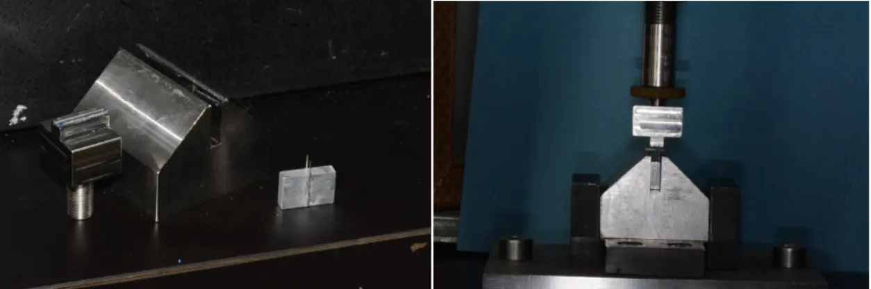
Figure 5. A) Specimen for SBS testing B) A specimen mounted in the Instron machine. ...33
Figure 6. A) Beams (~ 1 mm x 1 mm x 6 mm) prepared for µTBS testing B)
A beam mounted in EZ-test machine. ...34
Figure 7. A) 4-Point Bending test assembly B) A beam mounted in the Instron machine...35
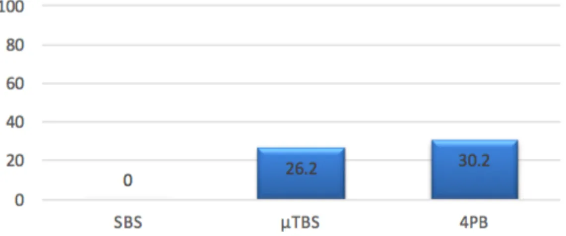
Figure 8. Percentage of pre-test failure by test method. ...36 Figure 9. Mean Dentin Bond strength of Fuji II LC by test method. ...37 Figure 10. Representation of types and relative percentages of modes of
1. Introduction
Different types of direct restorative materials are used in routine dental practice. The
most common are amalgam, resin composites, and glass-ionomer (GI) restorative materials.
Amalgam has a proven, long-term successful clinical performance record, is inexpensive and
easy to handle.62 The main drawback of dental amalgam is its poor esthetics and the concern
about mercury vapors. Resin-based composites are esthetic materials with satisfactory
physical properties. However, material cost and technique sensitivity, along with increased
risk of the development of secondary caries (compared with amalgam), may be considered as
relative disadvantages of this material.62 Glass ionomer (GI) materials that can bind with
calcium ions of mineral tissues are generally tooth colored and may be used in a wide range
of clinical applications.
The clinical application of GI materials depends on the ratio of powder/liquid, which
can be identified through the following classification:55
Type I: Luting cement with a low powder content.
Type II: Restorative material with high powder content and therefore improved physical properties.
Type III: Cavity lining material (low powder content), or cavity base material (high powder content) with physical properties enabling its use as a dentin substitute.
Glass Ionomer Cement (GIC) is the generic name for the dental restorative material that is
the dental market in the early 1970s.2 Dental researchers and clinicians have an ongoing
interest in GICs because these materials 1) are water based and are able to bond to a moist
surface, 2) are able to develop a stable chemical bond with the mineral phase of tooth
structure, 3) contain fluoride ions that are released over time with a resultant decrease in the
critical pH of immediately adjacent tooth structure, 4) are able to absorb topically applied
fluoride for subsequent release 5) are able to seal the cavity and resist microleakage and 6)
are biocompatible and not irritating to the pulp.3,6,57,60,61,70 However, poor mechanical
properties such as, brittleness (decreased fracture toughness) and low wear resistance, as
compared with composite resin restorative materials, limit their use in dentistry as a
permanent filling material in stress-bearing areas.52 Moreover, GICs have been found to be
sensitive to moisture contamination during initial set, are sensitive to subsequent desiccation
and have poor polishability.3,61,64 Modifications in the composition of GICs have resulted in
the steady improvement of material properties over time. The addition of light-polymerizable
resins to GICs, now generally referred to as resin modified glass ionomer (RMGI)
formulations, has been found to improve mechanical/physical properties of the materials
without compromising the bond to the mineral phase of tooth structure.5 The most notable
physical property of GI material is the ability to form a stable, adhesive bond with
hydroxyapatite. The strength and durability of this bond has been evaluated in the laboratory
and by means of clinical trials.
The in vitro research studies note that the relative bond strength value, obtained by various test methods, is not considered an inherent material property of a restorative material but is
helpful in comparison of various dental material formulations.36 Laboratory methods used to
Bond Strength (SBS) test and the Microtensile Bond Strength (µTBS) test. A recently
developed laboratory method, referred to as the Four-Point Bending Bond Strength (4PBBS)
test, has been used to assess the adhesive attachment of composite resin materials to tooth
structure but has not been used to evaluate the strength of RMGI materials to tooth
structure.10,14
It has been reported that SBS tests may be inadequate for the evaluation of the
restorative material bond strength to dental hard tissues as shear stress is not uniformly
distributed along the interface. SBS tests result in increased tendency for cohesive RMGI
failures.11,12,50,65 Cohesive RMGI failures represent the inherent strength of the material and,
therefore, the measured SBS is not a good representative of interface bond strength between
the dentin and the restorative material.9- 12
Micro-Tensile Bond Strength tests are able to assess a tensile force at the adhesive
interface, which may more closely mimic clinical forces.9 Other potential advantages of
µTBS tests over SBS tests include ability to measure the restorative bond to various regions
in dentin as well as the production of more specimens from a given tooth.8 However, µTBS
tests have been found to be laborious and technique-sensitive. One particular draw back of
the µTBS test is the potential for the bonded specimen (especially when GIC materials are
being tested) to break apart before testing (Pre-test failures). Furthermore, some studies
reported cohesive fractures in the RMGI materials being tested.15,16
Recent studies have suggested use of the 4PBBS test as a more optimal means of
assessing dentin bond strength of dental restorative materials in general.10,14 The reason for
this suggestion is that all specimens tested failed adhesively (i.e. none failed cohesively in
interface.10,14 The materials tested in the 4PBBS studies where composite resin in nature. It
was observed that bond failures were immediately proceeded by fracture at the interface.
The authors suggested that this means of bond failure may provide more accurate
information about the material behavior at the adhesive interface.10 However, currently there
are no studies in the literature that have utilized the 4PBBS test to evaluate the bond strength
2. Objectives
The objectives of this study were to assess whether the in vitro dentin bond strength
values of a resin-modified glass ionomer restorative material (RMGI) are affected by
different in vitro test methods and to assess the modes of bond failure for each type of test.
2.1 Specific Aims: 2.1.1 Primary Aim:
To assess the dentin bond strength values of a RMGI restorative material using SBS,
µTBS and 4PBBS tests.
2.1.2 Secondary Aim:
To use optical stereomicroscopy to qualitatively assess the mode of bond failure
(adhesive, cohesive or mixed) for each specimen tested.
2.2 Null Hypotheses:
Primary Null Hypothesis: There is no difference in the mean dentin bond strength
values of a RMGI restorative material when using SBS, µTBS and 4PBBS tests.
3. Literature Review
3.1 Resin Modified Glass Ionomer (RMGI) Restorative Material
Clinical limitations of early GICs have forced the development of new hybrid GI
materials (see section 3.1.3). Resin monomers (as well as initiators necessary to affect resin
polymerization) have been added to these materials in various formulations. As such, these
particular GICs are referred to as Resin-Modified Glass Ionomer (RMGI) materials.5
3.1.1 Setting Reaction of RMGI Materials:
Glass-ionomer materials have a slow acid-base setting reaction with resultant
sensitivity to contamination and desiccation and a need for delayed finishing.40 The setting
process occurs by reaction of an aqueous polyacrylic acid with finely powdered
fluoroaluminosilicate glass (Figure 1).41 RMGI materials, in addition to the acid-base
reaction, undergo polymerization as a result of exposure to visible light. The polymerization
reaction occurs through interaction of water-soluble resin monomers and methacrylate groups
attached to the glass-ionomer acid chains (Figure 2).40 Some manufacturers (Fuji II LC, GC
America) report additional polymerization of HEMA molecules, thus claiming a triple-curing
mechanism. Others (Vitremer, 3M ESPE) report a separate setting reaction which is initiated
by oxidation and reduction catalysts in the material. This “self-curing” aspect of the RMGI
may help to ensure complete cure of the material even in areas that curing light energy
cannot reach.49 Furthermore, ongoing polymerization may occur following exposure to the
curing light, as in composite resins. Polymerization of RMGI continues for up to 24 hours
Light activation allows a longer working time and shorter setting time than are possible with
conventional glass-ionomer materials, which results in placement and finishing procedures
that are less complex.40,42
3.1.2 Adhesion of RMGI Materials:
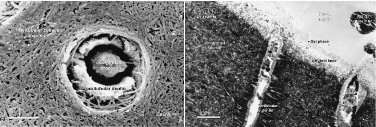
Glass-ionomers are generally considered to be the only materials that self-adhere to
tooth structure.6,7,19 Clinical protocol recommends that the tooth cavity be pretreated by weak
acid solution, Polyacrylic acid (PAA), which removes surface debris (commonly referred to
as the "smear layer"), and effects a partial demineralization of the cavity walls, without
extensive surface hydroxyapatite removal. This increases the surface area available for
chemical interaction of the polyacrylic acid with residual hydroxyapatite (enamel and dentin)
and exposes collagen for subsequent micro-mechanical interlocking (hybridization), 6,19
(Figure 3 A).
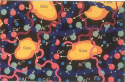
The adhesion of RMGIs to dentin surfaces is considered twofold:6 1) a calcium
chelation bond with the calcium hydroxyapatite phase of both dentin and enamel.7 This
chemical bond may be paly a role in the resistance to hydrolytic degradation.18 2)
Micro-mechanical interlocking of RMGI with the fibril network of the matrix results in a shallow
hybrid layer, about .5-1 micrometers deep.6,43 (Figure 3 B)
3.1.3 Properties of RMGI Materials:
RMGIs have been developed to overcome the limitations of early GIC formulations.5
Even though there are individual differences from brand to brand, RMGIs generally exhibit
1) greater early flexural strength and diametral tensile strength,52 2) less sensitivity to
variation in levels of moisture55 3) improved finishability,3 4) improved translucency,3 and 5)
release fluoride over extended periods of time.61 However, when compared with composite
resin based restorative materials, RMGIs have much less wear resistance, less rigidity, and
are less esthetic.53 Although RMGIs material have higher coefficient of thermal expansion
over conventional GICs, their thermal expansion compares favorably to tooth structure.61
Most RMGI formulations contain 2-hydroxyethyl methacrylate (HEMA), which is a
hydrophilic component. The presence of HEMA causes these materials to act like a mild
hydrogel with rapid water uptake over the first 5-7 days after placement. The uptake of
water results in a small amount of expansion of the restoration and increased risk of staining
from diet related pigments.56 Therefore it is recommended that newly placed RMGIs be
covered immediately with a layer of varnish or light-activated unfilled resin material.59
3.2 Evaluation of the Bond Strength of Dental Materials 3.2.1 Clinical Studies:
Randomized, controlled clinical trials are considered the ultimate means by which to
collect scientific evidence on the effectiveness of restorative treatment.4,9,18 Clinical studies
assessing the durability of restorative material adhesive interfaces have primarily focused on
restoration of non-carious cervical lesions (NCCLs) of the human dentition. Reasons for use
of NCCLs include: 1) little to no macro-mechanical retention is naturally present, 2) margins
of the that are located in enamel as well as dentin, 3) natural occurrence is wide spread in the
population, 4) reasonable access for restoration and evaluation procedures and 5) minimum
or no requirement for preparation prior to restoration. 9,18,38 The presence of various levels of
dentinal sclerosis (hypermineralized dentin) in NCCLs may complicate development of the
Many external variables influence the outcome of clinical studies. Among these are
patient related factors such as age, oral hygiene, occlusal loading, intra oral temperature and
the degree of dentin sclerosis.18,38,46 Other variables include operator skill, specific restorative
material properties, type and effectiveness of the light curing unit, type and effectiveness of
isolation, and finishing methods and instrumentation. 18 Interpretation of clinical findings
(i.e. restorative outcomes of relative clinical success or failure) must consider all known
variables. Identification of which variable(s) contributed most to clinical outcomes may be
difficult.18 The best clinical performance with regard to retention in NCCLs has so far been
achieved by glass ionomer restorative materials with an average annual failure rate of 1.9%.73
3.2.2 Laboratory Bond Strength Studies:
The goal of laboratory bond strength testing is to measure the amount of force
required to separate two bonded materials in a way that is clinically relevant. This
quantification of the strength of the interface configuration depends on use of an appropriate
test machine9. The in vitro assumption is that the higher the actual bonding capacity of a material, the better it will clinically withstand similar force vectors i.e. the longer the
restoration will survive in vivo.4
In general, laboratory bond strength tests have many practical advantages over the
clinical studies. These advantages include, (1) testing is relatively quick and easy, (2) better
control of study variables, (3) ability to directly compare the performance of a new and/or
experimental material/technique with that of the current ‘gold-standard’, (4) ability to
simultaneously test multiple materials within one study set-up and (5) relatively low financial
investment is required. 4
evaluated with the same testing method. Experimental factors that influence the results of
laboratory bond strength tests, include variation within a group of researchers at the same
institution (intra-institute variability) and between different research institutions
(inter-institute variability). Reported intra-(inter-institute bond strength values varied between 30 and
50% and inter-institute bond strength values varied between 20 & 40 %.8,9 Additional
variability in reported bond strengths may result from the adhesive system being evaluated,
the origin of the substrate (bovine or human), how the substrate has been prepared (which
would include specifics such as bur type and bur speed) and even the flexural modulus of the
dental material that has been bonded to the tooth substrate. Research teams use laboratory
tests designed to measure the adhesive bond that occurs at the interface between the dental
material and the tooth substrate. When the test results in bond separation at the interface it is
referred to as an adhesive failure, which represents the best approximation of the strength of
the adhesive bond to the substrate. However, testing does not always result in adhesive
failure. If the prepared specimen separates within the substrate, then it is referred to as a
substrate cohesive failure. If the prepared specimen separates within the restorative material, then it is referred to as a restorative material cohesive failure. Neither of the types of cohesive failure provide anappropriate approximation of the strength of the adhesive bond.
At times both adhesiveand cohesive failures occur in a single specimen, which is referred to
as a mixed failure. It is necessary for researcher to carefully note and report the various types
and relative percentages of adhesive, cohesive and mixed failures.
The presence of experimental variables, and the resultant lack of consistent test
results, severely limits direct comparison of the reported absolute bond strength test values.18
be useful in predicting minimal levels of clinical performance of a test material. For example,
an adhesive that performed poorly in several independent laboratory studies was also found
to be less clinically effective.18 Ultimately, the objective of laboratory testing of bond
strengths is to use carefully designed methods in such a way that prediction of eventual
clinical outcomes becomes more reliable.19
The bond strength test methods that have been developed for dental materials are
generally categorized based on the size of the bonded area under evaluation. The two basic
categories are known as Macro- and Micro-bond strength tests.4
3.3 Macro-Bond Strength Tests:
Testing of any bonded area larger than 3mm2 is considered to be a Macro-bond
strength test. This test is used in protocols designed to measure shear-, tensile- and push-out
bond strengths.4
3.3.1 Shear Bond Strength (SBS) Test:
It has been reported that 46 % of bond strength studies utilize the SBS test.9 The
reason for its high popularity, as compared with other test methods, is the simplicity of
specimen preparation and that no further processing is required after the adhesive interface
has been created.19 The SBS test applies a force designed to slide or twist one material across
another, parallel to the interface (Figure 5).1 This test has been criticized because forces
applied to the interface result in non-uniform stresses within the interfacial zone that are
unlikely to create pure shear stress. 9
The SBS test results in a high percentage of restorative material cohesive failures,
especially in the case of weak restorative materials. Current studies consistently report that
it has been reported that SBS testing of a bonded composite cylinder revealed that the
bending moment resulted in compression on the side of the cylinder but failed to create
adequate tensile forces at the interface.20,21 Therefore, SBS test results depend on the physical
properties of the material. SBS testing of materials with relatively low physical properties, or
materials (with relatively high physical properties) that contain some type of flaw (or crack)
will guide crack growth into the material that propagates toward a region of high stress near
the base of the interface resulting in a cohesive failure.36 However, if the strength of the
adhesive interface is low enough, then the failure will begin at the interface in the region of
maximum local stress. In another words, in the case of weak dental materials, the higher the
adhesive bond strength, the higher the rate of cohesive failure.4,36
Effort have been made to standardized the SBS test protocol so that it is consistent
with the ISO Technical Specification (TS) number 11405 entitled “Testing the adhesion to
tooth Structure”.28 ISO TS 11405 requires restriction of the bonding area to a limited size.
Therefore, in order to control the bonded surface area, specific jigs have been designed
accordingly. These include the Ultradent jig (Ultradent, Salt Lake City, UT, USA) and the
more recent SDI jig (SDI, Bayswater, Victoria, Australia).4 Despite such standardization
attempts, SBS testing is influenced by crosshead speed of the testing device and the means by
which the shear force is actually applied to the specimen by wire loops, points and knife
edges.4 It has been found that SBS test results have a positive correlation with the modulus of
elasticity of the restorative material. 27,36 Therefore, it is preferable to use the same dental
3.3.2 Tensile Bond Strength (TBS) Test:
The TBS test was first used for assessing adhesion of dental materials in by Bowen in
1965.26 The force in the TBS test results in elongation of the bonded specimen.1 The TBS test requires that the specimen aligned exactly perpendicular to the tensile force vector.28
Misalignment of the specimen will result in flexure while under tension and the interfacial
stress generated will not be a pure tensile stress.1 Moreover, other variables, such as
inconsistent geometry of the specimens or use of materials with different elastic moduli may
lead to non-uniform interface stress application among the test specimens. 36
3.3.3 Push-out Bond Strength(PoBS) Test:
The PoBS test uses a compressive force to push a material out of a ring made of
another material. The force vector results in a shear stress at the interface.23,37 In dentistry,
this test has been used primarily for measuring the bond strength of posts luted to root
dentin.24,25 Complexities of specimen preparation and test methodology have limited the
universal use of the PoBS test especially when compared to the traditional SBS testing
process.19
3.4 Micro-Bond Strength Test:
Micro-bond strength tests utilize specimens that are approximately 1 mm X 1 mm in
cross section. A reported advantage of testing a smaller specimen is that it more readily
allows measurement of the restorative bond to various regions in dentin. In addition, a
smaller specimen size may allow opportunity of increased test numbers per tooth. 19
Generally, it has been noted thatthe smaller the bonding surface, the higher the bond strength
values.8 Therefore it is essential that the type of bond strength test method be noted when
include microtensile, micro-shear, fracture toughness, and four-point bending bond strength
tests.
3.4.1 Microtensile bond strength (µTBS) Test:
The µTBS test was first reported by Sano and others in 1994. 13 Review of the current
literature reveals that approximately 60% of research studies assessing adhesive bond
strength used the µTBS test.4,18 Improved design of the specimen mounting jig, which often
has a toggle or freely rotating attachment, allows for tensile forces to be more accurately
aligned with the long axis of the specimen.1 The resultant alignment allows for better tensile
stress distribution at the adhesive interface. The resultant mean bond strength values may
allow for more accurate comparison and ranking of various adhesive systems. Those systems
with consistently higher bond strengths may have a greater likelihood of clinical success.
This is in contrast to the results of SBS testing which reveal more variation among studies. It
may be that this is part of the reason why the findings of µTBS testing correlate more closely
with the reported clinical retention rates of cervical restorations than those identified by SBS
testing.9 Indeed it has been reported that µTBS test results correlate more accurately with the
clinical findings, which suggests that the µTBS test discriminates more effectively between
different adhesive systems than SBS and other bond strength tests. 8,9
As with any bond strength test, there are important factors to consider so as to obtain
meaningful research findings. These factors include the geometry of the beam, type of jig,
specimen trimming, loading speed, specimen alignment and specimen shape. 28,29,32,33
Dental researchers at the University of Iowa have created a specimen former (the
Iowa Micro-Specimen Former) which helps to ensure the tensile stress is maximally
cylindrically constricted at the interface. It has been reported that the smaller bonding surface
area reduces the development of cohesive failures (i.e. increases the tendency to development
adhesive failures).19 Hence, some have recommended that this µTBS-testing protocol
become the standard for measure bonding effectiveness in the laboratory.19
3.4.2 Micro-shear Bond Strength (µSBS) test:
The µSBS test was introduced in 2002 by Shimada and others.34,35This test sought to
benefit from the relative simplicity of the SBS while, at the same time, enable increased
numbers of specimens per tooth.4 It has been found that there are no advantages of the µSBS
test over the macro-shear test. The reason is that the same problem of non-uniform stress
distribution is still present and may be even more pronounced.4 Specimen formation is more
technically demanding as it is very difficult to confine the adhesive to the area that is to be
tested.4 In addition, the µSBS test results revealed similar percentages of the modes of failure
as compared with µTBS test results, while, at the same time, the bond strength values
detected were lower. 35
3.4.3 Fracture Toughness (KIC) Bond Strength Test:
The Fracture Toughness Bond Strength test was introduced in 1993 by Tam and
Pilliar.29 It has been used in bond strength studies to characterize the ability of the adhesive
interface to resist further fracture propagation from a crack that has been artificially created.29
Specimen preparation for this test is laborious, time consuming and technique sensitive. The
test is particular problematic when testing materials, such as glass ionomer restorative
materials, that are already prone to fracture.4,64 It is not a routine test and any correlation to
toughness of the bonding interface may be regarded as a supplementary test to the
macro-tensile test.9
3.4.4 Four-point Bending Bond strength (4PBBS) Test:
The 4PBBS test measures the net effect of simultaneous tensile, compressive, and
shear stress on an adhesive interface.1 The specimen to be tested must be positioned in the
test jig so that a constant stress may be loaded at the central point of the beam which also
must be the location of the adhesive interface.1 Tensile stress concentrates at the interface on
the lower aspect of the specimen beam. Compressive stress forms at the adhesive interface on
the upper surface of the beam and a natural zone occurs in the center of the interface.1,10
Additionally, a shear stress is produced close to the supporting ends of the specimen that
does not play a significant role in the failure process.1 Studies suggest that this test may
provide more accurate information about the material behavior at the adhesive interface.10,14.
Even so, this bond strength test has not been commonly used in dentistry to date. Currently
there are no studies in the literature that have utilized the 4PBBS test to evaluate the bond
strength of a RMGI to dentin.
3.5 Dentin Bond strength of RMGI Restorative Materials
SBS and µTBS test methods have traditionally been used to measure the strength of
RMGI materials to dentin. 4,18 However, the failure mode of the SBS studies has been
predominantly cohesive in the RMGI material. Therefore, the SBS values associated with
cohesive failures are not representative of the interface bond strength between the RMGI and
dentin.11,12,50,65 This indicates that the bond strength of the RMGI material is higher than the
inherent strength of this material.11 Although the stress in µTBS testing is more uniformly
specimen to break apart before testing (pre-test failure) is high.15,16 Furthermore, cohesive
failures have still been reported in some µTBS studies.15.16 High levels of variation in
reported RGMI dentin bond strength values, as well as failure modes being predominantly
cohesive in nature, suggests potential benefit from exploring the use of an alternative, more
recently developed, testing strategy. Therefore, this study sought to assess the human dentin
bond strength values of a RMGI restorative material (Fuji II LC, GC America) using SBS,
4. Materials and Methods
The UNC Institutional Review Board determined that this study was not human
subjects research and exemption from review was granted (IRB # 15-2558). A commercially
available RMGI restorative material was obtained (Table 1). Freshly extracted, non-carious
human third molars were obtained from the UNC Oral and Maxillofacial Surgery clinics and
placed immediately in a 0.1% aqueous thymol solution and stored at a temperature of 2-5°C
until preparation of the dentin surface. Crowns of 36 teeth were separated from the roots
using an Isomet diamond saw (Buehler Ltd., Lake Bluff, IL, USA) under running water. The
roots were sectioned and all pulp tissue was removed (Figure 4 A, B). The occlusal surfaces
of the crowns were ground mechanically (Ecomet 3, Buehler Ltd.) under running water with
180-grit silicon carbide (SiC) paper to obtain a flat dentin surface. The exposed flat dentin
surface was examined to be free of enamel using 2.5X magnification loupes (HiRes™ 2,
Orascoptic, WI, USA). The dentin surface was then polished mechanically using 320-grit
SiC and 600-grit SiC paper under running water for 10 s to create a standardized smear
layer.17 Specimens were then randomly assigned to SBS, µTBS and 4PBBS test methods in
three groups of 12 specimen.
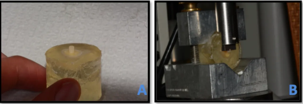
4.1 Shear bond strength (SBS) test:
The prepared dentin specimens were embedded in a block of polymethyle
methacrylate acrylic resin (Great Lakes Orthodontics, NY, USA). The RMGI (Fuji II LC
Capsule, GC America) was bonded to the dentin according to manufacturer’s instructions
5A). The specimen former created one 2.38 diameter RMGI cylinder per tooth. Light-curing
was accomplished using Demi plus (Kerr, CA, USA) for 20s with a light output 1200
mW/cm2. Specimens then were stored in 100% humidity at 37°C for 24 hours. Shear bond
strength tests were then performed using a model 4411 universal testing machine (Instron
Corporation, Norwood, MA, USA) with a crosshead speed of 0.5 mm/min using Ultradent
notched crosshead contact (Figure 5B). Bond-strength values were calculated by dividing the
peak break force (N) by the cross-sectional area of the bonded interface and were expressed
in MPa units.
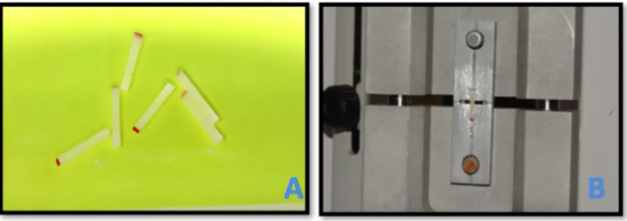
4.2 Micro tensile bond strength (µTBS) test:
The RMGI was applied to the dentin surface to a thickness of 5 mm following
manufacturer instructions (Table 1). Specimens were then immediately stored in 100%
humidity at 37°C for 24 hours. Specimens were then sectioned mesiodistally using an Isomet
diamond saw (Buehler Ltd., Lake Bluff, IL, USA) under running water to obtain around 1
mm thick sections. The specimens were then further sectioned faciolingually to obtain 1 mm
X 1 mm rods that were approximately 6 mm long (Figure 6A). The dentin-RMGI interface
was located at the center of the rods. Each specimen was fixed to a Geraldeli Jig (EZ-Test,
Shimadzu, Kyoto, Japan) using a cyanoacrylate-based adhesive (Figure 6B). The specimens
were carefully placed on the jig so that the RMGI-dentin interface was exactly perpendicular
to the long axis of the testing assembly. The microtensile bond strengths of all specimens
were tested using a universal testing machine (EZ-Test, Shimadzu) with a crosshead speed of
0.5 mm/min. The bond strength of each specimen was determined as the failure load (N)
divided by the cross-sectional area of the bonded interface and expressed in MPa units.
4.3 Four-point Bending Bond Strength (4PBBS) test:
The RMGI was applied to the dentin surface to a thickness of 5 mm following
manufacturer instructions (Table 1). Specimens were then immediately stored in 100%
humidity at 37°C for 24 hours. Specimens were then sectioned mesiodistally using an Isomet
diamond saw (Buehler Ltd., Lake Bluff, IL, USA) under running water to obtain around 1
mm thick sections. The specimens were then further sectioned faciolingually to obtain 1 mm
X 1 mm rods that were approximately 10 mm long. The dentin-RMGI interface was located
at the center of the rods. A custom stainless steel device was fabricated such that the distance
between loading points would be 1.8 mm for upper and 7.2 mm for lower members of the
test device (Figure 7A). Prepared specimens were then subjected to the 4PBBS test and
loaded to failure (Figure 7B). The test was performed using a model 4411 universal testing
machine (Instron Corporation, Norwood, MA, USA) with a crosshead speed of 0.5 mm/min.
Bond strengths σb (in MPa), were computed using the standard relationship (ASTM
E855/1984): 𝜎𝑏 =$%&'() .
Where P is the maximum load (in N), a is the spacing (in millimeters) between upper and
lower loading points, b and h are, respectively, the specimen width and thickness (in
millimeters.
4.4 Qualitative Assessment of the RMGI - Dentin Fracture Interfaces
After bond strength testing, all specimens were examined using optical
stereomicroscopy (Nikon SMZ18, Tokyo, Japan), at 8X magnification for SBS and 13.5X
magnification for µTBS and 4PBBS, to determine the mode of failure at the fracture
interface. The number of “adhesive”, “cohesive” and “mixed” failures in each test group was
4.5 Statistical Analysis
The mean (+/- SD) SBS, µTBS and 4PBBS test values from each tooth (12 teeth per
group) were calculated. Mean bond strengths from multiple beams from each tooth in the
µTBS and 4PBBS were calculated so as to provide an average bond strength for each tooth.
Specimens showing pre-test or cohesive dentin failures were excluded from the statistical
analysis as these do not represent measurements of adhesion.8 Bond-strength data were
analyzed using one-way ANOVA. Data were further analyzed using the Tukey’s post-hoc
test. Modes of failure were analyzed using Pearson’s Chi-squire test after taking into account
the range of failures for each tooth that had more than one beam. All statistical tests were
performed at the 95% confidence level. Stereomicroscopy images were analyzed and
5. Results
The 12 teeth assigned to the SBS group provided 12 cylinders for SBS testing (Table
2). The 12 tooth specimens provided 61 beams for µTBS testing and 43 beams for 4PBBS
testing respectively (Table 2). The number of pre-test failures for each test group is presented
in Table 2 and Figure 8. Approximately 1/3rd of the beams prepared for µTBS and 4PBBS
tests developed pre-test failures. The mean values and standard deviations were as follows:
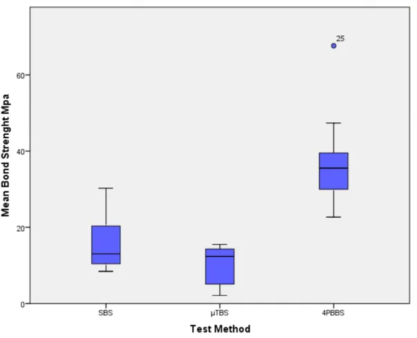
SBS = 15.7 +/- 7.1 MPa, µTBS = 9.7 +/- 5.3 MPa, 4PBBS = 37.3 +/- 12.8 MPa (Table 2 and
Figure 9). There was a statistically significant difference between bond strength values of
both SBS and µTBS tests and the 4PBBS test (p<0.0001). There was no statistically
significant difference between SBS and µTBS test results (p=.24).
There was a statistically significant difference among the bond strength tests in the
mode of failure (p=.006) (Table 3). Failure occurred predominantly at the adhesive/dentin
interface for SBS (83%) while the majority of µTBS and 4PBBS failures were mixed (69%
and 47% respectively). The 4PBBS test showed higher cohesive failure rate than the µTBS
(33.3% and 11.1 respectively) (Figure 10). Only one beam (2.2%) of µTBS specimens failed
6. Discussion
The present study evaluated the ability of three different test methods (shear-,
microtensile-, and 4-point bending bond strength test) to determine the bond strength
between a RMGI restorative material and human dentin. Efforts were made to limit
intra-institutional variation in methods while conducting the study. Many studies can be found in
the literature that compare different bond strength methodologies using dentin as the main
substrate. However, no currently reported research studies have compared these
methodologies when using RMGI bonded to dentin and no studies have used the 4PBBS test
as a method to measure the dentin bond strength of RMGI.
The first null hypothesis was rejected indicating that there was a difference in the
strength value among three testing methods. Our data showed that the 4PBBS tests resulted
in significantly (p<0.0001) higher mean bond strength values than the µTBS and the SBS
tests (Figure 9). The finding of higher bond strength values for the 4PBBS test is consistent
with other investigations that used this test.10,14 For example, 4PBBS testing found that the
mean bond strength of a self-etching primer (Clearfil SE Bond, Kuraray, Osaka, Japan) to
dentin was 90.6 ± 2.5 MPa. This value is considerably higher than the mean values of the
same adhesive system using the SBS and the µTBS test (47.1±7.6 and 60.5 ± 7.0 MPa
respectively)47,48. It is not appropriate to compare the absolute mean bond strength test values
among different test methods as each test has different variables.
have reported values in the range of ~ 5 – 22 MPa.11,49,50,69 The wide range of SBS values
may be related to variation in specimen geometry, test load configuration and increased
potential for the presence of pre-existing cracks in the RMGI material. Other reasons for high
scatter among the mean SBS values may be due to the inclusion of test results from cohesive
failures as well as pre-testing failures into the statistical analyses.8
The mean µTBS value of Fuji II LC in this study (9.7 ± 5.3 MPa) was lower than the
range of values reported in the current literature. Recent µTBS studies report the µTBS value
of the dentin/Fuji II LC to be in the range of ~ 18.5 -31 MPa.15,16,66.67 The wide range of
µTBS values may be related to differences in beam geometry, jig type, trimming methods,
loading speed, specimen alignment, degrees of dehydration, variations in dentinal tubules,
and specimen shape.28,29,32,33 All of these factors are critical and influence final µTBS test
outcomes.Because no internationally recognized standardized test protocol for the testing of
adhesive systems is yet available, it can be easily noticed that the absolute values differ
widely even when testing the same material and with the same test method. This highlights
the influence of the test institute on the bond strength values.8,9
The secondary null hypothesis was rejected, since modes of failures of RMGI
restorative material were found to be statistically significantly different between the test
methods used in this study (p=0.006). There is no clear consensus in the literature regarding
classification of failure modes.8 In this study the modes of failure of the tested specimens
were classified as adhesive at the interface, cohesive in the material or in the dentin, and
mixed (Figure 10).23 In the present study, SBS test showed a higher percentage of adhesive
failure. This finding is in contrast to other SBS studies that assessed the adhesive bond of
shear bond strength of RMGI material to the dentin. Based on Schreiner & others, SBS
values higher than 20 MPa, will likely result in more cohesive failures.44 The mean SBS
value of 15 MPa found in this study may reveal the adhesive failure during SBS testing.
Reduced tendency for cohesive failure within the Fuji II LC may also be secondary to
ongoing improvement in the material properties of this RMGI. The SBS findings of this
study are similar to others who reported predominantly adhesive failures for this material. 69
The failure mode of the µTBS test was predominantly mixed. Microscopic
assessment of the specimens revealed a half-moon pattern on the border of the bonded area in
69% of the specimens. This type of failure pattern may result from the inherent brittleness
and the presence of voids in the RMGI material. Additionally, specimen misalignment during
jig assembly and/or testing may also have contributing to increased percentage of mixed
failures.
The 4PBBS test showed the highest percentage of RMGI cohesive failure (33%). The
majority of this type of failure occurred in the first 8 tested beams. It may be that these beams
where not properly aligned in the test apparatus, which requires that the RMGI/dentin
interface be exactly in the middle of the apparatus. In general, the majority of the area of the
mixed failures of 4PBBS and µTBS tests was located at the adhesive interface with small
areas (≤20% of the total surface) of cohesive failure within the RMGI.
SBS testing of Fuji II LC resulted in no pre-test failures. This may be due to the
relatively large size of the bonding area and that no post-bonding specimen processing is
required. Both µTBS and 4PBBS tests had pre-test failures that occurred at the interface.
This finding suggests that these tests may not be appropriate for assessing the dentin bond
specimen bonding area (about 1mm2 or less) and/or post bonding processing required in
these tests.4 The level of RMGI brittleness may limit the usefulness of the µTBS method.
Brittle materials are generally considered unsuitable for µTBS testing.68
Limitations should always be considered for results of in vitro bond strength studies.
Clinical conditions cannot be fully simulated in vitro.63 Aspects such as the internal pulpal
pressure (and resultant dentinal fluid movement into the bonding interface), tooth stress
dynamics and the 3-dimensional nature of cavity preparations can not be fully replicated
using in vitro protocols.71 Obviously this study was not able to replicate or even simulate any
of these clinical realities. Further limitations of the study include that specimens were tested
only at 24 h after the RMGI was placed. Storage time is also known to influence bond
strengths. Extended periods of water storage cause mechanical and morphological
degradation,72 which leads to a decrease in bond strength and might better simulate in vivo conditions.4.8,71
SBS testing demonstrated the highest percentage of adhesive failure, less technique
sensitivity, and a lower amount of test material for specimen preparation. It is interesting to
note that specimen preparation for SBS testing required less Fuji II LC as compared with the
specimen preparation for µTBS and 4PBBS (Table 4). One Fuji II LC capsule was used to
create two SBS specimens, while µTBS and 4PBBS specimen preparation required at least
two capsules to create one specimen. However, SBS testing creates a non-uniform shear
stress at the interface. Test values may not represent a true measurement of the adhesive bond
strength of the RMGI. The µTBS test may allow better stress distributed at the interface as
compared to SBS methods but the specimen preparation was found to be laborious and
for RMGI restorative materials secondary to the difficulty of specimen preparations,
orientation, and the resulted cohesive failures in the material. The 4PBBS may be applicable
7. Conclusions
Within the limitations of this in vitro study, the following conclusions may be made:
1- The relative in vitro dentin bond strength value of a resin-modified glass ionomer
restorative material is greatly affected by the test method.
2- The SBS test method demonstrated the highest percentage of adhesive failure and
lowest technique sensitivity when testing a RMGI material.
3- The majority of µTBS and 4PBBS failures were mixed and therefore the µTBS
and 4PBBS tests may not be optimal for comparison of the relative bond strength
values of RMGI materials to dentin.
4- Use of the SBS test may allow more controlled comparison of the adhesive dentin
bond among various RMGI formulations, whether already commercially available or
FIGURES
Figure 3: Adhesion of RMGI material to dentin. A) The effect of a polyacrylic acid
TABLES
Table 1: Materials, composition and application directions followed in this study:
HEMA: 2-hydroxyethyl methacrylate. MMA: methyl methacrylate
Material Manufacturer Composition Application
Fuji II LC
Capsule GC America Powder:
fluoroalumino-silicate glass Liquid: polyalkenoic acid, HEMA, dimethacrylate, camphorquinone, water Apply cavity
conditioner to dentin surfaces and leave undisturbed for 10s; rinse with water for 10s; gently air-dry for 5s, leaving a moist surface. Automatically mix capsules for 10s; apply to dentin surfaces; light-cure was accomplished using Demi plus (Kerr) for 20s with a light output 1200 mW/cm2.
Apply a final coat of EQUIA Coat and light cure for 20s.
Cavity conditioner GC America 20% Polyacrylic
acid, 3% Aluminum chloride hydrate, 77% Distilled water
EQUIA Coat GC America 25–50% MMA, 1–
Table 2: Mean (+/- SD) dentin bond strengths values of the RMGI by test method. The number of pre-test failures were not included in the calculation of the mean bond strength values:
Test Teeth
Number of Cylinders or Beams Tested
Number of Pre-test Failures
Mean (+/-SD) Bond Strengths
in MPa
SBS 12 12 cylinders 0 15.7 ±7.1
µTBS 12 61 Beams 16 9.7 ± 5.3
Table 3: Number (specimen number, percentage) of modes of RMGI bond failure by test method as observed by Optical Stereomicroscopy:
Test Adhesive
(N, %)
Cohesive in the Material
(N, %)
Cohesive in the Dentin
(N, %)
Mixed (N, %)
SBS 10 (83) 0 (0) 0 (0) 2 (17)
µTBS 8 (17.8) 5 (11.1) 1 (2.2) 31 (68.9)
Table 4: Comparison between SBS, µTBS and 4PBBS tests based on the results of this study:
SBS µTBS 4PBBS
Test
Mechanics Sliding of two surfaces. Stretch/elongation of the bonded specimen
Perpendicular loading in the central area of an unsupported span. Stress Nature Not uniformly distributed at the interface. Improved stress distribution at the
interface.
Combination of Compressive, Tensile and Shear Stress. More tensile at
the interface Mode of failure Mostly adhesive with no cohesive failures. Mostly mixed with some cohesive
failures.
Mostly mixed with more cohesive failures
than µTBS
Technique
Easy, fast, with no pre-test
failures
Difficult, time consuming, with high % pre-test
failures
REFERENCES
1) Anusavice K Phillip’s Science of Dental Materials, 12th edition. Philadelphia, PA: Saunders; 2013.
2) Wilson AD and Kent BE. The glass-ionomer cement, a new translucent dental filling material. Journal of Applied Chemistry and Biotechnology. 1971, 21: 313–313.
3) Mount GJ. Glass ionomers: a review of their current status. Oper Dent 1999;24: 115–24.
4) Van Meerbeek B, Peumans M, Poitevin A, Mine A, Van Ende A, Neves A. Relationship between bond-strength tests and clinical outcomes. Dent Mater. 2010;26: e100–21.
5) Mitra SB. Photocurable ionomer cement systems. European Patent Application 323120,1988.
6) Van Meerbeek B, Yoshida Y, Inoue S, De Munck J, Van Landuyt K, & Lambrechts P. Glass-ionomer adhesion: the mechanisms at the interface. Journal of Dentistry. 2006;34: 615–617.
7) Yoshida Y, Van Meerbeek B, Nakayama Y, Snauwaert J, Hellemans L, Lambrechts P, Wakasa K. Evidence of chemical bonding at biomaterial-hard tissue interfaces. Journal of Dental Research. 2000;79: 709–14.
8) Scherrer SS, Cesar PF, Swain MV. Direct comparison of the bond strength results of the different test methods: a critical literature review. Dent Mater. 2010; 26: e78–93.
9) Heintze SD. Clinical relevance of tests on bond strength, microleakage and marginal adaptation. Dent Mater. 2013; 29: 59–84.
10 Staninec M, Kim P, Marshall GW, Ritchie RO, Marshall SJ. Fatigue of dentin-composite interfaces with four-point bend. Dental Materials. 2008; 24: 799–803.
11) Fritz UB, Finger WJ, Uno S. Resin-modified glass ionomer cements: Bonding to enamel and dentin. Dent Mater. 1996;12: 161–166.
12) Sidhu SK, Sherriff M, Watson TF. Failure of resin-modified glass-ionomers subjected to shear loading. J Dent. 1999;27: 373–381.
13) Sano H, Shono T, Sonoda H, Takatsu T, Ciucchi B, Carvalho R, Pashley DH. Relationship between surface area for adhesion and tensile bond strength evaluation of a micro-tensile bond test. Dent Mater. 1994;10:236–40.
15) Tanumiharja M, Burrow MF, Tyas MJ. Microtensile bond strengths of glass ionomer (polyalkenoate)cements to dentine using four conditioners. J Dent. 2000;28: 361–6.
16) Coutinho E, Cardoso M V, De Munck J, Neves AA, Van Landuyt KL, Poitevin A, Peumans M, Lambrechts P, Van Meerbeek B. Bonding effectiveness and interfacial characterization of a nano-filled resin-modified glass-ionomer. Dent Mater. 2009;25:1347– 57.
17) Pashley DH, Tao L, Boyd L, King GE, Horner JA. Scanning electron microscopy of the substructure of smear layers in human dentine. Arch Oral Biol. 1988;33:265–270.
18) De Munck J, Van Landuyt K, Peumans M, Poitevin A, Lambrechts P, Bream M, Van Meerbeek B. A Critical Review of the Durability of Adhesion to Tooth Tissue: Methods and Results. J Dent Res. 2005;84:118–132.
19) Van Meerbeek B, De Munck J. Adhesion to enamel and dentin: current status and future challenges. Oper Dent. 2003;28:215–235.
20) De Hoff PH, Anusavice KJ, Wang Z. Three-dimensional finite element analysis of the shear bond test. Dent Mater. 1995; 11:126–31
21) Versluis A, Tantbirojn D, Douglas WH. Why do shear bond tests pull out dentin? J Dent Res. 1997;76:1298–307.
22) Pashley DH, Sano H, Ciucchi B, Yoshiyama M, Carvalho RM. Adhesion testing of dentin bonding agents: a review. Dent Mater.1995; 11:117–25.
23) Otani A, Amaral M, May LG, Cesar PF, Valandro LF. A critical evaluation of bond strength tests for the assessment of bonding to Y-TZP. Dent Mater. 2015;31:648–656.
24) Bergoli C, Amaral M, Valandro LF. The disk-specimen height does not influence the push-out bond strength results between fiber post and root dentin. J Adhes. 2012;88: 212–23.
25) Zicari F, Couthino E, De Munck J, Poitevin A, Scotti R, Naert I, Van Meerbeek B. Bonding effectiveness and sealing ability of fiber-post bonding. Dent Mater. 2008; 24:967– 77.
26) Bowen RL. Adhesive bonding of various materials to hard tooth tissues. Methods of determining bond strength. Journal of Dental Research. 1965; 44:690-5
27) Leloup G, D’Hoore W, Bouter D, Degrange M, Vreven J. Meta-analytical review of factors involved in dentin adherence. J of Dental Research. 2001;80:1605–14.
29) Goracci C, Sadek FT, Monticelli F, Cardoso PEC, Ferrari M. Influence of substrate, shape, and thickness on microtensile specimens’ structural integrity and their measured bond strengths. Dent Mater. 2004;20:643–654.
30) Frankenberger R, Krämer N & Petschelt A. Fatigue behavior of different dentin adhesives. Clin Oral Investig, 1999;3:11-7
31) Tam LE, Pilliar RM. Fracture toughness of dentin/resin-composite adhesive interfaces. Journal of Dental Research 1993; 72:953–9.
32) Poitevin A, De Munck J, Van Landuyt K, Coutinho E, Peumans M, Lambrechts P, Van Meerbeek B. Influence of three specimen fixation modes on the micro-tensile bond strength of adhesives to dentin. Dent Mater. 2007;26:694–9
33) Pashley DH, Carvalho RM, Sano H, Nakajima M, Yoshiyama M, Shono Y, Fernandes CA, Tay F. The microtensile bond test: a review. J Adhes Dent 1999;1:299–309.
34) ISO. Dental materials – testing of adhesion to tooth structure. Technical specification no. 11405; 2003.
35) Barkmeier WW, Erickson RL, Latta MA. Fatigue limits of enamel bonds with moist and dry techniques. Dent Mater. 2009;25:1527-31
36) Van Noort R, Noroozi S, Howard IC, Cardew G. A critique of bond strength measurements. J Dent. 1989;17:61–67.
37) Armstrong SR, Keller JC, Boyer DB. The influence of water storage and C-factor on the dentin-resin composite microtensile bond strength and debond pathway utilizing a filled and unfilled adhesive resin, Dent Mater. 2001;17: 268- 276
38) Van Meerbeek B, Perdigão J, Lambrechts P, Vanherle G. The clinical performance of adhesives. J Dent. 1998;26:1-20.
39) Lohbauer U. Dental Glass Ionomer Cements as Permanent Filling Materials? – Properties, Limitations and Future Trends. Materials (Basel). 2009;3:76–96D.
40) Swift EJ, Perdigao J, Heymann HO. Bonding to enamel and dentin: a brief history and state of the art. Quintessence Int. 1995;26:95–110.
41) Wilson AD, Kent BE, Clinton D, Miller RP. The formation and microstructure of dental silicate cements. J. Mater Sci. 1972;7:220–238.
43) Inoue S, Van Meerbeek B, Abe Y, Yoshida Y, Lambrechts P, Vanherle G, Sano H. Effect of remaining dentin thickness and the use of conditioner on micro-tensile bond strength of a glass-ionomer adhesive. Dent Mater. 2001; 17:445-455.
44) Schreiner RF, Chappell RP, Glaros AG , Eick JD. Microtensile testing of dental adhesives. Dent Mater. 1998;14: 194-201.
45) Van Meerbeek B, Braem M, Lambrechts P, Vanherle. Morphological characterization of the interface between resin and sclerotic dentine. J Dent. 1994;22:141-146.
46) Gale MS and Darvell BW. Thermal cycling procedures for laboratory testing of dental restorations. J Dent. 1999;27:89-99.
47) Walter R, Swift EJ, Boushell LW, Braswell K. Enamel and dentin bond strengths of a new self-etch adhesive system. J Esthet Restor Dent. 2011;23: 390–6.
48) Sheikh H, Heymann HO, Swift EJ, Ziemiecki TL, Ritter AV. Effect of Saliva
Contamination and Cleansing Solutions on the Bond Strengths of Self-Etch Adhesives to Dentin. J Esthet Restor Dent. 2010;22:402–410.
49) Swift EJ, Pawlus MA, Vargas MA. Shear bond strengths of resin-modified glass-ionomer restorative materials. Oper Dent. 1995;20:138–143.
50) Thean HP, Mok BY, Chew CL. Bond strengths of glass ionomer restoratives to primary vs permanent dentin. ASDC J Dent Child. 2000;67:112–6, 82.
51) Wilson AD. Resin-modified glass-ionomer cements. Int J Prosthodont.1990;3:425–9.
52) Xie D, Brantley WA, Culbertson BM, Wang G. Mechanical properties and microstructures of glass-ionomer cements. Dent Mater. 2000;16(2):129–138.
53) Braem MJA, Lambrechts P, Gladys S, Vanherle G. In vitro fatigue behavior of restorative composites and glass ionomers. Dent Mater. 1995;11:137—41.
54) McCabe JF. Resin-modified glass-ionomers. Biomaterials. 1998;19:521–527.
55) Mount GJ. An Atlas of Glass-Ionomer Cement: A Clinician's Guide, 3rd edition, London: Martin Dunitz. 2002
56) Nicholson JW, Anstice HM & Mclean JW. A preliminary report on the effect of storage in water on the properties of commercial light-cured glass-ionomer cements. British Dental Journal. 1992; 173 98-101.
58) Hume WR, Pulpal responses to glass-ionomers. In Glass-ionomers: the Next Generation; Proceedings of the Second Symposium on Glass-Ionomers edited and published by Peter Hunt, Philadelphia.
59) Earl MS, Mount GJ & Hume WR. The effect of varnishes and other surface treatments on water movement across the glass-ionomer cement surface: II. Australian Dental Journal. 1989; 34:326-329.
60) McLean JW. Dentinal bonding agents versus glass-ionomer cements. Quintessence Int. 1996;27:659–67.
61) Burgess J, Norling B, Summitt J. Resin Ionomer Restorative Materials: The New Generation. J Esthet Restor Dent. 1994;6:207–215.
62) Opdam NJM, Bronkhorst EM, Loomans B a C, Huysmans MCDNJM. 12-Year Survival of Composite Vs. Amalgam Restorations. J Dent Res. 2010;89:1063–7.
63) Kelly JR. Evidence-based decision making: Guide to reading the dental materials literature. J Prosthet Dent. 2006;95(2):152–60.
64) Nicholson JW. Glass ionomer dental cements: update. Mater Technol. 2010;25:8–13.
65) Pereira PNR, Yamada T, Tei R, Tagami J. Bond strength and interface micromorphology of an improved resin-modified glass ionomer cement. Am J Dent. 1997;10:128–132.
66) Cook NB, Feitosa SA, Patel A, Alfawaz Y, Eckert GJ, Bottino MC. Bonding Ability of Paste-Paste Glass Ionomer Systems to Tooth Structure: In Vitro Studies. Oper Dent. 2014;40:304–312.
67) Pereira PN, Sano H, Ogata M, Zhang L, Nakajima J, Tagami J, Pashley DH. Effect of region and dentin perfusion on bond strengths of resin-modified glass ionomer cements. J Dent. 2000;28(5):347–354.
68) Sadek FT, Monticelli F, Muench A, Ferrari M, & Cardoso PE. A novel method to obtain microtensile specimens minimizing cut flaws. J Biomed Mater Res B Appl Biomater.
2006;78: 7-14.
69) Imbery AT, Namboodiri A, Duncan A, Amos R, Best AM, Moon PC. Evaluating dentin surface treatments for resin-modified glass ionomer restorative materials. Oper Dent. 2013;38:429–38.
71) Oliveira GCB, Oliveira GMS, Ritter AV, Heymann HO, Swift EJ, Yamauchi M. Influence of tooth age and etching time on the microtensile bond strengths of adhesive systems to dentin. J Adhes Dent. 2012;14:229–34.
72) Shono Y, Terashita M, Shimada J, Kozono Y, Carvalho RM, Russell CM, Pashley DH. Durability of resin-dentin bonds. J Adhes Dent. 1999;1:211-218.