THE IMPACT OF ACUTE AEROBIC EXERCISE ON NATURAL KILLER CELL, CATECHOLAMINE, AND CORTISOL RESPONSES IN BREAST CANCER
SURVIVORS
Elizabeth Serex Evans
A dissertation submitted to the faculty of the University of North Carolina at Chapel Hill in partial fulfillment of the requirements for the degree of Doctor of Philosophy in the School of
Medicine (Interdisciplinary Program in Human Movement Science).
Chapel Hill 2012
Approved by:
Claudio Battaglini, PhD A. C. Hackney, PhD, DSc Robert McMurray, PhD
ii ©2012
iii ABSTRACT
ELIZABETH SEREX EVANS: The Impact of Acute Aerobic Exercise on Natural Killer Cell, Catecholamine, and Cortisol Responses in Breast Cancer Survivors
(under the direction of Dr. Claudio Battaglini)
PURPOSE: The purpose of this investigation was to compare the effect of acute moderate intensity aerobic exercise on natural killer (NK) cell, catecholamine, and cortisol responses between breast cancer survivors and matched healthy controls. Additionally, relationships between post-exercise changes in NK cell responses and post-exercise changes in
catecholamines and cortisol were examined. PARTICIPANTS: Data were collected from 9 women who had been treated for Stage I-III invasive breast cancer 3-6 months prior to enrollment and 9 healthy sedentary women without a history of cancer treatment. Subjects exercised for 30 minutes on a cycle ergometer at 60% of VO2peak. Blood samples were obtained pre-exercise, immediately exercise, 2 hours exercise, and 24 hours post-exercise. METHODS: NK cell counts and NK cell cytotoxic activity (NKCA) were measured via flow cytometric analysis. Plasma catecholamines (epinephrine and
iv
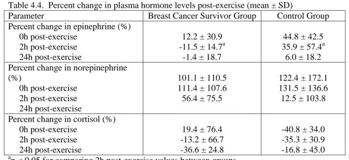
epinephrine was somewhat lower in the breast cancer group immediately post-exercise and significantly lower in breast cancer survivors at 2 hours post-exercise (p < 0.05). Cortisol levels were somewhat higher in breast cancer patients immediately post-exercise, and percent change in cortisol immediately post-exercise increased in breast cancer survivors but
decreased in controls (p < 0.05). Change in NK cell counts was only significantly correlated with change in cortisol at 2 hours post-exercise in the control group (p < 0.05).
v
ACKNOWLEDGEMENTS
When I started writing the proposal for this dissertation project in January 2011, I don’t think I fully understood how complex and intricate this endeavor was going to be. There were many aspects of this project that I had to learn completely from scratch, and much of the data collection phase required multiple hands-on-deck so that I could successfully and efficiently obtain the samples I needed for analysis. I could never have done any of this completely on my own, and so there are numerous individuals to whom I would like to express my sincerest thanks for helping me to complete the various aspects of this dissertation process.
Firstly, I would like to thank the 18 women who so willingly volunteered to
participate as subjects in this study. Each one of them gave approximately 7 hours of their time (and time off work) over the course of 3 laboratory sessions apiece. I thoroughly enjoyed getting to know each one of them, and without them, I would not have even had a project of which to speak. Secondly, I would like to thank the five members of my
vi
this project, as well for provision of financial resources to fund the ELISA experiments. I thank Dr. Mac for his thoroughness in reviewing my manuscripts, Dr. Muss for his insights into the clinical aspects of this project, and Dr. Randell for his guidance regarding the cell culture aspects of this project and for the use of his laboratory for all of my NK cell and ELISA experiments.
I must give a profound thanks to my student colleagues who helped me collect data: undergraduate EXSS major Coleman Mills; Exercise Physiology MA students Deanna Babcock, Miles Bartlett, Stephanie Bomberger, Kristen Koltun, Cecily Lehman, Carly Shatten, Jacob Allen, Dustin Buttars, Sarah Fulz, Rachel Graff, Dangaia Sims, and Mary Woessner; Exercise Physiology PhD student Eric Sobolewski, and Post-Doctoral Fellow Dr. Denise Spector. I would like to thank Dr. Dru Henson from Appalachian State University for answering many of my initial questions on how to measure NK cell activity, as well as to Dr. Elinor Fondell, Dr. Hans Gaines, and Kristina Franck from the Swedish Institute for
vii
well as to Amy DePue and Pat Decator at the NC Cancer Hospital for additional help with angiocatheter placements and physical examinations for my subjects.
viii
TABLE OF CONTENTS
LIST OF TABLES...xi
LIST OF FIGURES………..………...xii
Chapter I. INTRODUCTION Background………1
Rationale and Purposes of the Study……….………4
Definition of Terms………...………...…….…….6
Aims and Hypotheses………...……..…...7
II. REVIEW OF LITERATURE Introduction...10
Role of Natural Killer Cells in Destroying Pathogens...10
Catecholamines...14
Cortisol...17
Acute Aerobic Exercise and NK Cell Response in Healthy Individuals...20
Aerobic Exercise and Immune Function in Cancer Patients and Survivors...53
ix
III. THE IMPACT OF ACUTE AEROBIC EXERCISE ON NATURAL KILLER CELL RESPONSES IN BREAST CANCER SURVIVORS
Summary...63
Introduction...64
Methods...66
Results...74
Discussion...79
IV. THE IMPACT OF CATECHOLAMINE AND CORTISOL RESPONSES IN BREAST CANCER SURVIVORS Summary...85
Introduction...86
Methods...88
Results...94
Discussion...98
V. THE RELATIONSHIPS BETWEEN CHANGES IN NATURAL KILLER CELL NUMBERS. CATECHOLAMINES, AND CORTISOL AFTER ACUTE AEROBIC EXERCISE IN BREAST CANCER SURVIVORS Summary...105
Introduction...106
Methods...107
Results...111
Discussion...115
x
Significance of the Study and Implication of Results……….121
Summary of Research Hypotheses…………..………...……124
Strengths and Limitations………..………...………..125
Future Research……….…………..………..126
APPENDICES A. Extended Methods………...……….………...…..128
B. Medical History Questionnaire……….150
C. Astrand Cycle Ergometer Maximal Test Protocol………155
D. Perceived Stress Scale……….…..157
E. Sample Preparation for NK Cell Counts………...159
F. Sample Preparation for NKCA Assay………...161
xi
LIST OF TABLES Table
3.1. Subject physical characteristics...75
3.2. Metabolic responses during the submaximal aerobic exercise session...76
3.3. Immune cell counts and plasma volume shifts across time...77
3.4. Percent change in NK cell counts...78
3.5. NK cell activity across time for six breast cancer survivors and one control subject...79
4.1. Subject physical characteristics...94
4.2. Details of cancer treatments received by breast cancer survivor group...95
4.3. Plasma hormone levels across time...97
4.4. Percent change in plasma hormone levels post-exercise...98
5.1. Comparison on subject characteristics, VO2, workload, and PSS scores...112
5.2. NK cell counts, plasma catecholamine, and cortisol concentrations at baseline (pre-exercise) and fold changes across time (baseline to immediately post-exercise [0h post-exercise] and baseline to 2 hours post-exercise [2h post-exercise])...114
xii
LIST OF FIGURES Figure
1. Conceptual model leading to research aims...8 2. Timeline of study events...131 3. Example of regression for determining workload corresponding to
1 CHAPTER I INTRODUCTION
Background
Breast cancer is the most common cancer among women in the United States, other than skin cancer, and it is the second-leading cause of cancer deaths in women, after lung cancer (1). Major treatments for breast cancer include surgery, chemotherapy, and radiation therapy, and these treatments can cause significant physical and psychological distress that may persist for months or years after the completion of therapy (2). Fatigue is the most common physical distress experienced by breast cancer patients, affecting up to 70% of patients during therapy and up to 30% of patients years after the completion of therapy (3). This type of fatigue is often severe and may limit activities of daily living; therefore, in the past, patients have been advised to rest and reduce their activity levels (3). However,
inactivity leads to muscular atrophy and decreased cardiorespiratory function, and prolonged rest can actually worsen fatigue, thus contributing to a cycle of further muscular atrophy, reduced cardiorespiratory functioning, and a down-regulation of daily activity levels (3-5).
2
including changes in muscular strength, body composition, functional quality of life, fatigue, anxiety, and self esteem (6). Many of these “first generation” studies have shown that exercise training appears tomitigate many treatment-related side-effects, that patients are able to makeimprovements in cardiorespiratory fitness, and that regular exercise seems to be safe, well-tolerated, and associated with few adverse events (6, 7). Therefore, the results of these studies thus far seem promising.
Physical exercise may also be an attractive adjunct therapy for cancer patients and survivors because of its potentially positive influence on biologic systems involved in disease protection and anti-cancer defense (9). In the field of Exercise Immunology, a “J-shaped” hypothesis has been postulated describing the inverse relationship between disease
risk/cancer susceptibility (9, 10). This hypothesis states that regular, moderate exercise may lead to enhanced immune function and therefore a decreased risk of disease and cancer, whereas repeated bouts of exhaustive exercise and overtraining could lead to
immunosuppression, and therefore an elevated risk of disease and cancer (9, 10).
One particular cellular component of the immune system, the natural killer (NK) cell, may be especially characterized by this J-shaped curve in response to exercise. NK cells are part of the body’s innate immune response, which is activated as part of the body’s
3
training, resting NK cell numbers and activity may become increased, while in response to periods of heavy exercise training, NK cell numbers and activity may become depressed (12-14). Although not all studies show entirely consistent outcomes, and the clinical implications of exercise-induced changes in NK cell responses are not entirely known, it is thought that exercise-related enhancements in NK cell function may confer a protective effect against pathogen invasion, while exercise-related decreases in NK cell function may lead to increased incidence of viral infection and potential illness (12, 13).
Research regarding immune system responses to exercise in the cancer patient population is limited. To date, 19 studies have examined the effect of exercise on some parameter of immune function (15-33). Of these, only 3 have examined the effect of exercise on NK cell function in the breast cancer patient population, and all were in response to a period of exercise training (15, 17, 23). The results of these 3 studies seem to indicate that exercise training either does not affect NK cell function, or may even improve NK cell function in breast cancer patients and survivors possibly beyond what is associated with normal recovery after cancer therapy, and could possibly be correlated with increased disease-free and overall survival (23). Studies investigating NK cell responses in patients with other types of cancer, as well as the studies investigating responses of other immune parameters to exercise have shown similar results; that exercise training either does not seem to negatively affect immune function in cancer patients and survivors, or may lead to
improved immune function (16-33).
4
patients/survivors and healthy controls has not been performed. Also, no study has comprehensively examined immune responses to an acute bout of exercise by profiling cellular responses pre-exercise, immediately post-exercise, and at multiple time points during recovery. Furthermore, no study in the cancer patient/survivors population has examined the relationship between cellular immune parameters and other biologic factors, many of which are influenced by exercise and are also important in anti-cancer defense (9). Knowledge and understanding of how cellular immune responses behave pre- to post-exercise and then during recovery may help exercise specialists target optimal ranges of exercise intensity, frequency, and duration that would ideally strengthen the immune system. Knowledge and understanding of how biologic mediators may influence cellular immune system responses may help investigators to more fully appreciate the role of exercise in the reduction of cancer risk/recurrence/second malignancies and in the increase of survival time post-treatment (9). Purposes of the Study
Breast cancer survivors who are enrolled in exercise rehabilitation programs often exercise multiple times per week, usually 3-5 times per week (3-5, 6, 7, 18, 22, 23, 35-39). These sessions may occur on alternate days, but occasionally they may occur on consecutive days. Previous studies describing exercise interventions for breast cancer patients have generally used moderate-vigorous intensities (50-85% of VO2max, VO2peak, heart rate reserve, or maximum heart rate) for durations of one hour or less per session (3-5, 6, 7, 18, 22, 23, 34-38). Current guidelines put forth by the American College of Sports Medicine (ACSM) are very similar to those for the general population; that individuals affected by cancer should be “as physically active as their conditions allow,” to “avoid inactivity,” and that “some
5
6
prescription guidelines, and in order to construct the most relevant and specific guidelines for the cancer patient/survivors populations, researchers and clinicians must understand how exercise affects all physiological systems, including the immune system, so that a cancer patient or survivor may reap the maximum health benefits of exercise without putting themselves at risk for further illness.
As NK cells are extremely responsive to acute exercise, and they provide the body with a first line of defense against pathogens and tumor cells, it is clinically relevant to understand how acute aerobic exercise may influence their cytolytic activity, as well as their entry into/exit from circulation. These parameters may give insight to whether the exercise is contributing to enhanced immune function or leading to periods of potential
immunosuppression. Therefore, the purposes of this investigation were: 1) to compare the NK cell response to an acute bout of moderate-intensity aerobic exercise in breast cancer survivors and healthy controls, and 2) to compare the relationships between changes in NK cell, catecholamine, and cortisol responses to the acute bout of moderate-intensity aerobic exercise in breast cancer survivors and healthy controls.
Definition of Terms
Major cancer treatment: surgery, chemotherapy, and radiation therapy.
Adjuvant hormonal therapy: drugs used to treat women with breast cancer, particularly in tumors that are receptive to the hormone estrogen. This class of drugs may also be referred to as selective estrogen receptor modulators (SERMs).
7
Breast cancer survivors: study group which includes women who have been diagnosed with Stages I-III invasive breast cancer and have completed all major cancer treatment 3-6 months prior to participation in the study.
Healthy controls: study group which includes women who are healthy, sedentary, and have never received treatment for cancer of any type.
Sedentary: not participating in regular physical activity for at least 1 year prior to enrollment in the study. Regular physical activity isconsidered as 30-minutes of moderate-vigorous activity, 3 days per week.
VO2peak: a subject’s peak aerobic capacity, measured during a VO2peak test. The units of VO2peak used in this study are milliliters of oxygen per kilogram of body mass per minute (mL/kg/min).
Moderate intensity aerobic exercise: aerobic exercise that is performed at an intensity of 40-60% of VO2peak.
Aims and Hypotheses
The purposes of this investigation were addressed by two Research Aims. Aim #1 compared the changes in NK cell counts in response to the acute bout of moderate-intensity aerobic exercise in breast cancer survivors and healthy controls. This Aim is addressed in Chapter III. Aim #2 compared the relationships between changes in catecholamine
8 Figure 1. Conceptual model leading to research aims.
Primary and Secondary Aims
Aim #1: To examine the effect of one bout of moderate-intensity aerobic exercise on NK
cell counts in breast cancer survivors and healthy controls
Hypothesis 1a: There will be a significant increase in NK cell counts from pre-exercise to immediately post-pre-exercise in both breast cancer survivors and healthy controls.
Hypothesis 1b: There will be no significant difference in NK cell counts from pre-exercise to 2 hours post-pre-exercise in both breast cancer patients and healthy controls.
Hypothesis 1c: There will be no significant difference in NK cell counts from pre-exercise to 24 hours post-pre-exercise in both breast cancer patients and healthy controls.
Hypothesis 1d: There will be no significant differences in NK cell counts between breast cancer survivors and healthy controls at any time point (pre-exercise, immediately post-exercise, 2 hours post-exercise, and 24 hours post-exercise).
Aim #2: To examine the relationships between changes in catecholamine responses,
cortisol responses, and changes in NK cell counts in breast cancer survivors and healthy
controls.
Hypothesis 2a: Changes in catecholamine levels will be positively correlated with changes in NK cell counts from pre-exercise to immediately post-exercise in both breast cancer survivors and healthy controls.
Acute Bout of Moderate Intensity Aerobic Exercise
Δ epinephrine Δ norepinephrine Δ cortisol
Δ NK cells counts Aim #2
9
Hypothesis 2b: Changes in cortisol levels will be positively correlated with changes in NK cell counts from pre-exercise to immediately post-exercise in both breast cancer survivors and healthy controls.
Hypothesis 2c: Changes in catecholamine levels will not be correlated with changes in NK cell counts from pre-exercise to 2 hours post-exercise in both breast cancer survivors and healthy controls because changes in catecholamine levels and NK cell counts may not be significantly different from pre-exercise to 2 hours post-exercise.
Hypothesis 2d: Changes in cortisol levels will not be correlated with changes in NK cell counts from pre-exercise to 2 hours post-exercise in both breast cancer survivors and healthy controls because changes in cortisol levels and NK cell counts may not be
significantly different from pre-exercise to 2 hours post-exercise. Exploratory Analyses
10
CHAPTER TWO REVIEW OF LITERATURE Introduction
The purposes of this investigation were to compare the NK cell response to an acute bout of moderate-intensity aerobic exercise and to compare the relationships between changes in NK cell and stress hormone responses to the acute bout of moderate-intensity aerobic exercise in breast cancer survivors and healthy controls. The following literature review begins with a discussion of the role of NK cells in the destruction of pathogens, the major immune system factors that mediate the NK cell response and the general NK cell response to exercise. The literature review then focuses on three stress hormones that are major mediators of the NK cell response to aerobic exercise: the catecholamines
(epinephrine and norepinephrine) and cortisol. In these sections, the general function of each hormone is described, as well as its response to acute aerobic exercise and effect on NK cells. Next, the literature review includes a discussion of the major studies which have examined the effect of acute aerobic exercise on NK cell function in healthy individuals, both sedentary and physically active. Finally, this literature review concludes with a discussion of the current literature investigating the effect of aerobic exercise interventions on immune function in the cancer patient population.
Role of Natural Killer Cells in Destroying Pathogens
11
marrow and are morphologically defined as large granular lymphocytes that do not express T cell receptors or surface immunoglobulins (40, 41). They represent approximately 5-20% of mononuclear cells in the blood and spleen and are not commonly found in other lymphoid organs (40, 41). NK cells are classified as lymphocyte subset, and are also referred to by their cluster of differentiation (CD) nomenclature as CD3-CD16+CD56+ (42).
Functionally speaking, NK cells are able to exhibit spontaneous cytolytic activity against a wide variety of virally-infected cells and tumor cells and are able to lyse target cells without any apparent previous sensitization (10). They are able to respond rapidly to a viral challenge and can mount a proliferative and cytolytic response days before more specific adaptive immune responses can be generated (10, 43). Once a target cell has been identified, NK cells secrete a protein called perforin, which creates pores in the target cell’s membrane (41). Enzymes called granzymes enter the target cell through these pores, thus triggering an endogenous pathway of apoptosis (10, 41).
Major Immune System Mediators of the NK cell Cytolytic Response
The proliferative and cytolytic activities of NK cells are mediated by soluble factors called cytokines, which are proteins that can regulate and coordinate many of the activities of the cellular components of the immune system (44). Cytokines act on other cells through transmembrane cell-surface receptors, thus activating intracellular signaling transduction pathways to induce gene transcription and synthesis of new cellular proteins (40). The major cytokines that influence proliferation and activation of NK cells are interleukins (IL)-2, 12, 15, and 18, as well as interferons (IFN)-α and β (41).
12
sequential acquisition of functional cell surface receptors (45). Once committed to the NK cell lineage, IL-15 stimulates receptors in these NK cell precursors, thus transforming them into mature NK cells (45). IL-2, IL-12, IL-18, IFN-α, and IFN-β, are involved in stimulating the activity of NK cells. In particular, IL-12, which is produced by other immune cells, is a powerful inducer of NK cell cytolytic activity, and IL-2 and IL-18 may act to augment the effect of IL-12 on NK cells (41). IFN-α and IFN-β also increase the cytolytic potential of NK cells, possibly through upregulation of IL-12 receptor expression, and thus NK cell responsiveness to IL-12 (41).
Once activated, NK cells also secrete a variety of cytokines. In particular, IL-12 stimulates NK cells to secrete IFN-γ which may then stimulate other immune cells called macrophages to kill phagocytosed pathogens (41, 45). NK cells may also secrete 1β, IL-2, IL-3, IL-4, IL-5, and IL-6; each of which plays its own role in activating inflammatory pathways and stimulating proliferation, differentiation and activation of other immune cell types (45, 46).
General NK Cell Response to Acute Aerobic Exercise
13
(10). The magnitude of the NK cell fluctuations relative to baseline are generally related to intensity and duration of exercise. Exercise bouts of higher intensity and/or longer duration typically produce a more pronounced biphasic response compared to exercise bouts of lower intensity and/or shorter duration (13, 47). Increases in NK cell counts and activity during exercise are primarily associated with increased cardiac output and increased catecholamine levels which cause NK cells to be released into circulation from their marginal pools in blood vessels, lungs, lymph nodes, spleen, and intestines (10, 42, 48-50).
During the recovery period, particularly after exercise of higher intensity (> 60% of VO2max) and longer duration (> 1 hour), decreases in NK cell counts and activity below pre-exercise resting values have been observed, which may persist for up to 4 hours post-pre-exercise (42). These decreases have been associated with several biological factors, including
14 Catecholamines
General Function of Catecholamines
The catecholamines include the two hormones epinephrine and norepinephrine as well as dopamine. Epinephrine and norepinephrine are secreted from the adrenal medulla and released into the blood under stimulation from the sympathetic branch of the autonomic nervous system (56, 57). The ratio of adrenal secretion of epinephrine to norepineprhine is approximately 4:1; however the circulating levels of norepinephrine exceeds epinephrine by a factor of 5-10, as it is also directly released by sympathetic neurons (56-58). During times of significant physiological stress when the adrenal medulla is highly stimulated, circulating epinephrine may increase by 10-20-fold (57). Epinephrine and norepinephrine function together to elicit physiological effects by binding to α and β receptors in tissues; thereby activating second messenger systems (56, 57). The physiological responses caused by the catecholamines are often referred to collectively as “fight or flight” responses, which generally include increased activity of the cardiovascular, respiratory, muscular, metabolic, and thermoregulatory systems with decreased activity of the gastrointestinal and excretory systems (56, 57).
Acute Aerobic Exercise Response of Catecholamines
Catecholamine release during acute aerobic exercise is mediated by increases in sympathetic nervous system activity and is mostly proportional to exercise intensity and duration (56). During low intensity exercise (< 50% of VO2max), increased plasma
norepinephrine levels may be observed, which is likely more related to increased sympathetic nervous system activity than increased adrenal medulla secretion (57, 59). In contrast,
15
when adrenal medulla activity is also increased (57, 59). Generally speaking, plasma catecholamine levels increase significantly when exercise intensity reaches approximately 40-60% of VO2max (55). For example, 20 minutes of exercise at 60% of VO2max may cause plasma catecholamines to increase 2-3 times pre-exercise levels (57, 59-61).
In response to high intensity aerobic exercise, plasma catecholamine levels generally increase very dramatically. Twenty minutes of aerobic exercise at 80% of VO2max may elicit a 350-500% increase in plasma catecholamines, while exercise at ≥ 100% of VO2max may elicit a 1000%-1500% increase in plasma catecholamines (57, 60, 61-63). Similar increases in catecholamine levels can be observed with increasing exercise duration, where 50 minutes of moderate intensity exercise (60-70% of VO2max) may elicit a 300-900% increase in
epinephrine and norepinephrine, while a 90 minute exercise session at the same intensity may result in a 10-11-fold increase (57). When considering an exercise bout at a moderate
intensity (50% of VO2max) until exhaustion, plasma catecholamines may increase 28-fold (57, 63-65). After the completion of exercise, clearance of catecholamines from circulation occurs rather quickly with a half-life of approximately 2-3 minutes (58).
16
systemic blood vessels to cause generalized vasoconstriction, while epinephrine activates β-adrenergic receptors in the arterioles of the heart and skeletal muscles to elicit vasodilation, the activities of which aim to decrease blood flow to non-essential tissues and increase blood flow to the exercising heart and muscles (57). In the skeletal muscles and other tissues associated with metabolism, epinephrine is especially important in the upregulation of metabolic processes. Epinephrine activates β-receptors in adipose tissue to cause the breakdown of triglycerides into free fatty acids to be used as energy (57, 59). Epinephrine also has major effects on glucose metabolism and blood glucose maintenance by activating β-adrenergic receptors in the liver to increase glycogenolysis and gluconeogenesis (56, 57). In the skeletal muscle, epinephrine activates β-adrenergic receptors to increase the activities of the enzyme phosphorylase which further upregulates glycogenolysis, as well as
phosphofructokinase which increases the rate of glycolysis (57). Additionally, epinephrine activates α-receptors in the pancreas to decrease the release of insulin and increase the
release of glucagon, further contributing to upregulation of glycogen and glucose metabolism (56, 57). Thus, increased catecholamine secretion during increasing exercise intensity and duration is essential for increasing cardiac function to deliver blood to working muscles, increasing energy usage from lipids (particularly important during prolonged exercise), increasing energy usage from carbohydrates (particularly important during high intensity exercise), and maintaining blood glucose levels (57, 59).
The Effect of Catecholamines on NK Cells
17
of NK cells into circulation (11, 13, 66, 67). The surface density of β-adrenergic receptors is high in NK cells, and the density of these receptors is further increased after exposure to increased levels of catecholamines (11, 66). When catecholamines bind to these β-adrenergic receptors, a second-messenger system involving the adenyl cyclase system and cAMP is activated (66). Changes in the configurations of one or more adhesion molecules, including CD11, CD18, CD31, CD43, CD44, CD62L, sVCAM-1, sICAM-1 and sE-selectin, causes reduced expression on the NK cell’s surface, thus leading to demargination of NK cells from small venules and the spleen into circulation (66). This effect is reversed after the cessation of the exercise bout. Falling catecholamine levels are associated with a drop in NK cell counts, likely as a result of a decrease in β-adrenergic receptor density, which may return to pre-exercise levels within 1 hour post-exercise (66). A detailed review of the literature examining the association between exercise-induced catecholamine response and NK cell response is discussed in the section entitled “Acute Aerobic Exercise and NK Cell Response in Healthy Individuals.”
Cortisol
General Function of Cortisol
18
mechanism; additionally, ACTH and cortisol levels fluctuate throughout the day due to circadian rhythms, eating, and exercise patterns, with the highest levels observed during the morning hours (56, 57, 68, 69).
Once released, the major targets of cortisol are the liver, skeletal muscle, and adipose tissue; i.e., tissues that are crucial in providing energy to the body and maintaining blood glucose levels (56, 57). Cortisol elicits its physiological effects through direct gene activation; it penetrates the target cell’s membrane, and binds to a cytoplasmic receptor protein (70). The steroid-receptor complex is then activated and translocated to the target cell’s nucleus, where target cell gene expression is modulated by either stimulation or
inhibition of specific mRNA production, thus manifesting as the cellular response specific to the hormone’s action (70). In the liver, cortisol stimulates gluconeogenesis, which results in increased blood glucose levels (57). In the liver and skeletal muscle, cortisol stimulates proteolysis (56, 57). In the adipose tissue, cortisol acts in conjunction with epinephrine to stimulate lipolysis (57). Additionally, large doses of glucocorticoids may lead to a reduced inflammatory response by inhibiting the production of pro-inflammatory cytokines such as IL-1, IL-6, and TNF-α, as well as by inhibiting the effects of these pro-inflammatory cytokines on target tissues (57, 68).
Acute Aerobic Exercise Response of Cortisol
19
plasma cortisol concentrations, and intensities below this threshold are either not stressful enough, or do not cause significant decreases in blood glucose levels (57). High intensity aerobic exercise seems to elicit a dramatic increase in plasma cortisol levels. For example, a study by Hill et al. (71) observed that 30 minutes of exercise at 80% of VO2max elicited an 83% increase in cortisol levels in moderately-trained men. During intense and prolonged exercise, plasma ACTH levels rise significantly, resulting in significant secretion or cortisol that may become particularly apparent after approximately 60 minutes of exercise (57, 70, 72).
During aerobic exercise activities undertaken by most individuals, the most significant effects of cortisol are to stimulate lipolysis in the adipose tissue and
gluconeogenesis in the liver, thus providing the body with free fatty acids for energy and allowing the body to maintain blood glucose levels (57). During very intense and prolonged exercise, energy derived from proteins may also become significant, and cortisol-induced muscle proteolysis may provide the body with this energy source (56).
The Effect of Cortisol on NK Cells
As reported in the literature, the effect of cortisol on NK cells is mixed and may be more relevant during exercise of prolonged duration (42, 72). Increased plasma cortisol levels have been associated with decreased NK cell counts and activity during recovery, and the major mechanisms of action are not completely understood, but may be related to
20
detailed review of the literature examining the association between exercise-induced cortisol response and NK cell response is discussed in the section entitled “Acute Aerobic Exercise and NK Cell Response in Healthy Individuals.”
Acute Aerobic Exercise and NK Cell Response in Healthy Individuals
NK cell response to exercise is commonly quantified using two main measurements: NK cell count and NK cell activity (NKCA). Both are measured from blood samples that are usually taken at various points pre-exercise and post-exercise. To obtain NK cell count for a blood sample, two measurements are generally needed: absolute number of lymphocytes in the sample and the proportion of NK cells in the sample (measured using flow cytometry). Absolute lymphocyte number and NK cell proportion are multiplied together to determine the NK cell count, which is expressed as number of NK cells per unit blood (for example; 1 x 10⁹cells/L, 1 x 10⁶ cells/mL, etc.). NKCA can be obtained using several different techniques, including a whole-blood or isolated-peripheral blood mononuclear cell (PBMC) radioactive chromium-51 (51Cr) release assay, fluorescence microscopy, or flow cytometry. These techniques measure the cytolytic capabilities of the NK cells in the sample by mixing NK cells (the “effector” cells) with target cells in various ratios, termed effector-to-target ratios (E:T ratios). Cells are radioactively- or fluorescently-labeled depending on the technique to be used, and NKCA is determined by measuring differences in radioactivity or fluorescence between live cells, dead cells, and control cells. NKCA is usually expressed as a percentage, commonly termed % cytotoxicity or % lysis.
21
others have chosen to report NK cell counts as proportions of peripheral blood mononuclear cells (PBMC). In the case of NKCA, most researchers have expressed results as %
cytotoxity or % lysis; however, not all researchers have used the same E:T ratios when measuring NKCA. Additionally, some studies have chosen to express NKCA in other units, such as lytic units per 107 mononuclear cells, lytic units per L blood, and % lysis per NK cell. Furthermore, some researchers choose to report results in table form, while others present their results in graphical form only.
This section of the literature review describes results for NK cell counts using percent changes of the absolute NK cell numbers from baseline whenever possible (i.e.,
[post-exercise NK cell count – pre-[post-exercise NK cell count)/pre-[post-exercise NK cell count]*100). NKCA will also be described using percent changes from baseline whenever possible (i.e., [post-exercise NKCA – pre-exercise NKCA)/pre-exercise NKCA]*100). The purpose for describing NK cell counts and NKCA in this fashion is to make comparisons of results between studies easier. In some cases, authors have directly reported percent changes in NK cell counts and NKCA, but in most cases, these have been calculated and estimated using data given in tables or graphs.
22
population, acute aerobic exercise responses in healthy individuals generally follow the same trend, with increases in NK cell counts and activity immediately post-exercise compared to pre-exercise levels, and decreases in NK cell counts and activity during the recovery period. The magnitude of these changes in NK cell function appears to be largely related to the intensity and/or duration of the exercise bout, with moderate intensities and/or shorter durations eliciting smaller changes from baseline compared to higher intensities and/or longer durations.
Effect of Acute Aerobic Exercise on NK Cell Count
Moderate Intensity Exercise and NK Cell Count
Several studies have exclusively examined the effect of an acute bout of moderate intensity aerobic exercise on NK cell counts in healthy individuals, with exercise intensities and durations ranging from 60-65% of VO2max and 37 minutes-4 hours, respectively. All studies measured NK cell counts from blood samples taken pre-exercise, immediately post-exercise, and at various points during the short-term recovery period (0-2 hours). One study measured NK cell counts further into the recovery period, at 24 and 72 hours post-exercise. Even at moderate intensities, NK cell counts have been shown to rise significantly
23
Huang et al. (74) investigated the effect of a short-duration, moderate intensity exercise bout on NK cell counts in 8 professional firefighters of sedentary-low fitness levels (VO2max = 36.9 ± 5.8 mL/kg/min). Subjects performed a cycle ergometry ride at 60% of VO2max for 37 minutes (an exercise intensity similar to what might be experienced during fire suppression activities). NK cell counts were measured 50 minutes pre-exercise, 30 minutes pre-exercise, immediately pre-exercise, immediately post-exercise, and every 15 minutes during recovery until 1 hour post-exercise. When comparing NK cell counts immediately pre-exercise and immediately post-exercise, NK cell counts increased significantly by approximately 120% (p < 0.001). At 1 hour post-exercise, NK cell counts were slightly below pre-exercise levels by approximately 19%; however, this difference was not significant.
24
Natale et al. (50) and Scharhag et al. (75) investigated the changes in NK cell count in response to long-duration, moderate intensity exercise bouts, and although their study
populations were different, the results were similar. Natale et al. (50) studied the NK cell response to exercise in 8 relatively sedentary healthy men who each performed a 2-hour cycle ergometry ride at 60-65% of VO2max. NK cell counts were measured pre-exercise, immediately exercise, 3 hours exercise, 24 hours exercise, and 72 hours exercise. NK cell counts increased significantly from pre-exercise to immediately post-exercise by over 200% (p < 0.05). By 3 hours post-post-exercise, NK cell counts had returned to pre-exercise values, and no further changes in NK cell counts were observed at 24 or 72 hours post-exercise. Scharhag et al. (75) examined the NK cell response to exercise in 12 male competitive triathletes and cyclists who each performed a 4-hour cycle ergometry ride at 60% of VO2max. NK cell counts were measured pre-exercise, immediately post-exercise, and during recovery at 1, 2, and 24 hours post-exercise. NK cell counts increased
significantly immediately post-exercise by approximately 76% over pre-exercise levels (p < 0.05). During the recovery period, NK cell counts decreased significantly below pre-exercise levels by approximately 66% at 1 hour post-exercise and 38% at 2 hours post-exercise (p < 0.05). At 24 hours post-exercise, NK cell counts had returned to near baseline levels.
25
exercise may act synergistically with increased catecholamine levels to affect vascular endothelial-lymphocyte adhesion, which would recruit NK cells from their marginal pools and other reservoirs (49, 75). NK cells may be especially responsive to increases in catecholamine levels during acute exercise because of their high density of β-adrenergic receptors. Additionally, changes in NK cell response after prolonged exercise may be related to changes in inflammatory cytokines, such as IL-6 and TNF-alpha (50). When comparing acute exercise responses before and after exercise training, Rhind et al. (49) explains that the increased NK cell response post-intervention may be related to exercise training-induced increases in the density of β-adrenergic receptors on NK cells, making them more responsive to the effects of catecholamines. During the recovery period, decreases in NK cell counts seem to be related to exercise-induced increases in catecholamines and cortisol; the latter of which may be mediated by changes in the inflammatory cytokine IL-6 (75).
High Intensity Exercise and NK Cell Count
The vast majority of studies examining the NK cell response to acute aerobic exercise have been performed using high intensity exercise, and of these, most have been performed using athletes and physically-active individuals. Many of these studies have exclusively examined the effect of an acute bout of high intensity aerobic exercise on NK cell response in healthy individuals, with intensities ranging from 70-100% of VO2max, and durations ranging from less than 10 minutes to nearly 5 hours. All studies measured NK cell counts
pre-exercise, immediately post-pre-exercise, and at various points 0-8 hours post-pre-exercise, although a few have measured NK cell counts 15-24 hours post-exercise.
200-26
meter rowing race in 7 female competitive rowers, and Wilson et al. (77) examined the effect of a 400-yard race-pace swim in 22 elite high school swimmers. The duration of the exercise sessions were 8.1 ± 0.3 minutes for the rowers and approximately 7 minutes for the
swimmers. NK cell counts were measured pre-exercise, immediately post-exercise, and in the Penkman et al. (76) study, NK cell counts were also measured 1 hour post-exercise. Even though the exercise sessions were quite short, NK cell counts increased significantly over pre-exercise values by approximately 629% for the rowing session (p < 0.05) and by approximately 373% for the swimming session (p < 0.0001). At 1 hour post-exercise, NK cell counts were decreased below pre-exercise values by approximately 35%, but this decrease below the pre-exercise level was not significant.
Four studies have examined changes in NK cell counts to high-intensity exercise of fairly short durations (18 minutes-slightly over 30 minutes). Moyna et al. (48) examined changes in NK cell counts in response to an 18-minute cycle ergometry ride in 32 healthy men and women. Simpson et al. (79) and Campbell et al. (80) investigated changes in NK cell counts in response to treadmill runs to volitional fatigue (33.8 ± 12.3 minutes and 24 ±11 minutes, respectively) in 8 endurance-trained healthy men. Lastly, Duester et al. (81)
27
approximately 53-543% (p < 0.001-0.05). At 1 hour post-exercise, NK cell counts were significantly decreased below pre-exercise resting levels by approximately 50-77% (p < 0.01) (79, 80). At 2 hours post-exercise, Moyna et al. (48) showed that NK cell counts had
returned to near baseline values.
Five studies have examined changes in NK cell counts to high intensity exercise of slightly longer durations (60-75 minutes). McFarlin et al. (51, 52) performed two studies investigating changes in NK cell counts in response to a 60-minute cycle ergometry ride in samples of 13 and 8 endurance-trained men, respectively. Pedersen et al. (53) examined changes in NK cell counts in response to a 60-minute cycle ergometry ride in 6 healthy untrained men. Braun et al. (54) studied changes in NK cell counts in response to a 60-minute treadmill run in 10 male distance runners. Lastly, Ronsen et al. (82) examined changes in NK cell counts in response to a 75-minute cycle ergometry ride in 9 male
28
slightly below pre-exercise levels by 25-35%, but the difference was not significant.
McFarlin et al. (52) also reported that NK cell counts were near pre-exercise levels at 2 hours post-exercise. However, McFarlin (51) reported a significant decrease in NK cell count at 2 hours post-exercise of approximately 53% below pre-exercise levels (p < 0.05). It seems though that these short-term decreases in NK cell counts appear to resolve by 4 hour post-exercise, and that NK cell counts do not seem to undergo further significant alteration between 4 and 24 hours post-exercise (50-52, 81).
29
counts during recovery at 2, 4, 6, 8, and 24 hours post-exercise, while Nieman et al. (85) measured NK cell counts during recovery at 1.5, 3, and 6 hours post-exercise. Results were varied among the studies. Briviba et al. (83) found that the proportion of NK cells increased pre-race to post-race in both the half-marathoners and the marathoners (17 ± 2% to 24 ± 6% and 16 ± 2% to 18 ± 2%, respectively) but the increases were not statistically significant. Kakanis et al. (84) found some rather unique results, in that NK cell counts decreased slightly immediately post-exercise compared to pre-exercise values by less than 10%, and the
difference was not statistically significant. At 4-8 hours post-exercise, NK cell counts had decreased significantly below pre-exercise levels, by approximately 40% (p < 0.05). By 24 hours post-exercise, NK cell counts were approximately 10% below pre-exercise levels (difference not significant), and had therefore returned to near baseline levels. Nieman et al. (85) showed results that were similar to previously-mentioned studies, that NK cell counts increased significantly immediately post-exercise by approximately 83% over pre-exercise levels (p < 0.05). At 1.5-3 hours post-exercise, NK cell counts were decreased by
approximately 50-60% below pre-exercise levels (p < 0.05). At 6 hours post-exercise, NK cell counts were approximately 30% below pre-exercise levels, but the difference was not statistically significant.
30
women before and after performing a graded cycle ergometry protocol. Simpson et al. (89) investigated changes in NK cell counts in response to a VO2max test in 16 healthy younger and older men. The exercise protocols used for each study were slightly different. In the Gabriel et al. (86) study, subjects performed an incremental graded cycle ergometry protocol. In the Woods et al. (87) study, subjects performed a modified Balke treadmill test. In the van der Pompe et al. (88) study, subjects performed a graded cycle ergometry protocol, with intensities progressing at 40%, 60%, 80%, and 100% of VO2max. In the Simpson et al. (89) study, subjects performed a walking protocol on the treadmill. All subjects exercised until volitional fatigue. In all 4 studies, NK cell counts were measured pre-exercise and
immediately post-exercise. During the recovery period, van der Pompe et al. (88) measured NK cell counts at 5, 15, and 30 minutes post-exercise, while Gabriel et al. (86) and Simpson et al. (89) measured NK cell counts at 1 hour post-exercise. All 3 studies showed significant increases in NK cell counts immediately post-exercise compared to pre-exercise levels. Gabriel et al. (86) found that NK cell counts increased by approximately 250% over pre-exercise values (p < 0.025). Woods et al. (87) found that the proportions of NK cells in circulation increased significantly immediately post-exercise both before and after the 6- month exercise intervention (~22 ± 3% to ~31 ± 5%, p < 0.05 before the intervention; and ~17 ± 2% to ~26 ± 2%, p < 0.05 after the intervention). No differences were observed when comparing the magnitude of the NK cell count responses pre-intervention and
31
approximately 286% in the younger group and 297% in the older group (p < 0.001). At 30 minutes post-exercise, van der Pompe et al. (88) showed that NK cell counts had returned to near baseline value. At 1 hour post-exercise, Gabriel et al. (86) showed that NK cell counts decreased significantly below pre-exercise values by approximately 50% (p < 0.01), while Simpson et al. (89) showed that NK cell counts were significantly decreased below pre-exercise values by approximately 79% in the younger group (p < 0.001) and 70% in the older group (p < 0.05).
The potential mechanisms which may be associated with the biphasic response of NK cell counts to these high intensity aerobic exercise bouts are similar to those described for moderate intensity aerobic exercise. The marked rise in NK cell counts immediately post-exercise is likely associated with increased cardiac output and catecholamines. Epinephrine in particular may induce selective recruitment of NK cells from marginal pools by changing the adhesive interaction between endothelial cells and NK cells (48, 53). During the
recovery period, cortisol may be a major factor mediating the NK cell cytopenia, possibly through alterations in cellular adhesive interactions (53, 84, 85). This may be of particular importance during exercise exceeding 60 minutes in duration, where increases in cortisol may overshadow the influence of epinephrine, thus facilitating the egress of NK cells out of circulation (85).
32
exercising for different durations (ranges of 1.5-2.6 hours for the half-marathon, and 3.4-4.8 hours for the marathon). Additionally, blood sample timing may have affected results, as pre-race NK cell counts were determined from samples taken 10 days pre-race, and post-race NK cell counts were determined from samples taken anytime between 0-20 minutes post-race. Kakanis et al. (84) did not offer any explanation of why a decrease in NK cell counts was observed immediately post-exercise. However, when the authors examined NK cells according to their subsets (CD56bright and CD56dim), they observed that the CD56bright NK cells significantly immediately post-exercise, compared to pre-exercise levels. This increase in the CD56bright NK cells have implications in enhancing other innate immune responses, particularly through inducing increases in TNF-alpha, which can lead to increases in neutrophil counts.
Kakanis et al. (84) also suggests that the NK cell cytopenia observed during the recovery period from 0-8 hours post-exercise may possibly support the “open-window” theory. As NK cells are a first line of defense against pathogens, decreased NK cell numbers during recovery could provide an opportunity for pathogens to invade the body, thus
potentially increasing susceptibility for infections such as upper respiratory tract infections (URTI). The observed decrease in NK cell counts 6-8 hours post-exercise may be of relevance when considering the implications of repeated bouts of exercise, as performing additional exercise bouts when NK cell numbers are suppressed may lead to chronic NK cell suppression, thus further increasing the susceptibility to infection (84).
Studies Comparing Exercise Intensities and NK Cell Count
33
In this section, seven studies are now discussed which have explored and compared the effects of different exercise intensities on changes in NK cell counts. These studies have examined and compared various combinations of exercise intensity, such as comparing the effects of low and high intensity, moderate and high intensity, or low, moderate, and high intensity.
Anane et al. (90) and Campbell et al. (91) compared changes in NK cell counts in response to short-duration aerobic exercise at low and high intensity in 11 and 13 healthy physically-active men, respectively. In both studies, subjects performed two 20-minute cycle ergometry rides, one at 35% of VO2max and one at 85% of VO2max. Anane et al. (90)
34
(91) found that NK cell counts were decreased below pre-exercise values by approximately 35%; however, the difference was not statistically significant. When comparing the
magnitude of the NK cell responses to the low and high intensity bouts, both studies
observed marked differences, where the high intensity session produced a greater change in NK cell counts immediately post-exercise (156% vs. 829%; p < 0.001, and 158% vs. 935%). The studies did not directly compare the NK cell count responses between intensities during the recovery period, although it is apparent that NK cell counts returned to near baseline levels within 15 minutes post-low intensity exercise whereas they were still elevated after high intensity exercise. At 1 hour into recovery, high intensity exercise had elicited a 35% decrease in NK cells, which was not seen after low intensity exercise (91).
Nieman et al. (92) examined changes in NK cell counts in 10 male runners following two 45-minute treadmill runs, one at 50% of VO2max and one at 80% of VO2max. NK cell responses (reported as percentages of lymphocyte counts) were measured pre-exercise, immediately exercise, and during recovery at 1 hour, 2 hours, and 3.5 hours post-exercise. Both exercise bouts elicited significant increases in NK cells during post-exercise. In response to the moderate intensity bout, the percentage of NK cells in circulation increased from 14.0 ± 2.4% pre-exercise to 19.1 ± 2.6% immediately post-exercise (p < 0.0125). In response to the high intensity bout, the percentage of NK cells in circulation increased from 17.8 ± 3.2% pre-exercise to 26.7 ± 3.2% immediately post-exercise (p < 0.0125). During the recovery period, the percentage of NK cells decreased significantly below pre-exercise values in response to both intensities. In response to the moderate intensity exercise bout, the percentage of NK cells was 9.6 ± 2.5%, at 1 hour exercise, while at 2 hours
35
3.5 hours post-moderate intensity exercise, the percentage of NK cells had risen to 12.7 ± 1.9%, which was still decreased below pre-exercise values, but the difference was not significantly significant. In response to the high intensity exercise bout, the percentage of NK cells was 7.8 ± 1.3% at 1 hour post-exercise, while at 2 hours post-exercise, the
percentage of NK cells was 9.2 ± 1.4%, (p < 0.003 compared to pre-exercise values). At 3.5 hours post-high intensity exercise, the percentage of NK cells was still slightly decreased below pre-exercise levels at 12.3 ± 1.9%, but the difference was not statistically significant. The only time-point at which changes in NK cell counts statistically differed among the moderate and high intensity exercises sessions was immediately post-exercise, where the percentage of NK cells was significantly greater for the high-intensity exercise session (26.7 ± 3.2% vs. 19.1 ± 2.6%; p < 0.0125).
36
make any direct comparisons between the moderate and high intensity exercise bouts, but it is apparent that the magnitude of NK cell increase in response to the high intensity exercise bout is markedly greater than the response to the moderate intensity bout. Likewise, the NK cell cytopenia during recovery was greater in response to the high intensity bout compared to the moderate intensity bout.
In the second study by Wang et al. (94), 30 subjects participated in a moderate intensity cycle ergometry ride at 60% of VO2max for 40 minutes. Additionally, subjects performed a graded VO2max test on a cycle ergometer, as well as a combination moderate-high intensity exercise bout where subjects cycled at 60% of VO2max for 20 minutes, rested for 30 minutes, and then performed a VO2max test. NK cell counts were measured pre-exercise and immediately post-pre-exercise. In response to the moderate intensity pre-exercise bout, NK cell counts increased slightly immediately post-exercise by approximately 13% (not statistically significant). In response to the high intensity and combined moderate-high intensity exercise bouts, NK cell counts increased significantly by approximately 403% and 285%, respectively (p < 0.05). No direct comparisons were made between exercise bouts, but again, it is apparent that the high-intensity and combined moderate-high intensity bouts elicited a markedly greater NK cell cytosis compared to the moderate intensity bout.
37
counts increased significantly immediately post-exercise by approximately 360% compared to pre-exercise levels, in response to both types of cycle ergometry rides (p < 0.05). At 2 hours post-exercise, NK cell counts decreased below pre-exercise levels by approximately 23% and 34% for the concentric ride and the eccentric rides, respectively; however, neither decrease was significantly different from pre-exercise levels. NK cell counts were not significantly different between the concentric and eccentric exercise bouts pre-exercise, immediately post-exercise, or during recovery.
38
examining the NK response to the short-duration moderate intensity bout (65% of VO2max for 30 minutes), only the low-fit group experienced a significant increase immediately post-exercise compared to pre-post-exercise levels (~175%, p < 0.005). When examining the NK cell response to the long-duration moderate intensity exercise bout (65% of VO2max for 120 minutes), the low-fit and high-fit groups experienced significant increases immediately post-exercise compared to pre-post-exercise values (~275% and ~533% respectively; p < 0.005). Lastly, when examining the NK cell response to the high intensity exercise bout (75% of VO2max for 60 minutes), all 3 fitness groups experienced significant increases immediately post-exercise compared to baseline values (~275-900%, p < 0.005). The post-exercise increases in NK cell counts were transient for all exercise bouts in all fitness-level groups, and all returned to pre-exercise levels by 30 minutes post-exercise with no further changes observed throughout the 24-hour recovery period.
In all 7 of these studies, the magnitude of NK cell response was markedly more pronounced for exercise bouts of higher intensities compared to exercise bouts of low and moderate intensities. The mechanisms leading to the observed results are similar to what has been described previously. Increases in NK cell counts during exercise may be associated with increases in cardiac output as well as catecholamines, particularly epinephrine, as NK cells possess a high number of adrenergic receptors, and epinephrine is a potent
39
prolonged exercise, as corticosteroids can elicit a redistribution of NK cells from the blood to other sites in the body (92, 94).
Two studies offer further explanations pertaining to their specific observed results. Campbell et al. (91) noted that during the recovery period after high intensity exercise, NK cell counts were decreased by approximately 35% below pre-exercise levels and that this decrease was not statistically significant. While the exercise intensity was quite high (85% of VO2max), the duration was rather short (20 minutes). Therefore, the exercise may not have been of sufficient duration in order to elicit a significant NK cell cytopenia during recovery (91). Kendall et al. (96) observed NK cell responses of varying magnitudes to the different exercise intensities and durations in the 3 fitness-level groups. The authors speculate that the wide array of responses may be due to differences in absolute exercise workloads rather than relative workloads, as well as differences in perceived stress within fitness groups (96). Effect of Acute Aerobic Exercise on NKCA
Moderate Intensity Exercise and NKCA
40
sample from the percentage of killed target cells in the test sample, and the number of NK cells needed to lyse one target cell were calculated as follows: needed NK cells = effector cells * % NK cells measured by flow cytometry / target cells * % specific lysis. The authors found that the number of NK cells needed to lyse one K562 target cell (i.e., single cell NKCA) did increase immediately post-exercise compared to pre-exercise values, but the increase was slight and not statistically significant. However, as described previously, the authors did observe a significant increase in NK cell counts immediately post-exercise by approximately 76% over pre-exercise levels (p < 0.05). These findings may indicate that significant changes often observed for NKCA in response to aerobic exercise may be more related to the numerical redistribution of NK cells rather than of single cell NKCA (75). High Intensity Exercise and NKCA
Eleven studies have exclusively studied the effect of acute high intensity aerobic exercise on changes in NKCA in healthy individuals using both short and long duration protocols. The 51Cr release assay was the most common method used to determine NKCA, either by whole-blood or isolated-PBMC techniques. However, for three of the more recent studies, flow cytometric or calcein-AM fluorescence techniques were used to determine NKCA.
41
0.5x10⁶ cells/mL, and 0.25x10⁶ cells/mL). NKCA was expressed in cytolytic units, calculated using an algorithm that includes the percentage specific lysis and the natural logarithm of the concentration of target cells. The authors found that NKCA increased significantly immediately post-exercise by approximately 100% over pre-exercise levels (p < 0.001). At 2 hours post-exercise, NKCA had returned to near baseline levels along with NK cell count. Percent change in NK cell count was significantly correlated with percent change in NKCA during exercise at 85% of VO2max (r = 0.60; p < 0.001). Percent change in NK cell count was also significantly correlated with percent change in NKCA at 2 hours
post-exercise (r = 0.31; p < 0.001).
Pedersen et al. (53), Braun et al. (54), and McFarlin et al. (51, 52, 97) examined NKCA responses to high intensity exercise of 60-75 minutes in duration. Pedersen et al. (53) examined the NKCA response to a 60-minute cycle ergometry ride at 80% of VO2max in 6 healthy untrained men. As was the case for NK cell counts, NKCA was measured pre-exercise, during the last few minutes of pre-exercise, 2 hours pre-exercise, and 24 hours post-exercise. A 51Cr release assaywas used to determine NKCA from isolated blood
42
returned to pre-exercise levels. NKCA returned to pre-exercise levels by 24 hours post-exercise as did the percentage of CD16+ NK cells.
43
McFarlin et al. (51, 52, 97) performed a series of 3 studies investigating the NKCA response to a 60-minute cycle ergometry ride at 70-80% of VO2max in 8-13 trained subjects. All 3 studies measured NKCA pre-exercise, immediately exercise, and at 2 hours post-exercise. Two of the studies (51, 52) also measured NKCA at 4 hours post-post-exercise. NKCA was determined using two different methods. McFarlin et al. (51, 52) used a whole-blood 51
Cr release assay. McFarlin et al. (97) used a modified method, where whole blood was mixed with varying concentrations of K562 target cells which had been labeled with Calcein-AM fluorescent dye instead of 51Cr. NKCA was expressed on a 1:1 E:T ratio (a “per-NK cell” basis) for both assays. All 3 studies showed significant increases immediately post-exercise by approximately 68-290% compared to pre-post-exercise levels (p < 0.05). At 2 hours post-exercise, NKCA decreased by approximately 40% (50), 44% (52), and 19% (97); however, only the 40% decrease was statistically significant (p < 0.05). At 4 hours post-exercise, NKCA had returned to near baseline levels (51, 52).
44
values, and then increased to approximately 40% below pre-exercise values at 3 and 6 hours post-exercise. When NKCA was expressed on a “per-NK-cell” basis, the time effect was erased, and NKCA did not differ significantly from pre-exercise values at any time point post-exercise.
Briviba et al. (83) examined the NKCA to running a marathon or half-marathon in 22 recreational runners. As was the case for NK cell counts, NKCA was measured 10 days pre-race and within 20 minutes post-pre-race. NKCA was measured using flow cytometric methods. Briefly, PBMC cells were isolated and mixed with fluorescently-labeled K562 target cells in E:T ratios of 50:1, 25:1, and 12.5:1. NKCA (% lysis) was determined by examining
differences in fluorescence for damaged and undamaged target cells. For individuals competing in the half-marathon, NKCA increased significantly pre-race to post-race (61 ± 6% lysis vs. 69 ± 7% lysis; p = 0.009). For individuals competing in the full marathon, NKCA increased slightly pre-race to post-race, but the difference was not significant (61 ± 7% lysis vs. 62 ± 5% lysis).
Kakanis et al. (84) examined changes in NK cell counts in response to a 2-hour cycle ergometry ride in 10 elite male cyclists exercising at 90% of their second ventilatory
threshold. As was the case with NK cell counts, NKCA was measured pre-exercise, immediately post-exercise, and during recovery at 2, 4, 6, 8, and 24 hours post-exercise. NKCA was determined using flow cytometric methods with isolated PBMC and an E:T ratio of 25:1. NKCA (% lysis) remained largely unchanged from pre-exercise values for all time points post-exercise.
45
intervention and 13 post-menopausal women, respectively. Both studies measured NKCA pre-exercise and immediately post-exercise, and van der Pompe et al. (88) also measured NKCA at 5, 15, and 30 minutes post-exercise. Both studies used the 51Cr release assay to determine NKCA. Woods et al. (87) used an isolated PBMC assay with E:T ratios of 50:1, 25:1, and 12.5:1, while van der Pompe et al. (88) used a whole-blood assay. Woods et al. (87) found that for the VO2max test performed before the 6-month intervention, NKCA (25:1 E:T ratio) increased significantly immediately post-exercise compared to pre-exercise levels by approximately 70% (p < 0.05). In contrast, NKCA increased by only ~10% in response to the VO2max test performed after the 6 month intervention (result not statistically significant). Similarly, van der Pompe et al. (88) found that NKCA only increased slightly during
exercise, and NKCA did not change significantly throughout the 30-minute recovery period. The mechanisms mediating the changes in NKCA in response to acute exercise are generally similar to those previously described for changes in NK cell counts. Increases in catecholamines during exercise may also be associated with increases in NKCA immediately post-exercise, while increases in cortisol levels may be associated with decreases in NKCA during recovery (53). Additionally, other biological factors may influence the biphasic response of NKCA, which may also be associated with the exercise-induced changes in stress hormone levels (51, 52). During exercise, activated T lymphocytes may release
46
Some authors have suggested that differences in techniques used to determine NKCA may influence results. Moyna et al. (48), Braun et al. (54), and McFarlin et al. (52, 53) suggest that whole-blood techniques may be preferable over isolated-PBMC techniques when evaluating NKCA. When isolated-PBMC assays are performed, cells must be washed and are therefore not incubated with other immune and endocrine factors that were present when the blood sample was initially obtained, and it is possible that NKCA determined using these methods may not completely represent what would actually be occurring in vivo (54). In contrast, whole-blood assays do not require washing of cells, so therefore, the “blood borne milieu” is retained, and therefore NKCA determined using these methods may more fully reflect the in vivo environment (48, 54). As previously described, Braun et al. (54) compared NKCA results using both isolated-PBMC and whole-blood assays, and found that both methods resulted in significant elevations in NKCA immediately post-exercise; however, only the whole-blood technique resulted in a significant decrease in NKCA during recovery. The authors do caution that it can be difficult to compare whole-blood techniques with isolated-PBMC techniques because of the inability to establish equivalent E:T ratios (54).
47
during recovery, may be more related to a redistribution of NK cell counts rather than an impairment in NK cell functionality (54, 75).
Several authors give further insight into their specific NKCA outcomes. Moyna et al. (48) observed that the exercise-induced increases in NK cell counts and NKCA were
markedly different in magnitude; NK cell counts increased by over 500% while NKCA increased by only ~100%. The authors hypothesize that the NK cells that were recruited into circulation during exercise may have had decreased cytotoxic function compared to the NK cells circulating pre-exercise. In addition to the variable changes observed in NK cell count responses, Briviba et al. (83) also observed variable NKCA responses to the half-marathon and marathon runs; likely related to the wide range of race times and therefore
time-dependent effects of catecholamines and cortisol on recruitment and cytotoxicity of NK cells. Finally, van der Pompe et al. (88) observed that while NK cell counts increased significantly during exercise, NKCA did not. The exact mechanisms driving this result are not known; however, the authors speculate that a component of the signaling pathways triggered by agonist stimulation of the β-adrenergic receptors on NK cells may be impaired in post-menopausal women (88).
Studies Comparing Exercise Intensities and NKCA


![Table 5.2. NK cell counts, plasma catecholamine, and cortisol concentrations at baseline (pre-exercise) and fold changes across time (baseline to immediately exercise [0h post-exercise] and baseline to 2 hours post-exercise [2h post-post-exercise])](https://thumb-us.123doks.com/thumbv2/123dok_us/8315932.2203254/126.918.123.846.146.652/catecholamine-cortisol-concentrations-baseline-immediately-baseline-exercise-exercise.webp)
