Concept 41.1: An animal’s diet must
supply chemical energy, organic
molecules, and essential nutrients
• An animal’s diet provides:
• Chemical energy, which is converted into ATP to
power cellular processes
• Organic building blocks, such as organic carbon and organic nitrogen, to synthesize a variety of organic molecules
• Essential nutrients, which are required by cells and
must be obtained from dietary sources
Essential Nutrients
• There are four classes of essential nutrients:
• Essential amino acids • Essential fatty acids
• Vitamins • Minerals
Figure 41.5
Mechanical
digestion Chemical digestion (enzymatic hydrolysis)
Nutrient molecules enter body cells
Undigested material
Elimination Absorption
Digestion Ingestion
How does the structure fit its
function?
• How do the villi and microvilli of the intestine increase digestion and
absorption?
• How does the epithelial lining of the intestine aid in the absorption of
Study Tip!
• How do pieces of food you ingest become reduced to
monosaccharides, amino acids, fatty acids, and glycerol?
• How are monomers able to enter cells? • How do enzymes work?
Fat digestion Nucleic acid digestion
Protein digestion Fat (triglycerides) DNA, RNA Nucleotides Pancreatic nucleases Pancreatic lipase
• The first portion of the small intestine is the
duodenum, where chyme from the stomach mixes with digestive juices from the pancreas, liver,
gallbladder, and the small intestine itself
Pancreatic Secretions
• The pancreas produces proteases trypsin and
chymotrypsin that are activated in the lumen of the duodenum
• Its solution is alkaline and neutralizes the acidic chyme
Bile Production by the Liver
• In the small intestine, bile aids in digestion and
absorption of fats
• Bile is made in the liver and stored in the gallbladder • Bile also destroys nonfunctional red blood cells
Secretions of the Small Intestine
• The epithelial lining of the duodenum produces
several digestive hormones
• Secretin
• A hormone that regulates water homeostasis
• regulating secretions in the stomach, pancreas, and liver
• Cholecystokinin (CCK)
• causes the release of digestive enzymes and bile from the pancreas and gallbladder
• acts as a hunger suppressant
• Most digestion occurs in the duodenum; the jejunum
and ileum function mainly in absorption of nutrients and water
Absorption in the Small Intestine
• The small intestine has a huge surface area, due to
villi and microvilli that are exposed to the intestinal lumen
• The enormous microvillar surface creates a brush
border that greatly increases the rate of nutrient absorption
• Transport across the epithelial cells can be passive or
active depending on the nutrient
Figure 41.13
Vein carrying blood to liver
Muscle layers Blood capillaries Villi Intestinal wall Epithelial cells Large circular folds Key Nutrient absorption
Absorption in the Large Intestine
• The colon of the large intestine is connected to the
small intestine
• The cecum aids in the fermentation of plant material
and connects where the small and large intestines meet
• The human cecum has an extension called the
appendix, which plays a very minor role in immunity
• A major function of the colon is to recover water that
has entered the alimentary canal
• The colon houses bacteria (e.g., Escherichia coli) that
live on unabsorbed organic material; some produce vitamins
• Feces, including undigested material and bacteria,
become more solid as they move through the colon
Concept 41.4: Evolutionary
adaptations of vertebrate digestive
systems correlate with diet
• Digestive systems of vertebrates are variations on a
common plan
• However, there are intriguing adaptations, often
related to diet
Dental Adaptations
• Dentition, an animal’s assortment of teeth, is one
example of structural variation reflecting diet
• The success of mammals is due in part to their
dentition, which is specialized for different diets
• Nonmammalian vertebrates have less specialized
teeth, though exceptions exist
• For example, the teeth of poisonous snakes are modified as fangs for injecting venom
Figure 41.16
Carnivore
Herbivore Omnivore
Molars Premolars
Canines Incisors
Stomach and Intestinal Adaptations
• Many carnivores have large, expandable stomachs • Herbivores and omnivores generally have longer
alimentary canals than carnivores, reflecting the longer time needed to digest vegetation
Figure 41.17
Small
intestine Stomach
Cecum
Carnivore
Colon (large intestine)
Small intestine
Mutualistic Adaptations
• Many herbivores have fermentation chambers, where
mutualistic microorganisms digest cellulose
• The most elaborate adaptations for an herbivorous
diet have evolved in the animals called ruminants
Reticulum
Esophagus
Omasum Abomasum
Intestine Rumen
1 2
3 4
Concept 41.5: Feedback circuits regulate
digestion, energy storage, and appetite
• The intake of food and the use of nutrients vary
with an animal’s diet and environment
Regulation of Digestion
• Each step in the digestive system is activated as
needed
• The enteric division of the nervous system helps to
regulate the digestive process
• The endocrine system also regulates digestion through
the release and transport of hormones
Figure 41.19 Liver Gallbladder Food Stomach Duodenum of small intestine
Gastric juices
Pancreas
Bile
Chyme
1 2 3
Gastrin
CCK
Key
Stimulation Inhibition
HCO3, enzymes
Regulation of Energy Storage
• The body stores energy-rich molecules that are not
needed right away for metabolism
• In humans, energy is stored first in the liver and muscle
cells in the polymer glycogen
• Excess energy is stored in adipose tissue, the most
space-efficient storage tissue
Glucose Homeostasis
• Oxidation of glucose generates ATP to fuel cellular
processes
• The hormones insulin and glucagon regulate the
breakdown of glycogen into glucose
• The liver is the site for glucose homeostasis
• A carbohydrate-rich meal raises insulin levels, which
triggers the synthesis of glycogen
• Low blood sugar causes glucagon to stimulate the
breakdown of glycogen and release glucose
Figure 41.20 Transport of glucose into body cells and storage of glucose as glycogen Breakdown of glycogen and release of glucose into blood Homeostasis:
70–110 mg glucose/ 100 mL blood
Stimulus: Blood glucose
level drops below set point.
© 2011 Pearson Education, Inc.
Regulation of Appetite and Consumption
• Overnourishment causes obesity, which results from
excessive intake of food energy with the excess stored as fat
• Obesity contributes to diabetes (type 2), cancer of the
colon and breasts, heart attacks, and strokes
• Researchers have discovered several of the
Figure 41.21
Satiety center
Ghrelin
Insulin
Leptin
PYY
• Hormones regulate long-term and short-term appetite
by affecting a “satiety center” in the brain
• Studies on mice revealed that the hormone leptin
plays an important role in regulating obesity
• Leptin is produced by adipose tissue and can help to
suppress appetite
Obesity and Evolution
• A species of birds called petrels becomes obese as
chicks; in order to consume enough protein from high-fat food, chicks need to consume more calories than they burn
© 2011 Pearson Education, Inc.
• The problem of maintaining weight partly stems from
our evolutionary past, when fat hoarding was a means of survival
• Individuals who were more likely to eat fatty food and
Study Tip!
• How are the foods we eat reduced to molecules that can cross into
Circulation and Gas Exchange
Chapter 42
Concept 42.1: Circulatory systems link exchange surfaces with cells throughout the body
• Diffusion time is proportional to the square of the
distance
• Diffusion is only efficient over small distances • In small and/or thin animals, cells can exchange
materials directly with the surrounding medium
• In most animals, cells exchange materials with the
environment via a fluid-filled circulatory system
General Properties of Circulatory Systems
• A circulatory system has
• A circulatory fluid
• A set of interconnecting vessels
• A muscular pump, the heart
• The circulatory system connects the fluid that surrounds
cells with the organs that exchange gases, absorb nutrients, and dispose of wastes
• Circulatory systems can be open or closed and vary in
the number of circuits in the body
Figure 42.3
(a) An open circulatory system
Heart
Hemolymph in sinuses surrounding organs Pores Tubular heart Dorsal vessel (main heart) Auxiliary hearts Small branch vessels in each organ Ventral vessels Blood Interstitial fluid Heart
• In a closed circulatory system, blood is confined to
vessels and is distinct from the interstitial fluid
• Closed systems are more efficient at transporting
circulatory fluids to tissues and cells
• Annelids, cephalopods, and vertebrates have closed
circulatory systems
Organization of Vertebrate Circulatory Systems
• Humans and other vertebrates have a closed circulatory
system called the cardiovascular system
• The three main types of blood vessels are arteries,
veins, and capillaries
• Blood flow is one way in these vessels
Figure 42.4
(a) Single circulation (b) Double circulation
Double Circulation
• Amphibian, reptiles, and mammals have double
circulation
• Oxygen-poor and oxygen-rich blood are pumped
separately from the right and left sides of the heart
Systemic circuit Lung capillaries Pulmonary circuit A V Left Right Systemic capillaries
Mammals and Birds
Concept 42.2: Coordinated cycles of heart
contraction drive double circulation in mammals
• The mammalian cardiovascular system meets the
body’s continuous demand for O2
Mammalian Circulation
• Blood begins its flow with the right ventricle pumping
blood to the lungs
• In the lungs, the blood loads O2 and unloads CO2
• Oxygen-rich blood from the lungs enters the heart at
the left atrium and is pumped through the aorta to the body tissues by the left ventricle
• The aorta provides blood to the heart through the
coronary arteries
• Blood returns to the heart through the superior vena
cava (blood from head, neck, and forelimbs) and inferior vena cava (blood from trunk and hind limbs)
• The superior vena cava and inferior vena cava flow into
the right atrium
© 2011 Pearson Education, Inc.
Superior vena cava
Pulmonary artery
Capillaries of right lung
Pulmonary vein Aorta Inferior vena cava Right ventricle Capillaries of abdominal organs and hind limbs Right atrium Aorta Left ventricle Left atrium Pulmonary vein Pulmonary artery Capillaries of left lung Capillaries of
head and forelimbs
• The heart contracts and relaxes in a rhythmic cycle
called the cardiac cycle
• The contraction, or pumping, phase is called systole • The relaxation, or filling, phase is called diastole
• The heart rate, also called the pulse, is the number of
beats per minute
• The stroke volume is the amount of blood pumped in a
single contraction
• The cardiac output is the volume of blood pumped into
the systemic circulation per minute and depends on both the heart rate and stroke volume
• Four valves prevent backflow of blood in the heart
• The atrioventricular (AV) valves separate each atrium
and ventricle
• The semilunar valves control blood flow to the aorta
and the pulmonary artery
• The “lub-dup” sound of a heart beat is caused by the
recoil of blood against the AV valves (lub) then against the semilunar (dup) valves
• Backflow of blood through a defective valve causes a
heart murmur
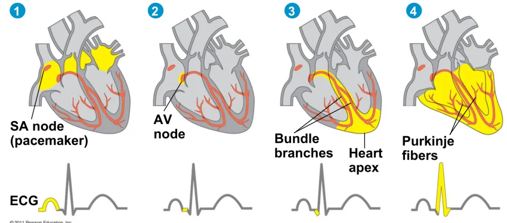
Maintaining the Heart’s Rhythmic Beat
• Some cardiac muscle cells are self-excitable, meaning
they contract without any signal from the nervous system
• The sinoatrial (SA) node, or pacemaker, sets the rate
and timing at which cardiac muscle cells contract
• Impulses that travel during the cardiac cycle can be
recorded as an electrocardiogram (ECG or EKG)
Figure 42.9-4
SA node (pacemaker)
AV
node Bundle
branches Heart apex
Purkinje fibers
ECG
• Impulses from the SA node travel to the atrioventricular
(AV) node
• At the AV node, the impulses are delayed and then
travel to the Purkinje fibers that make the ventricles contract
• The pacemaker is regulated by two portions of the
nervous system: the sympathetic and parasympathetic divisions
• The sympathetic division speeds up the pacemaker • The parasympathetic division slows down the
pacemaker
• The pacemaker is also regulated by hormones and
temperature
Changes in Blood Pressure During the
Cardiac Cycle
• Systolic pressure is the pressure in the arteries during
ventricular systole; it is the highest pressure in the arteries
• Diastolic pressure is the pressure in the arteries during
diastole; it is lower than systolic pressure
• A pulse is the rhythmic bulging of artery walls with each
heartbeat
Regulation of Blood Pressure
• Blood pressure is determined by cardiac output and
peripheral resistance due to constriction of arterioles
• Vasoconstriction is the contraction of smooth muscle in
arteriole walls; it increases blood pressure
• Vasodilation is the relaxation of smooth muscles in the
arterioles; it causes blood pressure to fall
Figure 42.17
Plasma 55%
Constituent Major functions Water Ions (blood electrolytes) Sodium Potassium Calcium Magnesium Chloride Bicarbonate Solvent for carrying other substances Osmotic balance, pH buffering, and regulation of membrane permeability Plasma proteins Osmotic balance, pH buffering Albumin Fibrinogen Immunoglobulins (antibodies) Clotting Defense
Substances transported by blood Nutrients Waste products Respiratory gases Hormones Separated blood elements Basophils Neutrophils Monocytes Lymphocytes Eosinophils Platelets
Erythrocytes (red blood cells) 5–6 million
250,000–400,000 Blood clotting
Transport of O2 and some CO2 Defense and immunity
Functions Number per L
(mm3) of blood
Cell type
Cellular elements 45%
Stem Cells and the Replacement of Cellular
Elements
• The cellular elements of blood wear out and are being
replaced constantly
• Erythrocytes, leukocytes, and platelets all develop from
a common source of stem cells in the red marrow of bones, especially ribs, vertebrae, sternum, and pelvis
• The hormone erythropoietin (EPO) stimulates
erythrocyte production when O2 delivery is low
Cardiovascular Disease
• Cardiovascular diseases are disorders of the heart and
the blood vessels
• Cardiovascular diseases account for more than half the
deaths in the United States
• Cholesterol, a steroid, helps maintain membrane fluidity
• Low-density lipoprotein (LDL) delivers cholesterol to
cells for membrane production
• High-density lipoprotein (HDL) scavenges cholesterol
for return to the liver
• Risk for heart disease increases with a high LDL to HDL
ratio
• Inflammation is also a factor in cardiovascular disease
Atherosclerosis, Heart Attacks, and Stroke
• One type of cardiovascular disease, atherosclerosis, is
caused by the buildup of plaque deposits within arteries
• A heart attack, or myocardial infarction, is the death of
cardiac muscle tissue resulting from blockage of one or more coronary arteries
• Coronary arteries supply oxygen-rich blood to the heart
muscle
• A stroke is the death of nervous tissue in the brain,
usually resulting from rupture or blockage of arteries in the head
• Angina pectoris is caused by partial blockage of the
coronary arteries and results in chest pains
Concept 42.5: Gas exchange occurs across
specialized respiratory surfaces
• Gas exchange supplies O2 for cellular respiration and
disposes of CO2
Partial Pressure Gradients in Gas Exchange
• A gas diffuses from a region of higher partial pressure to
a region of lower partial pressure
• Partial pressure is the pressure exerted by a particular
gas in a mixture of gases
• Gases diffuse down pressure gradients in the lungs and
other organs as a result of differences in partial pressure
Respiratory Media
• Animals can use air or water as a source of O2, or
respiratory medium
• In a given volume, there is less O2 available in water
than in air
• Obtaining O2 from water requires greater efficiency than
air breathing
Respiratory Surfaces
• Animals require large, moist respiratory surfaces for
exchange of gases between their cells and the respiratory medium, either air or water
• Gas exchange across respiratory surfaces takes place by
diffusion
• Respiratory surfaces vary by animal and can include the
outer surface, skin, gills, tracheae, and lungs
Lungs
• Lungs are an infolding of the body surface
• The circulatory system (open or closed) transports gases
between the lungs and the rest of the body
• The size and complexity of lungs correlate with an
animal’s metabolic rate
Mammalian Respiratory Systems: A Closer Look
• A system of branching ducts conveys air to the lungs
• Air inhaled through the nostrils is warmed, humidified, and sampled for odors
• The pharynx directs air to the lungs and food to the
stomach
• Swallowing tips the epiglottis over the glottis in the
pharynx to prevent food from entering the trachea
Figure 42.25 Pharynx Larynx (Esophagus) Trachea Right lung Bronchus Bronchiole Diaphragm (Heart) Capillaries Left lung
Dense capillary bed enveloping alveoli (SEM)
• Air passes through the pharynx, larynx, trachea,
bronchi, and bronchioles to the alveoli, where gas exchange occurs
• Exhaled air passes over the vocal cords in the larynx to create sounds
• Cilia and mucus line the epithelium of the air ducts and
move particles up to the pharynx
• This “mucus escalator” cleans the respiratory system
and allows particles to be swallowed into the esophagus
• Gas exchange takes place in alveoli, air sacs at the tips
of bronchioles
• Oxygen diffuses through the moist film of the epithelium and into capillaries
• Carbon dioxide diffuses from the capillaries across the
epithelium and into the air space
• Alveoli lack cilia and are susceptible to contamination • Secretions called surfactants coat the surface of the
alveoli
• Preterm babies lack surfactant and are vulnerable to
respiratory distress syndrome; treatment is provided by artificial surfactants
Concept 42.6: Breathing ventilates the lungs
• The process that ventilates the lungs is breathing, the
alternate inhalation and exhalation of air
How a Mammal Breathes
• Mammals ventilate their lungs by negative pressure
breathing, which pulls air into the lungs
• Lung volume increases as the rib muscles and
diaphragm contract
• The tidal volume is the volume of air inhaled with each
breath
Figure 42.28
Rib cage expands.
Air
inhaled. Rib cage getssmaller. Airexhaled.
1 2
Lung
• The maximum tidal volume is the vital capacity
• After exhalation, a residual volume of air remains in the
lungs
Control of Breathing in Humans
• In humans, the main breathing control centers are in
two regions of the brain, the medulla oblongata and the pons
• The medulla regulates the rate and depth of breathing
in response to pH changes in the cerebrospinal fluid
• The medulla adjusts breathing rate and depth to match
metabolic demands
• The pons regulates the tempo
• Sensors in the aorta and carotid arteries monitor O2 and
CO2 concentrations in the blood
• These sensors exert secondary control over breathing
Homeostasis:
Blood pH of about 7.4
CO2 level
decreases. Stimulus:
Rising level of CO2 in tissues lowers blood pH. Response:
Concept 42.7: Adaptations for gas exchange
include pigments that bind and transport gases
• The metabolic demands of many organisms require that
the blood transport large quantities of O2 and CO2
Coordination of Circulation and Gas Exchange
• Blood arriving in the lungs has a low partial pressure of
O2 and a high partial pressure of CO2 relative to air in the alveoli
• In the alveoli, O2 diffuses into the blood and CO2 diffuses
into the air
• In tissue capillaries, partial pressure gradients favor
diffusion of O2 into the interstitial fluids and CO2 into the blood
Exhaled air Inhaled air
Pulmonary arteries
Systemic
veins Systemicarteries Pulmonary veins Alveolar capillaries Alveolar spaces Alveolar epithelial cells Inhaled air 160 120 80 40 0 Heart 8 1 2 3 4 6 7
CO2 O2
Systemic capillaries CO2 O2
Body tissue
5
(a) The path of respiratory gases in the circulatory system
(b) Partial pressure of O2 and CO2 at different points in the circulatory system numbered in (a)
4 3
2
1 5 6 7
Fick’s Law
• Explains scenario greatest rate of diffusion
• Ventilating surface is thin
• Surface area is large
• Partial-pressure gradient is large
Hemoglobin
• A single hemoglobin molecule can carry four molecules
of O2, one molecule for each iron- containing heme group
• The hemoglobin dissociation curve shows that a small
change in the partial pressure of oxygen can result in a large change in delivery of O2
• CO2 produced during cellular respiration lowers blood
pH and decreases the affinity of hemoglobin for O2; this is called the Bohr shift