doi.: 10.21839/jaar.2017.v2i1.47
http://www.phoenixpub.org/journals/index.php/jaar ISSN 2519-9412 / © 2017 Phoenix Research Publishers Research Article – Biological and Bio-medical Sciences
Nanoparticle-assisted Polymerase Chain Reaction (NanoPCR): Optimization of
PCR detection of
Leifsonia xyli
subsp.
xyli
by the addition of nanoparticles
Patrícia Virgínia Padilha Dantas1*, Caio Figueiró Melo2, Laureen Michelle Houllou3, Giovanna Machado4
1
Laboratório de Diagnose Fitossanitária, Centro de Tecnologias Estratégicas do Nordeste, Brazil
2
Universidade Federal de Pernambuco, Brazil
3
Laboratório de Pesquisas Aplicadas Biofábrica, Centro de Tecnologias Estratégicas do Nordeste, Brazil
4
Laboratório de Nanotecnologia, Centro de Tecnologias Estratégicas do Nordeste; Universidade Federal de Pernambuco, Brazil
Abstract
Leifsonia xyli subsp. xyli (Lxx) causes ratoon stunting disease (RSD) in sugarcane, and is one of major causes of production losses. The detection of Lxx bacteria in sugarcane is made mainly through molecular biology techniques, especially polymerase chain reaction (PCR). However, PCR presents some barriers to provide reliable results. The present work brings a Nanoparticle-assisted Polymerase Chain Reaction (NanoPCR) assay for the detection of Lxx in its latent infection on micropropagated sugarcane. This assay was based in the addition of Gold and Titanium dioxide nanoparticles to conventional PCR and evaluation of its effects. It was observed that the reactions performed with Titanium dioxide nanoparticles provided the formation of singular well-defined bands under electrophoresis, consistent with the expected molecular weight, without occurrence of non-specific bands or presenting false negatives occurrence, negative effects that were observed in the control assay. While the performed NanoPCR adding AuNP also provided the formation of well-defined bands, been able to inhibit the occurrence of false negatives, but wasn’t able to eliminate the occurrence of non-specific amplifications. The results indicate that NanoPCR by the addition of Gold and Titanium dioxide nanoparticles to conventional PCR increased the detection of Lxx.
Key words: AuNP; Lxx; ratoon stunting disease; Saccharum spp.; TiO2NP
Introduction
Ratoon stunting disease (RSD) is one of the most important sugarcane diseases because of its damages to the production yield and its widespread presence in all growing regions. Efforts to establish an efficient protocol for Leifsonia xyli subsp. xyli (Lxx) diagnosis are based on the fastidious behaviour of Lxx, known as a restricted bacteria living in the sugarcane xylem system. Lxx is difficult to isolate with fastidious growth, even in selective culture media, and its cells are difficult
to see using optical microscopy techniques (Tiwari et al., 2012).
Due to the difficulty of detecting Lxx, many techniques have been developed and implemented for this purpose, including phase contrast and immune fluorescence microscopy (to detect the presence of the bacterial cell in xylem extracts), serological methods such as tissue blot enzyme immunoassay (TBEIA) or dot-blot enzyme immunoassay (EIA) (Croft et al., 2012), and molecular methods (polymerase chain reaction - PCR) which have high reliability and detection specificity (Zhou et al., 2012; Houllou et al., 2015).
Received: 12-01-2017; Accepted 06-02-2017; Published Online 10-02-2017
٭Corresponding Author
Molecular diagnosis using PCR is one of the most relevant techniques used for plant health certification. However, some substances in
Patrícia Virgínia Padilha Dantas, Laboratório de Diagnose Fitossanitária, Centro de Tecnologias Estratégicas do Nordeste, Brazil
plant tissues (polysaccharides, proteins, phenolic compounds and other plant secondary metabolites) can interfere with PCR amplification, making it difficult to establish a diagnostic routine test for some plant diseases. The rapid heating-cooling response in the PCR machines is restricted by the large thermal flux (heating and cooling rates). Thus, to solve this limitation, a thermocycler was enhanced to increase the surface/volume ratio and decrease the content volume. These changes have increased the efficiency of thermal cyclers, but the thermal efficiency of appliances has reached its threshold, so that maximum efficiency in alternating temperature of the blocks should be combined with techniques that allow the reaction fluid distribute the heat and cool faster and with more homogeneity. Despite all these improvements, routine procedures still present a non-specific response (Li et al., 2005a; Lagally et al., 2011; Schori et al., 2013).
Lxx detection by PCR is characterized by the incidence of false negatives and non-specific amplicons, probably because the analysis is performed using DNA extracted from plant tissues. This phenomenon does not occur when the reaction is performed with DNA obtained from pure Lxx cultures. The occurrence of false negatives is attributed to some substances (attached to plant tissue) which tend to interfere in the reaction. These substances are not easy to identify or remove from the extraction process of genomic DNA (Martinelli et al., 2015). As Lxx isolation from plant tissues is difficult, the diagnosis of field crops from pure bacterial cultures (isolated from these samples) becomes unfeasible. Thus, the optimization of the PCR technique is necessary to perform reactions without the presence of non-specific DNA amplification.
Nano-PCR is an improved technique strategy that consists in adding nanoparticles to the polymerase chain reaction (PCR) to enhancing its efficiency and specificity (Khaliq et al., 2011). Since the first test, different kinds of nanoparticles (metallic, oxide, carbon nanotubes) have been used in several investigations (Fadhil et al., 2014). Metal nanoparticles, especially gold (AuNP) and titanium dioxide (TiO2NP) have recently
demonstrated an optimization effect on the conventional PCR technique. These nanoparticles contribute to increased specificity in the amplification,
probably because the heat conductivity and diffusivity are more efficient in solutions (e.g. PCR mix) with these particles. Heat transfer efficiency can generate a greater amount of amplification products (amplicons), translated into more intense and well-defined products after PCR when viewed by electrophoretic techniques (Li et al., 2005a; Martinelli et al., 2015).
The present work aimed to evaluate if the incorporation of gold and titanium dioxide nanoparticles in conventional PCR would be able to increase the detection of the pathogenic bacteria Leifsonia xyli subsp. xyli after obtaining DNA from sugarcane tissues.
Material and Methods
Preparation of Nanoparticles
Tetrachloroauric acid (HAuCl4.3H2O; 49.0%
Au basis) and sodium citrate were purchased from Aldrich, and used as received. Initially, a 0.88*10-3M HAuCl
4 solution was prepared
in water (13.3684 mg HAuCl4 in 20 mL deionized
H2O) under vigorous stirring a second solution of
trisodium citrate (Na3C6H5O7.2H2O) 5.0*10-3 M
(58.82 mg Na3C6H5O7 in 20 mL deionized H2O)
was added to prepare a final volume of 40 mL. This solution was stirred for 15 minutes and then heated in a microwave oven (CEM- ISCOVER SP) at 300 W power for 6.5 min after it reached 95°C. After the reaction, the colour of the solution changed immediately from light yellow to cherry red, indicating the formation of gold nanoparticles.
The TiO2 colloidal solution was prepared from
TiO2 (STS-100, Mw = 80 g mol−1, 15.4 wt% in
titanium; Ishihara Sangyo Kaisha Ltd.); a solution with a concentration of 0.0375 mol.L−1 was used. The morphological characterization of Au and TiO2 nanoparticles was verified by TEM, UV-Vis
and XRD. The absorbance spectra of colloidal solutions of TiO2 and Au were measured using a
CARY 300 (Agilent Technologies) spectrophotometer over a wavelength range of 200−800 nm. To ensure a more stable signal, measurements were made using a double light beam configuration.
The Transmission electron microscopy (TEM) images were obtained on a FEI TECNAI 20. The treatments for TEM analysis were prepared by placing a drop of the as synthesized colloids onto a carbon coated Cu grid followed by slow
evaporation of solvent at ambient condition. The average particle size (Av) and standard deviation (SD) were calculated by counting 600 particles from more than five TEM images of different areas of the Cu grid.
The X-ray diffraction patterns were obtained using an X-ray diffractometer (model BRUCKER ADVANCED 8- XRD) with Cu Kα radiation (λ = 1.5406 Å) with a scan range of 10° ≤ 2θ ≤ 50°, a step size of 0.02°, and a measuring time of 2 s per point. Diffraction patterns were defined by comparing them with the crystallographic Joint Committee on Powder Diffraction Standards (JCPDS) No. 4- 0784 for Au and the Inorganic Crystal Structure Database (ICSD) no. 78-2486 for TiO2 100% anatase.
Nano PCR Conditions
Samples were collected from in vitro cultivated Saccharum spp. (RB92579) plant, in which Lxx colonization was previously detected. Leaves were collected (25 mg for each sample) from shoot tips and frozen in liquid nitrogen for subsequent grinding. After pre-processing, plant material was submitted to genomic DNA extraction with the Wizard® Genomic DNA Purification Kit (Promega, Wisconsin, USA). The material was used in PCR for Lxx genome amplification.
The PCR was performed using a master mix that contained the primers LxxW.1 (CAGAGCACCATCGTGAAGAC) and LxxW.2 (AAGGACAAGTCCACCAAGGA) for RSD with 182 base pairs (Houllou et al., 2015). The PCR reaction was carried out in a volume of 50 μL containing 10 μL of DNA sample, primers at the final concentration of 0.5 μM each, 1.5 mM of MgCl2, 1X PCR buffer, 0.2 mM dNTP and 0.05
unit of Taq DNA polymerase, with the addition of sterile ultrapure Milli-Q® water to complete the reaction volume. In order to test the hypothesis that the nanoparticles would improve the PCR reaction, 1 μL of nanoparticle solution, i.e. 0.08 mM AuNP and 2.0 mM TiO2NP, were separately added
to the original master mix.
The program was performed under the following conditions: denaturation step at 94°C for 5 minutes, followed by 35 cycles of denaturation at 94°C for 45 seconds, annealing at 56°C for 30 seconds, extension at 72°C for 60 seconds, and
a final extension hold of 10 minutes at 72°C. At the end of the PCR reaction, the amplicons were submitted to electrophoresis on a 2% agarose gel and visualized under ultraviolet light. The generated amplicons were visualized in the form of bands and were considerate as positive when a 182 bp fragment was observed, compared with the pattern of the 100 bp molecular weight marker.
In the present work, three different treatments were established, assessed in triplicate. The control treatment was the assay where the amplification was performed without the addition of the metallic nanoparticles. Treatments corresponding to Nano PCR were performed with the addition of the nanoparticles, AuNP and TiO2NP, respectively.
Results and Discussions
Nanoparticles Analysis
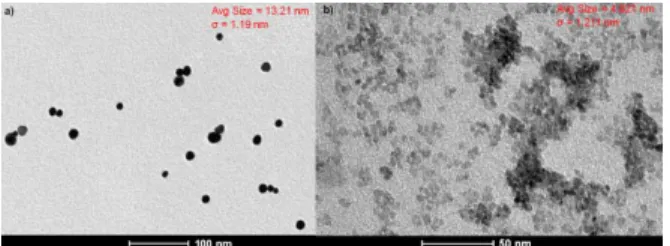
Nanoparticles prepared in synthesized passed through spectroscopic characterizations. As a local probe analysis, Transmission Electronic Microscopy (TEM) of the particles was carried, and TEM images of Au (Figure 1a) and TiO2 (Figure 1b) nanoparticles
presented spherical shape nanoparticles, in both analysis, which were highly dispersed in the colloids with an average particle size of 13.21 nm ± 1.19 nm and 4.821 ± 1.21 nm for Au and TiO2,
respectively, which was consistent with higher surface area for TiO2NP.
Figure 1. Images of transmission electron microscopy showing the morphlogy and sizes of gold (a) and titanium dioxide (b) nanoparticles. Avg size refers to the average size of the nanoparticles produced in the synthesis, whereas σ corresponds to the standard deviation regarding the shown average size.
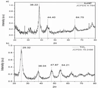
As a bulk sensitive analysis, X-ray based Diffraction (DRX) was carried and showed the diffractograms for Au (Figure 2a) and TiO2
(Figure 2b) nanoparticles in which the phases were defined comparing them with the crystallographic Joint Committee on Powder Diffraction Standards
(JCPDS) No. 4-0784 for Au and the Inorganic Crystal Structure Database (ICSD) no. 78-2486 for 100% anatase TiO2.
Figure 2. X-Ray Diffraction patterns of gold (a) and titanium dioxide (b) nanoparticles. The results agree with 4-784 and 78-2486 JCPDS cards for gold and titanium respectively
Also UV-visible spectroscopy (UV-Vis) was performed, and results obtained confirm plasmonic band for AuNPs (Figure 3a) in 520 nm and absoption band for TiO2NP (Figure 3b) in 264 nm.
Figure 3. UV-Vis images shown the absorption peak for: (a) gold nanoparticlesat 521 nm and (b) titanium dioxide nanoparticles at 263 nm
Nano PCR Analysis
The DNA extracted to provide a PCR template showed that the DNA quality for PCR amplification was adequate. All samples with or without nanoparticles presented DNA amplification using the specific primers (LxxW.1 and LxxW.2). However, the quality and specificity of DNA amplification was influenced by the nanoparticles. Figure 4. DNA amplification of sugarcane samples infected with Lxx though primers LxxW.1 and LxxW.2. First left column corresponds to the molecular weight marker, followed at right by triplicates of control amplification without nanoparticles, amplification with AuNP and amplification with TiO2NP. Each triplicate is
followed at right by its blank reaction.
The control test analysis (DNA + mix without nanoparticles) showed the presence of non-specific DNA amplification, as shown in the control treatment (Figure 4). Visual analysis of the amplified products on the gel shows that the samples corresponding to the control tests presented a lack of amplification uniformity. The amplification of the control treatment showed non-specific bands, with low, but visible, definition intensity. These non-specific bands showed distinct molecular weights, different to that expected for the PCR product with specific primers. Analysing the AuNP treatment, it was possible to observe the formation of a distinct band; however, this band did not correspond to the molecular weight of the amplified product expected based on the parameters of this assay. This treatment presented non-specific bands amplified in smaller quantities than those in the control assay. From the comparison between the three treatments, it is visually noticeable that only the TiO2NP treatment
revealed an amplification result with only the specific band for detecting Lxx. However, even that band was a little heavier than expected.
As both treatments containing nanoparticles showed higher uniformity and a better standard of visual presentation of the bands when regards to the control treatment, and presented bands of the same molecular weight, the two nanoparticles show the ability to inhibit false negatives, besides an increase in PCR amplification specificity. However, TiO2NP showed the most reliable
results, as it did not show the occurrence non-specific amplifications.
The explanation for this could be due to concentrations of TIO2NP treatment being higher
than the tested AUNP treatment. To evaluate the actual effect of concentration on the obtained results a new TiO2NP sample was tested with the same
concentration of gold nanoparticles (1.6 g.L-1).
The result confirmed what was obtained initially demonstrating the improved efficiency of the TiO2
nanoparticles. Discussion
The variation in the ability to detect Lxx bacteria in extracts of infected sugarcane tissues has been the main obstacle in terms of reliability in the diagnosis of healthy plants using this technique. The occurrence of false negatives is the main point of contention that the conventional PCR technique has to overcome, and in the present work inconsistencies in detection are evident by comparing the three replicates in the control tests (Urashima and Grachet, 2012). While the PCR technique is one of the sums of tools for the diagnosis of microscopic pathogens in plants, the technique has bottlenecks long been known with respect to compounds inherent to plant tissue which inhibit the amplification reaction, in addition to it the difficulty in quantifying DNA of this pathogen in samples originating from extractions performed in plant tissues with varying conditions of colonization (Cubero et al., 2001; Schrader et al. 2012; Green and Sambrook, 2012). As such, optimizing the specificity of amplification is extremely relevant to implement a PCR diagnosis routine test; the addition of nanoparticles may be an effective alternative to this application. All these non-specific bands may be a result of some unidentified substance, present in the sugarcane tissue, once secondary metabolites may interfere in PCR reactions in extracts from plant material (Schori et al., 2015).
These results could be related to the fact that DNA polymerase exhibits many functional differences in complex samples and reagents (Li et al., 2005a). There are many substances in plant tissues, such as polysaccharides, proteins, phenolic compounds and other plant secondary metabolites that can interfere with PCR amplification (Schori et al., 2015). DNA quality, obtained after the extraction procedure, can be also a problem because the mentioned inhibitors can degrade the genomic DNA kept stored for long time, demanding especial protocols of extraction (Valledor et al., 2014), but the present work used DNA samples recently extracted and with high quality. Additionally, components used for DNA isolation (e.g. buffers) can affect PCR reactions in different ways. These substances can inhibit the PCR reaction, change Ct values and decrease PCR reaction efficiency (Cankar et al., 2006). Thus, it is possible that some unidentified substance, present in the DNA obtained from sugarcane samples, interfered with the PCR process and caused the presence of non-specific bands.
The reaction established on PCR, especially when the target DNA is extracted from complex samples, can be inhibited by a number of compounds present in the sample that are not efficiently removed in the extraction process, so that the efficiency of amplification is reduced, and the detection sensitivity has considerable losses, reaching even the occurrence of false negatives results(Schrader et al., 2012). Attempts to remove these inhibitors from plant samples using various methods are currently applied, but the alternatives to remove inhibitors can make the processing of samples expensive or protracted (Rådström et al., 2004). However, nanoparticle synthesis, as described, can be performed easily, and the present study shows that TiO2NP, which a cheap nanoparticle to
synthetize, provided the best results in terms of inhibiting false negatives and non-specific amplifications. The application of nanoparticles is a rapid, inexpensive and efficient alternative to optimize the detection of Lxx from sugarcane tissues.
The results show that there was an improvement in the DNA yield and purity when the amplification was performed with nanoparticles. Positive results using nanoparticles in PCR were also reported by different works using TiO2NP, and AuNP
(Lin et al., 2013; Fadhil et al., 2014). Both studies underscore the value of the nanoparticles to increase the specificity of PCR and highlight the ability of nanoparticles to increase the heat conductivity of the reaction fluid. Consequently, nanoparticles could provide gains not only in the steps involving annealing temperatures of the primers, which are essential for oligonucleotide initiator specificity, but also for the whole cycling. As PCR is a technique that essentially depends on temperature alternation between cycling steps, the use of metal or metal oxides nanoparticles increases the efficiency of the DNA amplification reaction.
Nanoparticles through their properties in efficiency of heat transfer can work as modulators to the PCR (Khaliq et al., 2010), and is reported that metal nanoparticles (Au) and oxides nanoparticles (TiO2) have been commonly used in improve PCR
efficiency since the first report of nanoparticle-assisted PCR was published (Pan et al., 2011), so that the metal nanoparticles can enhance PCR specificity by applying the appropriate concentration and provide better bands of the target product. These properties are extremely useful in the case of Lxx detection in plant tissue, since most of the inconsistencies in the method are related to non-occurrence of the reaction, observed as no bands on the running gel, or when the PCR is effective but specific, leading to the formation of non-specific bands. It has been shown in research evaluating Nano PCR containing titanium dioxide that these nanoparticles interact distinctively with the PCR mastermix and with the DNA itself, so
that in a comparison between TiO2NPs and AuNPs
is described that PCR got improved thermal fluid
efficiency using TiO2NPs been the same property
previously recorded in literature for AuNPs,
however the TiO2NPs also promote greater efficiency
in the denaturation of genomic DNA strand, first stage of cycling, from which the entire reaction is developed, as well as the AuNPs was credited with the property of interacting directly with the DNA strand also generating gains the reaction (Khaliq
et al., 2010). In addition to the aspects of the
thermal efficiency in the reaction, the use of nanoparticles, 35 nm ZnONP, 100 nm TiO2NP
(Fadhil et al., 2014) and 13 nm AuNP (Li et al., 2005b), may also contribute to reduction of the PCR reaction time, thus the Nano PCR technique can not only offer better results, as can still allow
savings in terms of time of use of the resistance of thermal cyclers who consume a lot of electricity. In this work, beyond what has already been mentioned on TiO2 NPs, they may have interacted
with the fluid with higher efficiency due to their smaller size compared to gold nanoparticles, which confer higher surface area of the TiO2 nanoparticles.
The use of AuNPs showed promise for amplification of gene classically reported as troublesome (Yang et al., 2013), however it was also demonstrated that the use of AuNPs can not always provide significant improvement of results in target DNA samples obtained from matrices containing inhibitors compounds capable of suppressing the activity of the enzyme Taq polymerase in the presence of Au colloids (Li et al., 2005b). This suggests that, despite the results observed for Lxx DNA amplification in sugarcane samples, specific optimization of PCR conditions for other tissue treatments could be necessary when using nanoparticles.
The poor efficiency of conventional PCR for the detection of Lxx and linked this to exogenous inhibitors not removed in the process of DNA extraction, which culminated in the occurrence of false negatives (Iglesia et al., 2003). In this regard, it is known that the addition of nanoparticles to the master mix promotes increased conductivity of the reaction fluid, and the present work shows that by adding metal nanoparticles, non-specific bands and false negatives were reduced. It is possible that the action of the substances involved in the inhibition of the reaction performed on DNA extracted from plant tissue may be connected to a decrease in the conductivity of the fluid, providing insight into a better understanding of the issues involved in generating false negative results.
Despite the reason behind non-specific amplification, the presence of nanoparticles seems to overcome the occurrence of non-specific amplification products in Lxx assays. PCR is a priceless tool for the detection of Lxx, as the detection of the pathogen will allow for development of control practices that reduce the spread of this bacterium to new fields, preventing a disease that can result in losses to the grower (Srivastava, 2016). In this sense, the application of metal nanoparticles may be an advance in the current Lxx detection methods and RSD diagnosis for use in the field.
Conclusion
AuNPs and TiO2NPs were able to optimize
PCR efficiency to detect Leifsonia xyli subsp. xyli extracted from sugarcane tissues. TiO2NPs were
able to eliminate the occurrence of non-specific amplification products as well as false negative events.
Acknowledgement
The authors thank the Conselho Nacional de Desenvolvimento Científico e Tecnológico, Fundação de Amparo à Ciência e Tecnologia de Pernambuco and Centro de Tecnologias Estratégicas do Nordeste by structural, material and financial resources that enabled this research. References
Cankar, K., Štebih, D., Dreo, T., Žel, J. and Gruden, K. (2006). Critical points of DNA quantification by real-time PCR – effects of DNA extraction method and treatment matrix on quantification of genetically modified organisms. Bmc Biotechnology, 6. Retrieved from http://bmcbiotechnol.biomedcentral.com/ articles/10.1186/1472-6750-6-37.
Croft, B.J., Green, J. and Braithwaite, K.S. (2012). Comparison of RSD assays for diagnosis and screening varieties for resistance. Proc Aust Soc Sugar Cane Technol., 34: 1-10.
Cubero, J., Graham, J. H. and Gottwald, T.R. (2001). Quantitative PCR method for diagnosis of citrus bacterial canker. Applied and Environmental Microbiology, 67: 2849-2852.
Fadhil, A.M.A., Al-Jeboory, M.R., Al-Jailawi, M. H. (2014). Improving and enhancement of PCR amplification by using ZnO and TiO2
nanoparticles. International Journal of Current Microbiology and Applied Sciences, 3: 549-557.
Green, M.R. and Sambrook, J. (2012). Isolation and Quantification of DNA. Molecular Cloning: A Laboratory Manual. New York: Cold Spring Harbor.
Henson, J. and French, R. (1993). The polymerase chain reaction and plant disease diagnosis. Annual Review of Phytopathology, 31: 81-109. Houllou, L.M., Torres, D.C.B.A., Medeiros, M. J. L., Dantas, P.V.P., Soares, A.B., Souza, R.A., Cavalcante, P.F. and Sobral, M.C.M. (2015).
Electrotherapy treatment for ratoon stunting disease (Leifsonia xyli subsp. xyli) elimination in sugarcane micropropagation. International Journal of Current Research, 7: 19888-19892. Iglesia, A., Fonseca, D., Díaz, M., Nuñez, O.,
González, R., Pazos, V. and Peralta, E.L. (2003). Detección de Leifsonia xyli subsp. xyli mediante la reación em cadena de polimerase (PCR). Revista Proteción Vegetal, 18: 23-27. Khaliq, A., Kafafy, R., Salleh, H.M., Faris, W.F.
(2012). Enhancing the efficiency of polymerase chain reaction using grapheme nanoflakes.
Nanotechnology, 23: 455106.
Khaliq, A., Sonawane, P.J., Sasi, B.K., Sahu, B.S., Pradeep, T., Das, S.K. and Mahapatra, N.R. (2010). Enhancement in the efficiency of polymerase chain reaction by TiO2 nanoparticles: crucial
role of enhanced thermal conductivity. Nanotechnology, 21: 255704.
Khaliq, R.A., Raed, M.K., Waleed, F.F. and Hamzah, M.S. (2011). Enhancement of polymerase chain reaction using graphene nano-flakes. Nanobiotechnol. J., 3: 978-983. Lagally, E.T., Simpson, P.C. and Mathies, R.A.
(2000). Monolithic integrated microfluidic DNA amplification and capillary electrophoresis 80 analysis system. Sensors Actuators B, 63: 138–146.
Li, H., Huang, J., Lv, J., An, H., Zhang, X., Zhang, Z., Fan, C. and Hu, J. (2005). Nanoparticle
PCR: Nanogold‐Assisted PCR with Enhanced
Specificity. Angewandte Chemie International Edition, 44: 5100-5103.
Li, M., Lin, Y.C., Wu, C.C. and Liu, H.S. (2005). Enhancing the efficiency of PCR using gold nanoparticles. Nucl. Acids Res., 33: e184-e184. Lin, Y., Li, J., Yao, J., Liang, Y., Zhang, J., Zhou, Q.
and Jiang, G. (2013). Mechanism of gold nanoparticle induced simultaneously increased PCR efficiency and specificity. Chinese Science Bulletin, 58: 4593-4601.
Martinelli, F., Scalenghe, R., Davino, S., Panno, S., Scuderi, G., Ruisi, P., Villa, P., Stroppiana, D.,
J. Appl. Adv. Res. ● Vol. 2 ● Issue 1 20 Boschetti, M., Goulart, L.R., Davis, C.E. and
Dandekar, A.M. (2015). Advanced methods
of plant disease detection. A review. Agronomy
for Sustainable Development, 35: 1-25.
Pan, D., Wen, Y., Mi, L., Fan, C. and Hu, J. (2011). Nanomaterials - based polymerase chain reactions for DNA detection. Current Organic Chemistry, 15: 486-497.
Schori, M., Appel, M., Kitko, A. and Showalter. A.M. (2013). Engineered DNA polymerase improves PCR results for plastid DNA. Applications in Plant Sciences, 1: 1-7.
Schrader, C., Schielke, A., Ellerbroek, L. and Johne, R. (2012). PCR inhibitors–occurrence, properties and removal. Journal of Applied Microbiology, 113: 1014-1026.
Srivastava, S. (2016). Molecular Diagnostics and Application of DNA Markers in the Management of Major Diseases of Sugarcane. In: Kumar, P., Gupta, V.K., Tiwari, A.K., Kamle, M. Ed. Current Trends in Plant Disease Diagnostics and Management Practices. Switzerland: Springer International Publishing.
Tiwari, A.K., Vishwakarma, S.K., Kumar, P., Pandey, N. and Lal, M. (2012). Ratoon stunting disease (Leifsonia xyli) of sugarcane. Plant Knowledge Journal, 1: 20-24.
Urashima, A.S. and Grachet, N.G. (2012). Detection method for Leifsonia xyli subsp. xyliand effect of thermotherapy on bud germination in sugarcane varieties. Tropical Plant Pathology, 37: 57-64.
Yang, W., Li, X., Sun, J. and Shao, Z. (2013).
Enhanced PCR amplification of GC-rich DNA templates by gold nanoparticles. ACS Applied Materials & Interfaces, 5: 11520-11524.