Effects of an Acute Bout of Resistance Training on Leukocyte and Leukocyte Subset Response in Breast Cancer Survivors
Rachel Graff
A thesis submitted to the faculty of the University of North Carolina at Chapel Hill in partial fulfillment of the requirements for the degree of Masters of Arts in the Department of Exercise
and Sport Science (Exercise Physiology).
Chapel Hill 2013
Approved By:
Claudio L. Battaglini, PhD Anthony C. Hackney, PhD, DSc Eric D. Ryan, PhD
ii ©2013 Rachel Graff
iii Abstract
RACHEL GRAFF: Effects of an acute bout of resistance training on leukocyte and leukocyte subset response in breast cancer survivors
(Under the direction of Dr. Claudio Battaglini)
The purpose of this study was to examine the immune response to a single bout of moderate-intensity resistance training in breast cancer survivors (BCS). BCS (n=4) and healthy controls (n=8) completed an exercise session consisting of 3 sets of 10 repetitions at 70% of one repetition-maximum on leg press, lateral pull down, leg extension, and seated row performed in a circuit fashion. Blood samples taken at baseline, immediately post (0h post), 2-hours post (2h post), and 24-hours post (24h post) exercise were assessed for total leukocyte, granulocyte, lymphocyte, and monocyte cell counts. Percent change (%Δ) scores from baseline-0h post,
baseline-2h post, and baseline-24h post were compared using mixed-model ANOVAs between groups and indicated that there were no significant differences between total leukocytes
iv
Acknowledgements
I would like to take this opportunity to thank all of those that made this thesis project possible. First and foremost, I would like to thank Dr. Claudio Battaglini and Dr. Anthony Hackney, who invested so much time and effort into virtually all aspects of the study and who put up with my endless amount of questions. Without their expertise, guidance, and mentorship, this thesis would not have been possible. I have thoroughly enjoyed working with and learning from both of them on a professional as well as a personal level. I would also like to thank Jacob Allen, as he served as my ‘partner-in-crime’ throughout the duration of the thesis process from beginning to end and an integral member of our research team. This project was definitely a team effort and these individuals are certainly all-stars!
I would like to extend a special thanks to Dr. Beth Evans, as the ‘back-bone’ of this project came from her doctoral dissertation and she served as an invaluable resource throughout all stages of the research process. Also, I would like to thank Dr. Eric Ryan, the final member of my thesis committee, for investing his time and effort into making this thesis the best it can be.
v
Table of Contents
List of Figures ... viii
List of Tables ... ix
Chapters I. Introduction ... 1
Statement of Purpose ... 4
Hypotheses (H) ... 5
Definition of Terms... 5
Assumptions ... 6
Limitations ... 7
Delimitations ... 7
Significance... 8
II. Review of the Literature ... 9
Breast Cancer Prevalence and Treatments ... 9
Physical Activity and Breast Cancer... 10
Resistance Exercise in Cancer Patients ... 12
Relationship Between Cancer and the Immune System ... 14
vi
Exercise and Immune Function: Implications for Breast Cancer Survivors... 20
Summary and Significance ... 23
III. Methodology ... 25
Subjects ... 25
Recruitment ... 26
Instrumentation ... 26
General Procedures ... 27
Statistical Analysis ... 32
IV. Results ... 33
V. Discussion ... 38
Immune Response to Resistance Training in Breast Cancer Survivors ... 40
Baseline – 0h Post-Exercise ... 40
Baseline – 2h Post-Exercise ... 45
Baseline – 24h Post-Exercise ... 48
Limitations of the Study... 50
Conclusion ... 51
Recommendations for Future Research ... 51
Appendices Appendix A ... 53
vii
Appendix C ... 69
Appendix D ... 74
Appendix E ... 77
Appendix F... 81
Appendix G ... 83
Appendix H ... 85
Appendix I ... 86
viii List of Figures
1. Protocol Timeline………31
ix List of Tables
1. Table 1: Subject Characteristics……….….33
2. Table 2: Subject Cancer Treatment Characteristics………34
3. Table 3: Leukocyte, Leukocyte Subset, and Cortisol Values……….35
4. Table 4: Mean Hematocrit (Hct) and Hemobglobin (Hb) Values………..36
Chapter I
Introduction
In the year 2012, more than 200,000 people in the United States alone are expected to be diagnosed with invasive breast cancer (American Cancer Society, 2012). Of these, 90% are likely to survive for at least five years after diagnosis (ACS, 2012). Many of these individuals will have to undergo surgery, radiation, chemotherapy, hormonal therapies, or some combination of one or more of these treatments in order to survive the disease (ACS, 2012). Cancer and its treatments have profound impacts on the physiology of its patients, drastically altering physical, psychological, and functional capacity and quality of life (Battaglini et al., 2006). Therefore, attempts to improve or reverse these negative physiological alterations that result from cancer and its treatments are paramount.
2
cardiovascular systems (Battaglini et al., 2006; Loescher et al., 1989; Schneider et al., 2007). Loss of skeletal muscle, in particular, is a very frequent and undesirable effect of cancer and its treatments, which can negatively impact various aspects of life in cancer survivors (Al-Majid & Waters, 2008; Courneya, 2003; Schneider et al., 2007).
In the last couple of decades, alternative interventions, often referred to
complementary alternative medicine (CAM) have been explored in oncology patients in an attempt to attenuate the severe side-effects commonly experienced by cancer patients during and post-completion of cancer treatments. One intervention that has gained a lot of attention the past few years is exercise training due its efficacious ability to positively impact many physiological systems that are altered during cancer treatment, its non-invasive nature, and relatively affordable price. Since the mid 1980s, studies have been conducted examining the effects of exercise on different physiological systems as well as in different cancer
populations, with most of the studies being conducted in breast cancer patients. Most of these initial studies focused on aerobic forms of exercise (Courneya et al, 2003; Courneya et al. 2006; Decker et al. 1989; Dimeo et al., 1997; MacVicar et al., 1986). It wasn’t until 2002 that other modes of exercise training began to be explored. Kolden et al. (2002) was the first research team to examine the effects of a combined aerobic and resistance exercise training intervention on multiple physiological parameters. In 2004, Drake and colleagues were one of the first groups to examine the effects of resistance training alone in cancer patients. This intervention was called for due to the negative effects of cancer treatments on body
3
muscle cells and metabolism, often leading to reductions in protein synthesis. Resistance exercise, however, has shown to attenuate and even reverse the loss of muscle mass that is associated with cancer and its treatments (Courneya et al., 2007; Drake et al., 2004; Schmitz et al. 2005; Schwartz, 2007, 2009; Segal et al., 2009).
The body of research regarding the specific role of progressive resistance training in cancer patients and survivors is relatively small. However, studies are demonstrating that resistance training holds promise in improving physical, psychosocial, and functional aspects in cancer patients (Cheema et al., 2007; Courneya et al., 2008; Schmitz et al., 2005; Segal et al., 2009). Even though these first generation studies provide important information on patient acceptability and tolerability for exercise training, safety and precise information on optimum exercise training regimens for cancer patients have yet to be determined.
A well functioning immune system plays an integral role in maintaining overall health and optimal physiological function. However, cancer and its treatments are associated with immunodeficiency, which often lasts well beyond the completion of treatments (Fairey et al., 2005; Hutnick et al., 2005). The adaptive immune system is particularly influenced by the disease, and is often slow to recover (Hutnick et al., 2005). Therefore, cancer survivors often experience an increased risk of secondary infection as well as a risk of cancer
recurrence as a result of their immunosuppressed condition (Hutnick et al., 2005). Some research has shown that exercise may boost immune function in cancer patients, improving overall health and potentially enhancing longevity (Fairey et al., 2002; Fairey et al., 2005; Hutnick et al., 2005).
4
Furthermore, most of these studies have looked at aerobic exercise modes and have only examined the chronic effects of exercise. Even though these studies have shown promising results, which demonstrate the potential that exercise training may have on improving the immune system of breast cancer survivors, nothing is currently known about the effects of one bout of exercise on the immune response of these patients. Examining markers of immunity such as total leukocyte and leukocyte subset counts may serve as an important initial step for the development of the body of knowledge regarding the response of the immune system to an acute bout of exercise, since these markers are routinely part of hematological profile analyses in cancer patients.
In order to maximize the benefits of exercise for cancer patients, understanding their immune response to different intensities and modes of exercise is a paramount initial step for the development of future exercise prescription guidelines for optimum exercise training regimens. Since the use of resistance training has grown in popularity among the scientific community due to its potential to alleviate many of the negative side-effects associated with body composition and overall functionality in cancer patients, examining the effects of an acute bout on the immune response would allow for the development of more precise future interventions aimed not only at maximizing training response, but also at improving the safety of patients engaging in this mode of exercise training.
Statement of Purpose
5
of moderate intensity resistance training, as well as the relationship between the cortisol and leukocyte and leukocyte subset response in both the breast cancer survivors and control groups.
Hypotheses (H)
H1. There will be a significantly greater difference in leukocyte, granulocyte, lymphocyte, and monocyte percent cell count changes from baseline to immediate post-exercise in the control group compared to the breast cancer survivor group.
H2. There will be a significantly greater difference in leukocyte, granulocyte, lymphocyte, and monocyte percent cell count changes from baseline to 2 hours post-exercise in the breast cancer survivor group compared to the control group.
H3. There will be a significantly greater difference in leukocyte, granulocyte, lymphocyte, and monocyte percent cell count changes from baseline to 24 hours post-exercise in the breast cancer survivor group compared to the control group.
Definition of Terms
Leukocytes: White blood cells that help protect the body via involvement in immune and inflammatory responses (Marieb & Hoehn, 2010).
Leukocytosis: An increase in the number of leukocytes (Marieb & Hoehn, 2010). Lymphocytes: Agranular leukocytes that form within the bone marrow and become functionally mature in the lymphoid organs (Marieb & Hoehn, 2010).
6
Granulocytes: a subset of leukocytes that includes neutrophils, basophils, and eosinophils (Marieb & Hoehn, 2010).
Granulocytosis: An increase in the number of granulocytes.
Neutrophil: Most abundant type of leukocyte (makes up about 60% of total leukocytes). A phagocytic cell that often serves as the body’s first line of defense again infection (Marieb & Hoehn, 2010; Walsh et al., 2011).
Neutrophilia: An increase in the number of neutrophils.
Monocyte: Large leukocyte that makes up 3-8% of total leukocytes (Marieb & Hoehn, 2010). Monocytosis: An increase in the number of monocytes.
Breast Cancer Survivors: Individuals diagnosed with stage I, II, or III breast cancer who have completed their major treatment(s), including chemotherapy, radiation, and/or surgery within the last 6 months and who may or may not be currently receiving hormonal therapy.
Progressive Resistance Training: A training program where muscles work against gradually increasing resistance; involving implementation of the overload principle (Powers &
Howley, 2009).
1 Repetition Maximum (1-RM): The heaviest load that an individual can lift through a full range of motion one time (Tan, 1999).
Assumptions
• All participants in both groups adhered to the pre-assessment guidelines prior to experimental sessions.
• All participants were honest in their responses to the medical history form
7
• Subjects did not change their diet between the familiarization (1-RM testing) and acute bout.
Limitations
• Subjects may not have complied with the dietary and exercise guidelines. To
encourage participant compliance, subjects were asked upon arrival to verify that they have not eaten within the last 2 hours nor exercised within the last 12 hours.
• The training status of the subjects prior to their enrollment in the Get REAL & HEEL breast cancer program was not controlled for. Therefore, training status may
confound or interact with the results of the resistance training exercise bout.
• The various anti-cancer treatments underwent by the subjects were not accounted for during analysis.
• Diurnal rhythms of cortisol were not accounted for.
Delimitations
• All subjects in the breast cancer survivor group had completed chemotherapy and/or radiation treatments within 12 months of the experimental sessions.
• Subjects in the breast cancer survivor group were not taking any medications that may influence the response of the immune system, other than hormonal therapy (i.e., Tamoxifen).
• All subjects in the control group were healthy with no contraindications to
participating in resistance exercise, and not taking any medications that may influence the response of the immune system.
8 Significance
It is necessary for exercise science professionals to account for the effects of exercise training sessions on immune function when designing training regimens for their clients. This includes being aware of the extent to which the immune system is suppressed following certain types of training, and making certain to allocate adequate recovery time before implementing another training session. In athletes, the continuous implementation of consecutive training bouts with inadequate recovery time is suggested to facilitate immunosuppression and the onset of the Overtraining Syndrome (Gleeson, 2002). While breast cancer survivors are not likely to be performing exercise at the same level or intensity as high-level athletes, it remains important that the implementation of exercise training in this population does not result in further chronic immunosuppression in addition to that caused by the various anti-cancer treatments.
To ensure that the suppression of the immune systems of breast cancer survivors is not being exacerbated by exercise training programs, it is essential for the timeline of
9 Chapter II
Review of the Literature
Introduction
This review of the literature is divided into 6 sections. Section 1 will discuss breast cancer and its treatments; section 2 will review physical activity in breast cancer patients; section 3 will discuss the current research on resistance exercise in cancer patients; section 4 will introduce the relationship between cancer and the immune system; section 5 will focus on the relationship between acute exercise and the immune response; and section 6 will summarize the potential implications of exercise, particularly resistance exercise, in breast cancer survivors.
Breast Cancer Prevalence and Treatments
The prevalence of breast cancer within the United States has continually increased over the last number of years. Today, 1 out of 8 women will be diagnosed with breast cancer at some point in her lifetime (American Cancer Society, 2012). Surgery, radiation,
chemotherapy, and hormonal therapies comprise the list of the various treatments that these individuals may have to endure, and they are each accompanied by a vast array of side effects, which can drastically influence physiological factors and quality of life (ACS, 2012; Courneya, 2003).
10
2003). Radiation treatment is associated with potential toxicity to irradiated normal tissue, pain and blistering, reductions in ROM, fatigue, diarrhea, lung fibrosis, and damage to cardiac myocytes (Courneya, 2003). Chemotherapy is often coupled with side effects such as fatigue, reduced appetite, nausea, anemia, reduction in neutrophil counts, peripheral
neuropathy, ataxia, and cardiotoxicity (Courneya, 2003). Finally, hormonal therapy typically results in weight gain, sarcopenia, muscle weakness, increases in adiposity (particularly within the torso and face), bone loss, fatigue, hot flashes, and immunosuppression
(Courneya, 2003). Following a breast cancer diagnosis, individuals will often undergo one or more of these treatments, which clearly exhibit monumental influences upon various aspects of human physiology.
Physical Activity and Breast Cancer
Until recently, participation in regular physical activity for individuals undergoing and recovering from cancer treatments was contraindicated by medical professionals (Battaglini et al., 2006). Within the last several years, however, there has been a growing body of evidence to suggest that participation in a regular exercise program can be extremely beneficial in mediating and even reversing many of the aforementioned side effects
associated with cancer treatments both during and following cancer treatments. Although the first study to incorporate resistance training in cancer patients was conducted in 2002, successive studies over the past 10 years have demonstrated that resistance training shows promise in enabling the improvement in a wider range of treatment-related side effects than cardiovascular training alone (Kolden et al., 2002).
11
In addition to metabolic effects, the time period following a cancer diagnosis is often coupled with an increase in inactivity, which contributes to further muscle loss (Al-Majid & Waters, 2008; Schneider et al., 2007). Metabolically, individuals diagnosed with cancer typically experience a decrease in protein synthesis and a subsequent increase in protein degradation (Al-Majid & Waters, 2008; Schneider et al., 2007). In other words, cancer patients’ bodies are unable to re-build skeletal muscle at the same rate at which it is being broken down. It is suggested that the reduction in protein synthesis may be due to one or more of the following: increased levels of tumor-released proteolysis-inducing factor, angiotensin II, disruption of the mTOR pathway, and a reduction in daily physical activity levels as a result of fatigue and muscle weakness (Al-Majid & Waters, 2008). It is also possible that the reduction in physical activity can play a role in exacerbating the metabolic effects on the reduction in protein synthesis (Al-Majid & Waters, 2008).
In addition, the increase in skeletal muscle protein degradation may be attributed to an activation of various proteolytic mechanisms. One such mechanism is the non-lysosomal calcium-dependent protease system that is responsible for the release of calpains, which break down the myofibrils that comprise skeletal muscle fibers. Another, is the ubiquitin-proteasome system, which breaks down damaged proteins in healthy individuals, but shows increased activity within the skeletal muscles of cancer patients. A third, is an increase in the amounts of pro-inflammatory cytokines such as tumor necrosis factor- alpha (TNF-α),
12
within the skeletal muscles of individuals with cancer serve to comprise the cachexia, or skeletal muscle wasting, experienced by cancer patients.
Resistance Exercise in Cancer Patients
Progressive resistance training in healthy individuals is responsible for providing a stimulus to increase the amount of skeletal muscle protein synthesis. This may be due, in part, to the anti-inflammatory effect of resistance training. Resistance training has
demonstrated a reduction in pro-inflammatory cytokine levels, which as previously
mentioned, are suggested to indirectly activate the ubiquitin-proteasome system, contributing to skeletal muscle breakdown (Al-Majid & Waters, 2008). By reducing the release of pro-inflammatory cytokines and thereby the indirect stimulation of the ubiquitin-proteasome system, protein degradation may be prevented. Further, resistance training has been shown to affect the mTOR pathway, which as previously mentioned, is another mechanism of protein synthesis that is oftentimes disrupted as a result of cancer and its treatments. In healthy individuals, resistance training significantly stimulates the mTOR pathway within skeletal muscle cells, thereby promoting skeletal muscle protein synthesis (Al-Majid & Waters, 2008; Spiering et al., 2008). Therefore, resistance exercise may be beneficial in cancer patients by opposing several cancer-related mechanisms that contribute to skeletal muscle loss.
13
cancer and its treatments, as it can improve various aspects of physiology and quality of life without the occurrence of any serious adverse effects (Cheema, et al., 2008; Drake et al., 2004; Galvao et al., 2006; Galvao et al., 2008; Schwartz et al, 2007; Schwartz & Winters-Stone 2009).
In a study by Segal et al., (2009), men currently receiving radiation treatment for prostate cancer were divided into resistance exercise, aerobic exercise, and non-exercise control groups. The exercise groups each trained 3 times a week for 24 weeks with
progression as participants became more physically fit. Results indicated that the resistance exercise group resulted in greater improvements in cancer-specific quality of life, muscular strength, triglycerides, and body composition than the aerobic training group. Both resistance and aerobic training groups had a positive effect on reducing levels of fatigue and in
improving aerobic fitness. Even though this study was conducted in men with prostate cancer who were undergoing radiation treatment, it suggests that resistance exercise could
potentially play an important role in individuals with other types of cancer who have undergone radiation therapy.
14
(VO2max), and health-related quality of life. In sum, these studies indicated that moderate to high intensity combined resistance and aerobic exercise training in cancer patients and survivors can improve muscular strength, performance, and quality of life as well as the functionality of cancer survivors.
Schmitz et al. (2005) and Courneya et al. (2007) both employed the use of resistance training in isolation in breast cancer survivors and breast cancer patients undergoing
chemotherapy, respectively. Both studies found significant improvements in body
composition and muscular strength, and were well tolerated by the participants. Schmitz et al. (2005) noted significant reductions in body fat percentages over the 6-12 month training period, which has important implications in preventing cancer recurrence. Courneya et al., (2007) reported that in addition to the improvements in body composition and muscular strength, resistance training was in fact superior to aerobic training in increasing self esteem and chemotherapy completion rate in participants over the course of the training intervention. Although research on resistance training alone in breast cancer patients and survivors is limited, it appears to have positive influences on many of the ailments caused by a cancer diagnosis and its subsequent treatments.
Relationship Between Cancer and the Immune System
15
The innate immune system is typically the body’s first line of defense against invading pathogens. The cellular components of the innate response include neutrophils (a subset of granulocytes, which comprise about 60% of all circulating leukocytes),
macrophages (which come from the maturation of monocytes), natural killer (NK) cells (a subset of lymphocytes), and dendritic cells (Koch, 2010; Walsh et al., 2011). These cells provide non-specific protection of the host against any foreign or non-self components that exist within the body (Walsh et al., 2011).
The cellular components of the adaptive immune system consist primarily of the various lymphocyte subpopulations with the exception of NK cells, which are part of the innate immune system. The basic lymphocyte subpopulations are divided into T-helper cells (CD4+), T-cytotoxic cells (CD8+), and B-cells, which can be further divided into additional subsets (Walsh et al., 2011). These cellular components are typically activated by the innate immune response to a particular antigen or abnormal body cell and undergo a cascade of events to enable the body to more effectively fight off the foreign invader or abnormal cell via antibody production or apoptosis (programmed cell death). Therefore, the duration of the adaptive response may take much longer than that of the innate response. Once the antigen or non-self component is effectively removed from the body, memory T-cells and B-cells remain so that upon secondary exposure, the body can respond more quickly and efficiently to wipe out the invader (Marieb & Hoehn, 2010).
16
all cancer treatments aside from tamoxifen and found that these individuals had lower proportions of NK cells and T-helper cells than those who were not fatigued. It is suggested that these lymphocyte subsets are particularly slow to recover following cancer treatment, and may be affected for up to five years after chemotherapy and radiation treatments (Bower et al., 2002; Hutnick et al., 2005).
The cellular components of the innate and adaptive immune systems have immense implications for cancer patients and survivors. The cells of both immune systems have
important roles in protecting the body from tumor formation as well as infection from outside elements (Marieb & Hoehn, 2010; Walsh et al., 2011). Both groups of cells are affected by cancer and anti-cancer therapies. Cancer and its treatments have been shown to reduce both the number and the functionality of lymphocytes (T-cells, B-cells, and NK cells), neutrophils, and macrophages (Fairey et al., 2005). Even after the completion of treatment, many of these components are unable to recover in a timely fashion (Fairey et al., 2005; Hutnick et al., 2005). Therefore, these individuals are more likely to develop additional infections or cancer recurrence. However, exercise has shown to play a mediating role in immune regulation and may potentially serve as a form of immunotherapy to help improve the immune systems of cancer survivors (Fairey et al., 2005; Hutnick et al., 2005).
Acute Exercise and the Immune Response
17
window” theory, which portrays the immune response to a single bout of acute exercise (Pederson, 1999). According to this theory, a single exercise bout can result in an initial up-regulation in immune parameters followed by a subsequent down-up-regulation of these parameters, which can last up to 72 hours post-exercise (Pederson et al., 1999). In other words, following a single exercise bout, immune function may increase immediately following the exercise session and then may decrease below baseline, thereby increasing susceptibility to infection. The magnitude of this immunosuppression is directly proportional to the intensity and duration of the exercise performed (Pedersen et al., 1999).
The majority of the research to date is focused on the effects of endurance training on the immune system. Therefore, much less is known about the impact of resistance training (Koch, 2010; Walsh et al., 2011). As a result of endurance exercise, an immediate and significant increase in neutrophils, a subset of granulocytes, is observed. This immediate neutrophilia is due primarily to catecholamine release and the shear stress exerted by an increase in cardiac output against the blood vessel walls that occur during exercise (Walsh et al., 2011). Several hours following the acute exercise bout, a secondary increase in
circulating neutrophils is typically observed. This is suggested to be mediated by cortisol, which stimulates neutrophil release from the bone marrow (Walsh et al., 2011).
18
from the spleen, lymph nodes, and gastrointestinal tract (Pedersen & Hoffman-Goetz, 2000). This recruitment is thought to be a response to increases in catecholamine levels as well as increases in cardiac output that typically accompany exercise sessions (Koch, 2010).
However, cortisol, which is often released during prolonged or intense exercise is associated with immunosuppression (Neves Jr. et.al., 2009). It has an inhibitory effect on the proliferative response of lymphocytes, suggesting that cortisol impairs the ability of
lymphocytes to respond to antigens (Nieman, 1997). The inhibitory effects of cortisol coupled with a redistribution of lymphocytes to respective body tissues are likely explanations for the decrease in circulating lymphocytes observed after acute exercise
(Gleeson, 2007; Pedersen & Hoffman-Goetz, 2000). An intensity threshold of 40% to 60% of VO2max has been established as the aerobic intensity threshold for initiation of
lymphocytopenia, or the reduction in circulating lymphocytes following the cessation of exercise (Koch, 2010).
Levels of circulating monocytes are also affected by acute endurance exercise (Pedersen & Hoffman-Goetz, 2000). However, increase in monocyte concentration is typically not observed until about 2 hours post-exercise (Walsh et al., 2011). This
19
substances (Marieb & Hoehn, 2010). Prolonged elevation in active macrophages can be indicative of chronic inflammation (Marieb & Hoehn, 2010).
As a result of moderate intensity endurance exercise, the leukocytosis observed immediately post-workout is often less pronounced than following intense endurance exercise (Koch, 2010). Further, the lymphocytopenia, or the reduction in circulating lymphocytes below baseline levels, is also of a smaller magnitude following moderate-intensity endurance exercise compared to high-moderate-intensity endurance exercise (Koch, 2010). It is suggested that the immune system responds similarly to resistance exercise as well, however, the lymphocyte response to resistance exercise has not been clearly established (Koch, 2010).
It appears that the immunosuppression following an acute bout of resistance exercise is less pronounced than that of the response following aerobic exercise (Neves Jr. et al., 2009). In the study by Neves Jr. et al. (2009), older women experienced an increase in circulating leukocytes following an acute bout of resistance training with no
immunosuppression observed during the recovery period. In fact, this study observed elevated lymphocyte levels at 3 hours post-exercise. The absence of observable immunosuppression was suggested to be due to a lack of cortisol elevation during the resistance exercise protocol, as cortisol is associated with initiating many of the immunosuppressive effects observed following endurance-type exercise sessions.
20
function (Neves Jr. et al., 2009). In a study by Kraemer et al. (1996), subjects underwent two identical high-intensity resistance training exercise sessions, with the only difference
between the two being the rest period between subsequent sets. One protocol required 3 minutes of rest in between sets, whereas the other limited recovery to 1 minute between sets. Results indicated that while cortisol was significantly elevated within the 1 minute rest protocol, it was not significantly elevated within the 3 minute rest protocol even though the absolute work performed during both protocols was the same. The authors further observed that in the immediately post-exercise blood sample, both protocols initiated a similar
leukocytosis. This is important because it suggests that the increase in circulating leukocytes is caused by factors unrelated to cortisol release during exercise, and that manipulating the rest period between subsequent sets of heavy resistance training exercises can influence whether or not cortisol is significantly elevated.
It is clear that exercise has a significant influence on the levels of circulating leukocytes. These effects seem to vary with differing training variables, such as intensity, duration, and mode of exercise. Even though the research exploring the influence of resistance training on these immune parameters is somewhat limited, the manipulation of various resistance training variables may have important implications in enabling the maximization of immune function, especially for those clinical populations, like cancer patients, to whom optimal immune function is essential to enhancing quality of life and preventing cancer recurrence.
Exercise and Immune Function: Implications for Breast Cancer Survivors
21
These programs included either aerobic only training, or a combined aerobic and resistance training regimen, with little emphasis placed on resistance-training only interventions (Fairey et al., 2002). Overall, training studies involving cancer survivors indicate that exercise
training can have a positive effect on immune function in these individuals (Fariey et al., 2002).
In addition, there are limited established exercise program design parameters for cancer populations (Schmitz et al., 2010). The American College of Sports Medicine has published a consensus regarding exercise guidelines for cancer survivors. However, large gaps remain in the prescription of certain training variables, such as frequency, duration, and intensity, particularly when pertaining to resistance training (Schmitz et al., 2010). Hence, the training intervention designs between studies are rarely homogenous, and consequently difficult to compare. Therefore, criteria for exercise prescription to improve immune function in cancer survivors must be derived from the limited data available, and often from studies involving alternate populations.
22
Taking this one step further, Hutnick et al. (2005) examined the effects of a mixed aerobic and resistance training intervention on lymphocyte function in breast cancer survivors. Before the initiation of the training program, all of the participants had mean lymphocyte subset values below the interquartile range for healthy individuals. After the 3 to 6 month training period, results indicated that circulating levels of T-helper cells had
increased proliferative capacity following the training period in the exercise group, but not in the non-exercising control group. The authors suggest that exercise training may emerge as a sort of immunotherapy for breast cancer survivors, aiding them in the prevention of
secondary infections as well as cancer recurrence.
Ramel et al. (2003) examined the immune response to an acute bout of resistance exercise in healthy men. Subjects performed a resistance exercise circuit consisting of 10 exercises focusing on the major muscle groups at 75% of 1 RM with 1 minute of rest in between each exercise and completed the 10-exercise circuit 2-3 times each. Results indicated that total leukocytes and leukocyte subset (NK cells, neutrophils, total
lymphocytes, and monocytes) values were increased during and immediately following the exercise bout. While neutrophils and monocytes remained significantly elevated throughout the 120 minute recovery period, lymphocyte values dropped below baseline after the
23
These conclusions suggest that resistance exercise may play less of an inhibitory role in immune function in healthy males, particularly that of lymphocytes whose fluctuations appear to be directly related to the magnitude of cortisol release. As lymphocyte levels and function appear to be sub-optimal in breast cancer survivors following cancer treatments, the implementation of resistance exercise may be beneficial in improving lymphocyte function in these individuals.
Summary and Significance
The current research in exercise science points to positive effects on the immune system as just one of the many benefits to be obtained from exercise. Knowledge regarding the influence of exercise on the immune system is integral to creating optimal program design for specific populations. For example, athletes, who typically employ the use of prolonged, high intensity training bouts must allow adequate time for their immune systems to recover before implementing a second intense training bout. Failure to allot adequate time for the recovery of the immune system may result in further immunosuppression with additional training and may eventually culminate in the overtraining syndrome, in which the athlete’s performance will regress to sub-optimal levels despite additional athletic training (Gleeson, 2002). Likewise, it is necessary for exercise professionals to be aware of the timeline in which breast cancer survivors’ immune systems recover from acute bouts of exercise when coming up with an exercise prescription for this population. This knowledge would allow for the establishment of guidelines regarding recovery periods between
24
precautions that should be taken against training these individuals to the point where their immune function may be further depressed by subsequent training bouts.
Breast cancer survivors, who are already immunosuppressed following anti-cancer treatments, comprise a population that can benefit immensely from regular participation in resistance exercise training. In addition to the variety of benefits initiated by resistance training on physiological variables including increased functional capacity, increased muscle mass, improved body composition, increased quality of life, reductions in muscle wasting and sarcopenia, and decreased risk of osteoporosis, resistance training is also showing promise as a means of potentially enhancing various aspects of the immune system. As such, implementing regular resistance exercise in the breast cancer survivor population may reduce the risk of secondary infection, reduce the risk of cancer recurrence, and ultimately improve length and quality of life.
25 Chapter III
Methodology Subjects
Participants of this study consisted of 4 female early stage breast cancer (stage I-III) survivors and 8 healthy controls. The breast cancer survivor inclusion criteria consisted of: being a female between 40 and 72 years old, being within 4-12 months post breast cancer treatment (having undergone chemotherapy treatment at minimum), not taking any anti-inflammatory medications, and having clearance from their oncologists to participate in the present study. Patients receiving adjuvant hormonal therapy or adjuvant trastuzumab were also eligible to participate in the study.
Inclusion criteria for the control group consisted of: apparently healthy, 40 to 70 year old females, no history of cancer diagnosis or treatment, no limitations in range of motion that would impede upon the ability to perform the prescribed resistance training exercises safely, not taking any anti-inflammatory medications, and clearance from their primary physician to participate in the study for subjects older than 65 years of age. All control subjects were matched by age (within 5 years) with the breast cancer group. Also, all
attempts were made to match control subjects with the breast cancer group based on physical activity level.
26
Brubaker, & Otto, 2006). Information regarding the exclusion criteria was obtained from each subject based on their responses to their medical history and from a medical and physical screening.
Recruitment
Participants for the breast cancer survivor group were recruited from UNC Hospital’s Breast Cancer Care Clinic and from the Get REAL & HEEL Breast Cancer Program at the University of North Carolina, Chapel Hill. Participants for the control group were recruited by the posting of flyers at various locations throughout the campus of the University of North Carolina, Chapel Hill, via informational emails sent out to the campus community at the University of North Carolina, Chapel Hill, as well as through word of mouth.
Instrumentation
Anthropometric Measures: Resting cardiac function was assessed using a GE CASE
Cardiosoft V. 6.6 ECG diagnostic system (Palatine, IL). Height was taken using a
Perspective Enterprises Stadiometer (Portage, MI). Weight was obtained via a Detecto Scale (Webb City, MO). A Discovery Dual Energy X-Ray Absorption (DEXA) scanner (Bedford, MA) was used to assess body composition.
Exercise Testing: Blood pressure was assessed before and after the exercise protocol
27
Medway, MA), lateral pull down machine (Cybex, Medway, MA), seated leg extension (Life Fitness, Schiller Park, IL), and a seated row (Life Fitness, Schiller Park, IL).
Blood Analyses: Blood was taken at 4 time intervals throughout the study. The first
three were done using a 22 gage Protectiv Plus catheter (Ethicon Endo-Surgery Inc., Cincinnati, OH). The fourth was done using standard venipuncture techniques. Blood was collected in 10.8 mg K3EDTA BD Vacutainers ® (Franklin Lakes, NJ). Total leukocyte, granulocyte, lymphocyte, and monocyte counts were determined using a Coulter AC T diff Hematology Analyzer (Block Scientific, Inc., Bohemia, NY). Blood cortisol values were determined using a cortisol ELISA Kit, KA 0981, Abnova Version 2, (Tapei City, Taiwan).
General Procedures
The general timeline for the experimental protocol is depicted in Figure 1.
Familiarization Session: Medical history forms and pre-assessment guidelines were
28
throughout the day to stay hydrated; No caffeine use for at last 12 hours prior to the lab visit; No alcohol use for at least 48 hours prior to the lab visit.
Height and weight were then taken, and subjects underwent a body composition assessment using the DEXA. Following this, they were fitted with a heart rate monitor and rested in the seated position for 10 minutes, at which time resting heart rate and blood pressure were recorded. Subjects then underwent a 5-10 minute warm-up consisting of unloaded cycling on a cycle ergometer, while keeping their heart rates around 40 - 50% of heart rate reserve (HRR), as calculated by the Karvonen formula.
After the warm-up, subjects were familiarized with the resistance training equipment that was used in the experimental protocol by undergoing approximately 10 repetitions of each exercise using minimal resistance. Following initial exposure to each of the resistance exercise machines in the same order in which they were used during the experimental protocol, each subject’s 1 RM values were obtained on the leg press, lateral pull down
(LPD), leg extension, and seated row exercises using the protocol as described by Levinger et al. (2009). The order of exercises during the experimental protocol was as follows: leg press, lateral pull down, leg extension, and seated row. Therefore, 1 RM values were taken in that order as well to account for any neuromuscular fatigue obtained from the preceding exercise. Heart rate was monitored continuously throughout the exercise session.
29
Experimental Session: The second lab visit took place between 1.5 and 14 days
following the familiarization session. Upon arrival, subjects put on a heart rate monitor and rested in the seated position, during which time adherence to the pre-assessment guidelines was reviewed. A Psychological Stress Scale (PSS) was also administered to determine if the subject was experiencing psychological stress, which could affect the release of the stress hormones which were measured during the study. A resting heart rate and blood pressure were taken. Again, if either of these values were not within normal range for the individual, the subject was asked to return to the lab on a different day.
The subjects then underwent catheter placement into an antecubital vein in the arm for blood sampling by a faculty member in the Department of Exercise and Sport Science who has been trained and certified in phlebotomy techniques. After this, subjects rested in the seated position for approximately 10 minutes, and then the first pre-exercise blood sample was collected. A 5 minute warm-up consisting of unloaded cycling on a cycle ergometer followed, again keeping subjects’ heart rates around 40 - 50% of HRR. Subjects then
performed several repetitions of each of the four resistance exercises with minimal weight to ensure that their muscles were adequately prepared for the resistance protocol. After the warm up, the resistance training protocol began. Subjects performed 10 repetitions at 70% of 1-RM of each exercise (leg press, lateral pull down, leg extension, and seated row,
respectively) with 30 to 45 seconds of rest between subsequent exercises, and 1 minute and 30 seconds to 2 minutes of rest between subsequent circuits. This circuit was completed three times. Heart rate and RPE were taken following each set to ensure subject safety and monitor their conditions. Subjects were encouraged to consume water at any point during and
30
Directly upon completion of the third circuit of exercises, subjects were seated and a blood sample was taken immediately post-exercise. Subjects were then sequestered for the next 2 hours, during which time they were encouraged to consume plenty of water and to relax. They were allowed to perform a brief series of light stretches. Following this 2 hour period, the 2 hour post-exercise blood sample was taken, after which the subjects were encouraged to eat a snack of their choice. The catheter was removed, any necessary bandaging was performed, and the subjects was reminded not to perform any kind of strenuous activity over the next 24 hours, and to adhere, once again, to the pre-assessment guidelines before their third and final lab visit.
24 Hour Blood Draw: Twenty-four hours following the completion of the resistance
exercise session, subjects returned to the lab for their 24 hour post-exercise blood draw. There was a 1-hour window at which this blood sample will be taken. In other words, subjects may come in for their third visit 30 minutes prior to the time of exercise completion session on the previous day, or 30 minutes after the time of completion of the exercise session on the previous day. Adherence to the pre-assessment guidelines was reviewed, and the blood sample was taken by a certified phlebotomist using a 22 gage needle. After
obtaining the blood sample, subjects have completed the experimental procedure. The blood-draw timeline is depicted in Figure 2.
31
32 Statistical Analysis:
Descriptive statistics are presented in the form of means and standard deviations. The independent variables for this analysis include subject condition (breast cancer survivor and healthy control) and the acute bout of resistance training. The dependent variables are the total leukocyte, granulocyte, lymphocyte, and monocyte counts at the various time intervals (immediately pre-exercise, immediately post-exercise, 2 hours post-exercise, and 24 hours post-exercise). All data was analyzed using SPSS version 20.0 (Chicago, IL). Statistical significance was set a priori at an alpha-level of p ≤ 0.05.
Hypotheses 1, 2, and 3 were analyzed using 2 x 3 mixed model ANOVAs. The between subjects factor is subject condition (breast cancer survivor or healthy control). The within subjects factor is the percent change of cell counts (% change = immediately exercise count – pre-exercise count/ pre-exercise count x 100; % change = 2 hours post-exercise count – pre-post-exercise count / pre-post-exercise count x 100; and % change = 24 hours post-exercise count – pre-exercise count / pre-exercise count x 100, respectively) for each dependent variable (leukocytes, granulocytes, lymphocytes, and monocytes cell count).
33 Chapter IV
Results
The primary purpose of this study was to compare the effects of an acute bout of resistance training on percent cell count changes in leukocyte and leukocyte subset values in breast cancer survivors and healthy controls. A secondary purpose was to conduct
exploratory analyses on cortisol to examine the cortisol response to an acute bout of
moderate intensity resistance training in both the breast cancer survivors and control groups, since cortisol has been shown to be a potential mediator of some cellular immune responses. Participants consisted of 4 breast cancer survivors and 8 healthy controls. Descriptive characteristics are presented in Table 1 below.
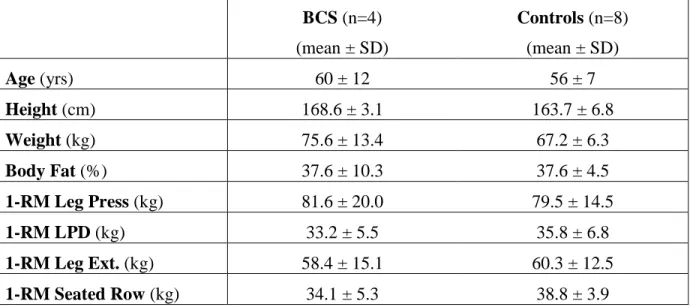
Table 1: Subject Characteristics and 1-RM Strength Assessment Results. No significant differences exist between the BCS and Control groups.
BCS (n=4) (mean ± SD)
Controls (n=8) (mean ± SD)
Age (yrs) 60 ± 12 56 ± 7
Height (cm) 168.6 ± 3.1 163.7 ± 6.8
Weight (kg) 75.6 ± 13.4 67.2 ± 6.3
Body Fat (%) 37.6 ± 10.3 37.6 ± 4.5
1-RM Leg Press (kg) 81.6 ± 20.0 79.5 ± 14.5
1-RM LPD (kg) 33.2 ± 5.5 35.8 ± 6.8
1-RM Leg Ext. (kg) 58.4 ± 15.1 60.3 ± 12.5
34
Table 2: Subject Cancer Treatment Characteristics for BCS Group
Means and standard deviations for total leukocyte, granulocyte, lymphocyte, and monocytes cell counts at each of the four measurement points (baseline, 0h post-exercise, 2h post-exercise, and 24h post-exercise) are presented below in Table 3. Immune parameter data at 0h post and 24h post-exercise for one control, and at 2h post-exercise for one BCS and one control subject were not obtained during the study due to technical measurement errors (i.e. blood clotting, catheter malfunction, inability to perform the venipuncture procedure, or inability of a subject to return to the lab for the 24h post-exercise blood draw). Mean
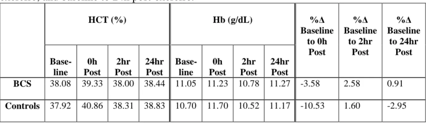
substitution procedures for the missing measurement data were performed for all variables of these subjects. Exploratory analyses on the effects of the acute bout of resistance exercise on cortisol were conducted and are also presented in Table 3. Similarly, mean substitution was performed for 2 control subjects at all four time points, and one breast cancer survivor at the 24h post time point. All reported cell and cortisol values have been adjusted for plasma volume shift (PV shift), which is depicted in Table 4.
Treatment Number of Subjects
(n=4) Surgery, Chemotherapy, Hormonal Therapy 2
Surgery, Chemotherapy, Radiation 1
Surgery, Chemotherapy, Radiation, Hormonal Therapy
35
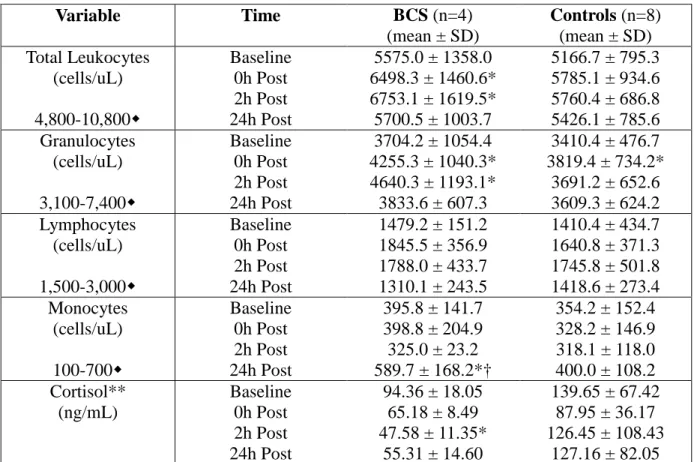
Table 3: Leukocyte, Leukocyte Subset, and Cortisol Values Adjusted for Plasma Volume Shift.
Variable Time BCS (n=4)
(mean ± SD)
Controls (n=8) (mean ± SD) Total Leukocytes (cells/uL) 4,800-10,800 Baseline 0h Post 2h Post 24h Post
5575.0 ± 1358.0 6498.3 ± 1460.6* 6753.1 ± 1619.5* 5700.5 ± 1003.7
5166.7 ± 795.3 5785.1 ± 934.6 5760.4 ± 686.8 5426.1 ± 785.6 Granulocytes (cells/uL) 3,100-7,400 Baseline 0h Post 2h Post 24h Post
3704.2 ± 1054.4 4255.3 ± 1040.3* 4640.3 ± 1193.1* 3833.6 ± 607.3
3410.4 ± 476.7 3819.4 ± 734.2*
3691.2 ± 652.6 3609.3 ± 624.2 Lymphocytes (cells/uL) 1,500-3,000 Baseline 0h Post 2h Post 24h Post
1479.2 ± 151.2 1845.5 ± 356.9 1788.0 ± 433.7 1310.1 ± 243.5
1410.4 ± 434.7 1640.8 ± 371.3 1745.8 ± 501.8 1418.6 ± 273.4 Monocytes (cells/uL) 100-700 Baseline 0h Post 2h Post 24h Post
395.8 ± 141.7 398.8 ± 204.9 325.0 ± 23.2 589.7 ± 168.2*†
354.2 ± 152.4 328.2 ± 146.9 318.1 ± 118.0 400.0 ± 108.2 Cortisol** (ng/mL) Baseline 0h Post 2h Post 24h Post
94.36 ± 18.05 65.18 ± 8.49 47.58 ± 11.35*
55.31 ± 14.60
139.65 ± 67.42 87.95 ± 36.17 126.45 ± 108.43
127.16 ± 82.05 Paired-samples and independent samples T-tests were performed for the evaluation of comparisons between post-exercise values and baseline values within as well as between the BCS and control groups at each assessment time. *Denotes statistical significance (p ≤ 0.05) when compared to baseline. †Denotes statistical significance (p ≤ 0.05) between BCS and control groups. Indicates normal cell ranges under resting conditions (Marieb & Hoehn, 2010). **For the cortisol analyses, only data for 6 control and 4 BCS subjects is presented.
Non-parametric analyses were conducted on the cortisol data, since the results were unequally distributed. A Mann-Whitney U analysis was conducted to test for between group differences, and no statistically significant differences were found (p=0.3938; p=0.6698; p=0.0550; p=0.5224 for baseline, 0h post-exercise, 2h post-exercise, and 24h post-exercise, respectively). A Friedman test was used to evaluate within-group differences, and a
36
Table 4: Mean Hematocrit (Hct) and Hemobglobin (Hb) values with corresponding plasma volume changes (Δ % changes) from baseline to 0h exercise; baseline to 2h post-exercise; and baseline to 24h post-exercise.
HCT (%) Hb (g/dL) %∆
Baseline to 0h Post %∆ Baseline to 2hr Post %∆ Baseline to 24hr Post Base-line 0h Post 2hr Post 24hr Post Base-line 0h Post 2hr Post 24hr Post
BCS 38.08 39.33 38.00 38.44 11.05 11.23 10.78 11.27 -3.58 2.58 0.91
Controls 37.92 40.86 38.31 38.83 10.70 11.70 10.52 11.17 -10.53 1.60 -2.95
A mixed-model ANOVA for each of the dependent variables (total leukocytes, granulocytes, lymphocytes, and monocytes) using the percent change (% Δ) scores from
37
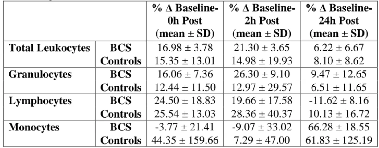
Table 5: Percent change (% ∆) scores for total leukocytes, granulocytes, lymphocytes, and monocytes depicting the % ∆ from baseline-0h post-exercise, baseline-2h post-exercise, and baseline-24h post-exercise.
% ∆ Baseline- 0h Post (mean ± SD)
% ∆ Baseline- 2h Post (mean ± SD)
% ∆ Baseline- 24h Post (mean ± SD)
Total Leukocytes BCS 16.98 ± 3.78 21.30 ± 3.65 6.22 ± 6.67
Controls 15.35 ± 13.01 14.98 ± 19.93 8.10 ± 8.62
Granulocytes BCS 16.06 ± 7.36 26.30 ± 9.10 9.47 ± 12.65
Controls 12.44 ± 11.50 12.97 ± 29.57 6.51 ± 11.65
Lymphocytes BCS 24.50 ± 18.83 19.66 ± 17.58 -11.62 ± 8.16
Controls 25.54 ± 13.03 28.36 ± 40.37 10.13 ± 16.72
Monocytes BCS -3.77 ± 21.41 -9.07 ± 33.02 66.28 ± 18.55
Controls 44.35 ± 159.66 7.29 ± 47.00 61.83 ± 125.19
No significant differences between the breast cancer survivor group and the control group were found between the %∆ scores for any of the dependent variables (total
38 Chapter V
Discussion
The examination of the effect of an acute bout of resistance training on the immune system’s response in breast cancer survivors can provide researchers with important
information for exercise prescription in a breast cancer population. Since resistance exercise has been shown to be a feasible and efficacious method for improving a plethora of
physiological and psychological parameters in individuals with breast cancer (Cheema et al., 2008; Courneya et al., 2007; Schmitz et al., 2005; Segal et al., 2009), it is of great importance for researchers to determine an exercise protocol designed to optimize these results. Further, it is essential that exercise programs for individuals with cancer do not impair the immune system because sub-optimal immune function can increase the risk of secondary malignancy and illness susceptibility (Hutnick et al., 2005). Although the body of research is small, current research in exercise oncology suggests that exercise may serve as a means of enhancing immune function in cancer patients, which can improve the chances of disease-free survivorship (Fairey et al., 2002; Fairey et al., 2005; Hutnick et al., 2005).
39
sessions in exercising individuals is suggested to result in chronic immunosuppression and impairment of overall health (Gleeson, 2001; Koch, 2010). Because breast cancer survivors often already have impaired immune function as a result of cancer treatments, further immunosuppression must be avoided. Since the acute immune response differs between the mode, intensity, and duration of exercise bouts (Koch, 2010), the aim of this study was to determine how the immune system in breast cancer survivors responded to a single bout of moderate-intensity resistance training.
To the authors’ knowledge, this is the first study to look at the immune response to a single bout of resistance training in a breast cancer population. The only other study to examine the effects of an acute bout of resistance exercise in cancer patients was a training study by Galvao and colleagues in 2008, where the immune response in prostate cancer patients was measured during and following a 20 week resistance training program. It is important to note that the acute response was not the main outcome variable of this study, as the authors’ primary objective was to evaluate the effect of chronic resistance training on various endocrine and immune parameters. The results of this study indicated that
immediately following an acute bout of resistance training, total leukocyte, neutrophil, lymphocyte, and monocyte cell counts in the prostate cancer patients responded similarly to those of healthy individuals. However, blood samples were not taken into the recovery period following the exercise bout. Therefore, the present study is the first to examine the timeline of immune response and recovery to an acute resistance training session in cancer patients.
40
exercise modalities even in healthy individuals have not been clearly established (Koch, 2010).
Immune Response to Resistance Training in Breast Cancer Survivors
The following discussion will be broken down into 6 sections. The first section will focus on the response from baseline to 0h post-exercise, the second will discuss the response from baseline to 2h post-exercise, the third will address the response from baseline to 24h post-exercise, the fourth will outline the limitations of the present study, the fifth will present concluding statements, and the sixth will provide recommendations for future research.
Baseline – 0h Post-Exercise
The immune response from baseline to 0h post-exercise in the BCS group is relatively consistent with the current literature in exercise immunology (Gleeson, 2007; Pedersen & Hoffman-Goetz, 2000; Walsh et al., 2011). Results indicated a significant increase in total leukocytes and granulocytes immediately following the exercise bout. Although the subset of granulocytes is comprised of neutrophils, basophils, and eosinophils, the majority of circulating granulocytes are made up of neutrophils, which are highly
responsive to acute exercise (Walsh et al., 2011). Further, neutrophils and lymphocytes comprise approximately 60% and 25%, respectively, of all circulating leukocytes , so transient changes in these cells will likely be reflected by the overall change in total leukocyte count as well (Gleeson, 2007; Marieb & Hoehn, 2010; Pedersen & Hoffman-Goetz, 2000; Walsh et al., 2011).
41
with the increased catecholamine secretion that happens upon engaging in physical activity cause a demarginalization of neutrophils into the circulation, thereby increasing the numbers of circulating neutrophils (Koch 2010; Pedersen & Hoffman-Goetz, 2000, Walsh et al., 2011). It is known that neutrophils contain a β-adrenergic receptor, which provides the basis for support that catecholamines play a partial role in mediating the neutrophil response to acute exercise (Pedersen & Hoffman-Goetz, 2000). Although the magnitude of
catecholamine release was not measured in this study, it is widely accepted that participation in physical activity is accompanied by sympathetic nervous system activation and the
secretion of epinephrine and norepinephrine into the circulation, which is likely a contributing factor to the observed neutrophilia.
The current research available on the immune response to resistance training is in accordance with the present findings, indicating that an acute bout of resistance training results in an increase in total leukocyte (Kraemer et al., 2006; Neves Jr. et al., 2009) and neutrophil counts (Galvao et al., 2008; Koch, 2010; Ramel et al., 2003) immediately following completion of the exercise bout. Although exercise has been shown to increase neutrophil mobilization into the circulation, research has suggested that these cells show a reduction in their responsiveness to bacterial lipopolysaccharide, which can last for several hours and is indicative of impaired functionality (Gleeson 2007; Nieman 1997). Neutrophil function was not measured in this study, therefore the capacity of these cells to respond to external stimuli following an acute bout of resistance training cannot be commented upon.
42
lymphocyte subsets and that optimal lymphocyte function, particularly that of NK cells, is related to overall survivorship (Hutnick et al., 2005; Fairey et al., 2002). No significant differences were observed in lymphocyte counts from baseline to 0h post-exercise in either the BCS or the control group. This is likely due to the nature of the exercise bout performed (moderate-intensity, relatively short duration; Nieman, 2003). Although lymphocyte counts did not increase significantly in either group, there were modest increases in the number of circulating lymphocytes immediately following the exercise bout in both BCS and control groups, which likely served as a contributing factor to the increase in total leukocytes observed immediately post-exercise. According to the literature, a post-exercise
lymphocytosis is observed due to the mobilization of these cells from a variety of organs, including the spleen, lymph nodes, and gastrointestinal tract (Pedersen & Hoffman-Goetz, 2000). Post-exercise lymphocytosis is depicted to occur in proportion to exercise intensity and duration, with intensity serving as the more prominent mediator of the lymphocyte response (Walsh et al., 2011).
Like neutrophils, T, B, and NK lymphocytes contain β-adrenergic receptors,
43
catecholamines initiate a down-regulation in adhesion molecule expression on endothelial cell walls (Walsh et al., 2011). As neither catecholamines or the specific lymphocyte subsets were measured as part of the present study, the magnitude of catecholamine secretion and its potential effect on the lymphocyte response cannot be commented upon.
Although derived primarily from studies examining the immune response to aerobic exercise, the upregulation of lymphocyte counts is supported in the study by Simonson and Jackson in 2004. This study demonstrated that the transient increase in the concentration of circulating lymphocytes immediately following an acute bout of resistance training was due primarily to increases in NK cell count, with less pronounced upregulation of CD8+ and CD4+ subsets. Conversely, Kraemer et al. (1996) and Neves Jr et al. (2009) did not report a significant increase in the numbers of circulating lymphocytes immediately following a single bout of resistance training in either healthy men or older women, respectively. This disparity in the lymphocyte response to resistance training may be related to the rest intervals allotted between subsequent exercises, but more research is needed to explore this possibility Therefore, the lymphocyte response to resistance training specifically remains controversial.
No significant difference in monocyte concentration was noted from baseline to 0h post-exercise in either the BCS or control groups. This appears to contrast with the current literature, which reports increases in monocyte counts in response to a single bout of resistance training (Galvao et al., 2008; Simonson & Jackson, 2004). The mechanism for post-exercise monocytosis is suggested to be in line with that of neutrophils and
44
monocytes into the circulation partially due to manipulation of the monocyte-endothelial interaction (Woods et al., 2000; Walsh et al., 2011).
Further, it is known that exercise affects the phenotype, cytokine expression, and cell surface proteins of circulating monocytes. Monocytes are responsible for secreting a variety of cytokines into the circulation. Also, monocytes can be classified as classical or as pro-inflammatory, which differ in their physiological function (Gleeson et al., 2011; Simpson et al., 2009). It has been shown that aerobic exercise results in an increase of the
pro-inflammatory type of monocytes into the circulation (Gleeson et al., 2011; Simpson et al., 2009). However, the clinical significance of exercise-induced changes in circulating monocyte counts has yet to be established, as monocytes are immature cells that ultimately become tissue macrophages (Walsh et al., 2011). It has not yet been determined whether changes in monocyte characteristics reflect changes within bodily tissues, nor whether they respond similarly to resistance training (Koch, 2010; Walsh et al., 2011).
Existing literature on the monocyte response to an acute bout of resistance training is relatively scarce. In the study by Simonson and Jackson (2004), there was a significant increase in monocytes immediately post-exercise. Further, Galvao and colleagues in 2008 saw a non-significant increase in monocytes following an acute bout of resistance training in prostate cancer patients at the halfway point of their 20-week exercise intervention, but a significant increase in circulating monocyte count following a single bout of resistance training at week 20. The properties of the monocytes in circulation before and following exercise in these, as well as the present, studies were not determined; therefore the
45
It is known that cancer and its treatments interfere with the function of the
hypothalamic-pituitary-adrenal axis (HPA axis; Bower, 2007). Therefore, many breast cancer survivors experience a disruption in the diurnal variations of cortisol even several years after completing treatment. Specifically, breast cancer survivors exhibit blunted diurnal variations in cortisol when compared to healthy counterparts, as their daily cortisol fluctuations are much less pronounced even at rest (Bower, 2007). As both BCS and controls experienced a modest, non-significant reduction in cortisol at 0h post-exercise, it is therefore assumed that the exercise session was not of a great enough intensity or duration to stimulate cortisol secretion.
Baseline – 2h Post-Exercise
Two hours into the recovery period following the acute bout of resistance training, total leukocytes and granulocytes were significantly elevated from baseline in the BCS group only, and not the control group. At the 2h post-exercise time point, the BCS group continued to undergo an upregulation in total leukocytes and granulocytes, whereas the control group began to plateau, or drop closer to baseline. The continued increase in granulocytes 2h into recovery, which is likely reflected by increases in neutrophils, is somewhat surprising as delayed secondary neutrophilia following an acute bout of exercise is typically attributed to an exercise-induced rise in cortisol (Walsh et al., 2011). However, cortisol actually decreased as a result of the exercise bout and continued to decrease 2h into recovery, reaching a
46
There were no significant changes from baseline and 2h post-exercise lymphocyte counts as a result of the single bout of resistance training in either the BCS or the control groups. However, the depiction of the lymphocyte response indicates a trend towards 2h post-exercise lymphocytosis in the control group, but a blunted response, or a slight
reduction in lymphocytisis in the BCS group. The literature, the majority of which focuses on the effects of endurance training on the acute immune response, consistently reports a
reduction in circulating lymphocytes below baseline when exercising individuals enter the recovery period, which can last up to several hours post-exercise (Gleeson, 2007; Nieman et al., 1991; Pedersen & Hoffman-Goetz, 2000; Walsh et al., 2011). Interestingly, in the study by Nieman and colleagues (1991), where healthy female subjects walked on the treadmill at the moderate intensity of 60% of VO2max, lymphocytes increased significantly immediately exercise, dropped slightly below baseline values (though non-significantly) at 1.5h post-exercise, and then returned to slightly elevated levels at both 3h and 5h into the recovery period. While this differs from the present study in exercise modality (aerobic vs. resistance), the moderate intensity of the exercise performed is similar between the studies. Because in the present study bloods were sampled at 0h and 2h post-exercise it is possible that due to infrequency of sampling, we might have missed the biphasic drop in lymphocytes that was seen in the study by Nieman (1991) where more frequent blood sampling took place.