Management of parapneumonic effusion and empyema
Sarvinder Singh, Santosh Kumar Singh, Ajai Kumar Tentu
Military Hospital, Namkum, Ranchi, Jharkhand, India
Received:1 December 2018 Accepted:27 March 2019
Parapneumonic effusions are pleural effusions that occur in the pleural space adjacent to a bacterial pneumonia. When bacteria invade the pleural space, a complicated parapneumonic effusion or empyema may result. Empyema is collection of pus in pleural cavity. If left untreated, complicated parapneumonic effusion/empyema leads to chronic encasement and pleural thickening. Simple parapneumonic effusions can be managed conservatively with appropriate antibiotics, but complicated parapneumonic effusions often require some kind of drainage along with antibiotics. Delay in treatment is associated with high morbidity and mortality. Clinically it is diagnosed with persistent fever, stony dull tender percussion, and absent breath sounds. Majority of cases are due to anaerobic infection. Gram-positive as well as Gram-negative organisms are also implicated. Many cases may have mixed organisms. Tuberculosis should be suspected if no organism is grown in empyema. Chest skiagram, thoracic ultrasound, and CT scan help in localization of effusion and detection of loculations. Confirmation is done by thoracocentesis and pleural fluid analysis, which shows exudate with polymorphonuclear leukocytosis. Management includes well-selected antibiotics and drainage by tube thoracostomy. Intrapleural fibrinolytics have been used in multiloculated complicated parapneumonic effusions with success. Advent of thoracoscopy and VATS has left very few cases requiring surgical decortication. Properly treated parapneumonic effusions have good prognosis.
KEYWORDS: Pleural effusion, empyema, chest tube thoracostomy, intrapleural fibrinolysis, VATS
I
NTRODUCTIONP
arapneumonic effusions are pleural effusions that occur in the pleural space with associated bacterial pneumonia. They are seen in approximately 40% of bacterial pneumonias.[1] The parapneumonic effusion is generally small and resolves with antibiotic therapy. A complicated parapneumonic effusion or empyema may result with bacterial invasion of the fluid. Most of the parapneumonic effusions may resolve without specific therapy but 10% of patients may require some intervention.Empyema has been a matter of concern for centuries. Around 500 B.C. Hippocrates recommended treating empyema with open drainage.[2] In 1923, Eggers at Walter Reed Hospital treated 99 patients of empyema with decortication and two-third of them healed well.[3] Tillett et al. used streptokinase and streptodornase for
intrapleural debridement in parapneumonic empyema in 1950.[4]Glenert in 1950 found pleural fluid glucose as an indicator for chest tube drainage.[5]Later, in 1972, Light et al.[6] suggested that a low pleural fluid pH was an indicator of tube drainage. The use of video-assisted thoracoscopic surgery (VATS) has become widespread in the treatment of loculated parapneumonic effusions in last one decade.[7]
A
BSTRACT
Address for correspondence: Lt Col Santosh Kumar Singh, Classified Specialist (MD) in Internal Medicine, Department of Medicine, Medical Division, Military Hospital, Namkum, Ranchi 834010, Jharkhand, India. E-mail: sksingh77@rediffmail.com
This is an open access journal, and articles are distributed under the terms of the Creative Commons Attribution-NonCommercial-ShareAlike 4.0 License, which allows others to remix, tweak, and build upon the work non-commercially, as long as appropriate credit is given and the new creations are licensed under the identical terms.
For reprints contact:reprints@medknow.com
How to cite this article:Singh S, Singh SK, Tentu AK. Management of parapneumonic effusion and empyema. J Assoc Chest Physicians 2019;7:51-8.
Access this article online Quick Response Code:
Website:
www.jacpjournal.org
DOI:
D
EFINITIONAny pleural effusion associated with bacterial pneumonia, lung abscess, or bronchiectasis is a parapneumonic effusion.[6] About 20 to 40% of patients hospitalized with bacterial pneumonia have a pleural effusion.[8]The morbidity and mortality in patients with pneumonia and pleural effusion are higher than in patients with pneumonia alone. An empyema is defined as pus in the pleural space. Generally 60% of empyemas are parapneumonic, whereas 20% arise after thoracic surgical procedures, and the remaining 20% due to complications of various conditions, such as thoracic trauma, esophageal perforation, thoracentesis, and subdiaphragmatic infection.[9]
P
ATHOPHYSIOLOGYThe evolution of a parapneumonic pleural effusion can be divided into three stages.
A)Exudative stage:In this stage, a focus of parenchymal infection leads to increased pulmonary interstitial fluid, which crosses the visceral pleura and causes the accumulation of fluid in pleural space. The pleural fluid in this stage is exudative, and primarily, polymorphonuclear leukocytes (PMN) are predominant with normal glucose level and a normal pH.
B) Fibropurulent stage: This stage is characterized by infection of pleural fluid with the bacteria. More pleural fluid accumulates in this stage and contains many PMNs, bacteria, and cellular debris. The fibrin is deposited as continuous sheets that cover both the visceral and the parietal pleura. As this stage progresses, there is a tendency for the fibrin membranes to partition the involved pleural space into multiple locules. The pleural fluid pH and glucose levels decrease, and the LDH level increases progressively in this stage.
C)Organization stage:In this stage, the fibroblasts grow into the exudate from both the visceral and the parietal pleural surfaces to produce an inelastic membrane. This membrane/pleural peel can encase the lung and hamper the re-expansion of the underlying lung when the pleural fluid is drained. If the underlying lung cannot re-expand, then decortication should be considered because it is difficult to eradicate the infection if the space persists after the fluid is drained. Once infection is controlled, the peel frequently resolves spontaneously over 3 to 6 months.
C
LINICALP
RESENTATIONThe clinical presentation of parapneumonic effusion or empyema depends on the time of presentation and virulence of the organisms causing infection. Patients with pneumonia and uncomplicated parapneumonic effusion present earlier in the course of their disease and those with empyema typically present later when
bacteria from the untreated pneumonia have entered the pleural space. Common clinical symptoms of bacterial pneumonia with parapneumonic effusion include cough, fever, pleuritic chest pain, dyspnea, and sputum production. Patients with empyema may have a longer course with several days of fever and malaise.
Physical examination may identify the presence of pleural fluid when the typical findings of consolidation, that is, fine or coarse crackles, bronchophony, and increased fremitus, are replaced by decreased breath sounds and decreased fremitus. Dullness on percussion is a clinical sign of lung consolidation from pneumonia and pleural effusion. These findings may be absent. Hence, X-ray of chest is a must for complete evaluation.
B
ACTERIOLOGYThe bacteriological features of parapneumonic effusions have undergone a change with the usage of antibiotics. Before the antibiotic era, the bacteriological species were predominantly Gram-positive species comprising of pneumococci and B hemolytic streptococci.[10] In a study conducted on 3000 cases of nontuberculous empyema before World War II, the organisms responsible were pneumococci (64%), B hemolytic streptococci (9%), and rest were Staphylococcus aureus (7%).[11] With wider use of antibiotics, bacteriology has changed to Gram-negative species such as Klebsiella pneumoniae, Pseudomonas aeruginosa, Proteussp., and Escherichia coli, especially if the patient is in the intensive care unit setting.[12]However, empyemas are seen in adult patients as a complication of community-acquired pneumonia, predominantly as a result of pneumococcal infectionvis-à-visfrank aspiration cases are more likely to contain anaerobes (usually bacteroides, peptostreptococci, fusobacterium), especially with alcoholism, epilepsy, depressed conscious levels, parietal paralysis, or incoordination with coexisting dental or oropharyngeal sepsis. Gram-negative enterobacilli infection is usually a result of infection pleura from below the diaphragm. In most cases of trauma or complicated hemothorax, S. aureus is implicated.[12] Moreover, S. aureus is the most common infective organism in infancy, accounting up to 92% empyemas in childhood, followed by P. aeruginosa and Haemophilus influenzae.[13-15] Anaerobic species are difficult to isolate by culture of fluid and/or blood.[16]Inoculation of pleural fluid directly into blood culture bottles may improve the microbiologic yield.[17]Putrid smell of empyema fluid is considered to be diagnostic of anaerobic infection.
Anaerobic bacteria have been cultured in 36 to 76% of human empyemas.[18] The predominant organisms isolated from anaerobic empyemas are Fusobacterium nucleatum, Prevotella species, Peptostreptococcus, and
the Bacteroides fragilis group.[19] Some centers have begun routine molecular analysis of parapneumonic effusions to detect Streptococcus pneumoniae infection by rapid antigen detection assays or broad-range 16S ribosomal DNA polymerase chain reaction. These centers report a much higher detection rate for S. pneumoniaethan historical case series.
Other bacteria that are commonly seen in empyema include Streptococcus milleri, S. aureus, and Enterobacteriaceae. Patients with diabetes mellitus are at increased risk of empyema, secondary to K. pneumoniae. Methicillin-resistant S. aureus (MRSA) often causes a necrotizing pneumonia that is complicated by pleural infection. Streptococcus group A pneumonia is also associated with a high rate of empyema. In patients with influenza, the major causes of bacterial superinfection and empyema have been S. aureus, S. pneumoniae, and S. pyogenes. Gupta in a study on parapneumonic effusions on rural population of Khammam district, Andhra Pradesh, showed that S. pneumoniae was the most frequently isolated organism (26.32%), followed by S. aureus (21.05%) and E. coli (15.79%).[20]
Tuberculous empyema should be considered in patients with risk factors for tuberculous infection. Tuberculous empyema is a disease in which pus is present in the pleural space and the predominant pleural cell is the PMN. Tuberculous empyema should be differentiated from tuberculous pleurisy, in which a lymphocytic effusion occurs from the immunologic response to tuberculous proteins. Pleural effusion may develop in patients on treatment for tuberculosis.
Diagnosis
The parapneumonic effusion should be considered during the initial evaluation of every patient with a bacterial pneumonia. The possibility of a parapneumonic effusion should also be suspected in patients who do not show satisfactory response to antimicrobial therapy. It is important to diagnose complicated parapneumonic effusion early, as delay in instituting proper pleural drainage in such patients increases morbidity.
X-Ray Chest
The blunting of costophrenic angle on X-ray chest PA view is appreciated only when fluid is more than 500 ml. The presence of pleural fluid is earliest picked up in the lateral chest radiograph. If both diaphragms are visible throughout their length and the posterior costophrenic angle is not blunted, then there is no significant fluid. If posterior CP angle is blunted, pleural effusion should be evaluated with bilateral decubitus chest radiographs, ultrasound of the pleural space, or computed tomography (CT) scan of the chest. Brixeyet al.[21]in a
study reviewed the chest radiographs of 61 patients with pneumonia who had a pleural effusion on CT scan. They reported that the sensitivities of the lateral, PA, and AP chest radiographs were 85.7, 82.1, and 78.4%, respectively. On the decubitus view with the suspect side down, free pleural fluid is indicated by the presence of fluid between the chest wall and the inferior part of the lung. Fluid collection of less than 10 mm on decubitus view is not a significant collection.
Ultrasound
Ultrasound is an excellent tool in the evaluation of pleural effusion. It helps not only in diagnosis but also in efficient drainage of the fluid from the pleural space. Ultrasound has two advantages. First, it is portable and can be performed easily in the intensive care unit, and second, it confirms whether the pleural fluid is septated. The amount of free pleural fluid can be semiquantitated by measuring the distance between the inside of the chest wall and the bottom of the lung either by decubitus radiograph or CT scan of the chest. This distance can also be measured with ultrasound. If this distance measures less than 10 mm, one can assume that the effusion is not clinically significant and thoracentesis is not indicated.
Thoracentesis
Thoracentesis is performed to guide further management of the effusion and to provide fluid for culture and sensitivity studies. In general, a parapneumonic effusion should be sampled if it meets any of the following criteria[22]:
• It is free-flowing but layers >10 mm on a lateral decubitus film.
• It is loculated.
• It is associated with thickened parietal pleura on a contrast-enhanced CT scan, a finding that is suggestive of empyema.
• It is clearly delineated by ultrasound.
P
LEURALF
LUIDA
NALYSISThe pleural fluid is examined grossly for color, turbidity, and odor. Fluid is analyzed for pleural fluid glucose, LDH, protein levels, pH, and ADA. Samples of pleural fluid are also sent for bacterial cultures, Gram stain, cytologic studies, and mycobacterial and fungal smears. Culture yields are higher if the pleural fluid is directly inoculated into blood culture bottles at the time of thoracentesis.[18] The pleural fluid cultures in patients with parapneumonic effusions are frequently negative. To identify the organism responsible for the pneumonia, nuclei acid amplification has been used. Maskell et al. performed this procedure on 404 pleural fluid specimens obtained during the First Multicenter lntrapleural Sepsis Trial.[23]They reported that the nucleic acid amplification
technique identified bacteria in 70 samples that were negative on culture.
The mimics of parapneumonic effusion are pulmonary embolization, acute pancreatitis, tuberculosis, and Dressler syndrome. The possibility of pulmonary embolization should always be considered if the patient does not have purulent sputum or a peripheral leukocytosis above 15,000/mm3. The pleural fluid with parapneumonic effusions varies from a clear, yellow exudate to thick, foul smelling pus. If the odor of the pleural fluid is feculent, the patient is likely to have an anaerobic pleural infection.[24] However, only 60% of anaerobic empyemas have a foul odor.
Novel biomarkers of infection (e.g., C-reactive protein, procalcitonin, STREM-1) have been evaluated for possible utility in distinguishing empyema from uncomplicated pleural effusions, but were found to be less useful than the more traditional pleural chemistries.[25]
Bad prognostic markers for parapneumonic effusions and empyema are as follows:
• Pus present in pleural space
• Gram stain of pleural fluid positive
• Pleural fluid glucose below 40 mg/dl
• Pleural fluid culture positive
• Pleura fluid pH<7.0
• Pleural fluid LDH>3× upper normal limit for serum
• Pleural fluid loculated
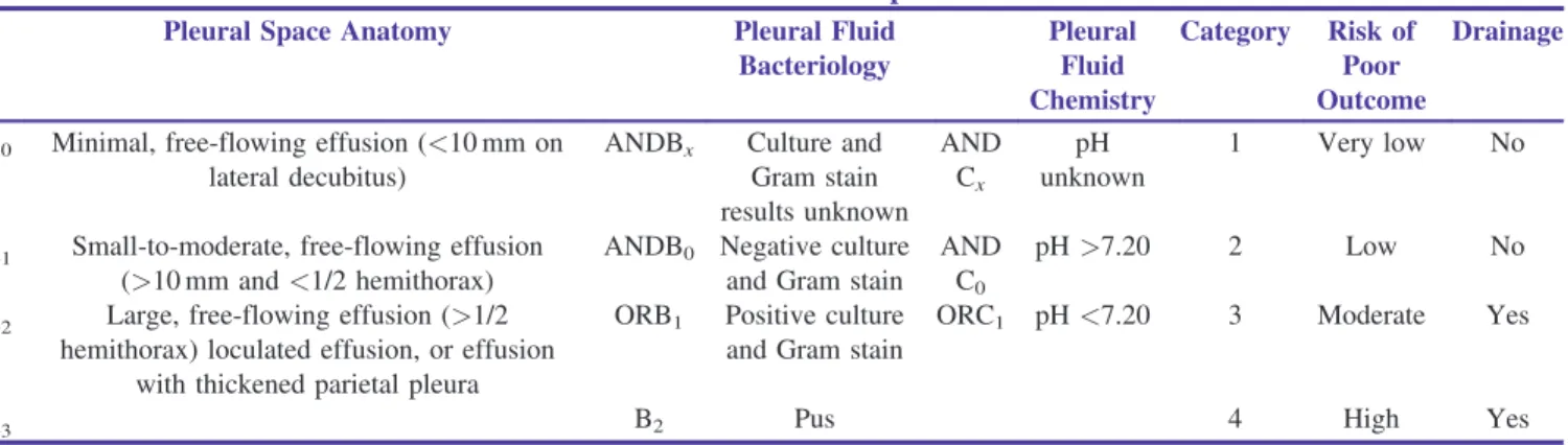
As per Light’s classification and treatment scheme for parapneumonic effusions and empyema, parapneumonic effusions are classified into seven classes [Table 1]. An expert panel from the American College of Chest Physicians has developed a new categorization of patients with parapneumonic effusions.[26] This categorization is modeled on the tumor–node–metastasis (TNM) classification of tumors and is based upon the anatomy of the effusion, the bacteriology of the pleural fluid, and the chemistry of the pleural fluid [Table 2].
M
ANAGEMENTThe management of parapneumonic effusions and empyemas involves the following:
(1) Appropriate antibiotic
(2) Management of the pleural fluid
Table 1: Light’s Classification and Treatment Scheme for Parapneumonic Effusions and Empyema
Event or State Number
Class 1
Nonsignificant pleural effusion
Small<10 mm thick on decubitus X-ray studyNo thoracentesis is indicated Class
2
Typical parapneumonic pleural effusion
>10 mm thickGlucose>40 mg/dl, pH>7.2LDH>3× upper limit normal and glucose>40 mg/ dlGram stain and culture negativeAntibiotics alone
Class 3
Borderline complicated pleural effusion
7.0<pH<7.20 and/orLDH>3× upper limit normal and glucose>40 mg/dlGram stain and culture negativeAntibiotics plus serial thoracentesis
Class 4
Simple complicated pleural effusion
pH<7.0 or glucose<40 mg/dl orGram stain or culture positiveNot loculated, no frank pusTube thoracostomy plus antibiotics
Class 5
Complex complicated pleural effusion
pH<7.0 or glucose<40 mg/dl orGram stain or culture positiveMultiloculated: tube thoracostomy plus fibrinolytics (rarely require thoracoscopy or decortication) Class
6
Simple empyema Frank pus presentSingle locule or free-flowingTube thoracostomy ± decortication Class
7
Complex empyema Frank pus presentMultiple loculesTube thoracostomy ± fibrinolyticsOften require thoracoscopy or decortication
Table 2: ACCP Classification of Parapneumonic Effusions
Pleural Space Anatomy Pleural Fluid
Bacteriology Pleural Fluid Chemistry Category Risk of Poor Outcome Drainage
A0 Minimal, free-flowing effusion (<10 mm on lateral decubitus)
ANDBx Culture and Gram stain results unknown AND Cx pH unknown 1 Very low No
A1 Small-to-moderate, free-flowing effusion (>10 mm and<1/2 hemithorax)
ANDB0 Negative culture and Gram stain
AND C0
pH>7.20 2 Low No
A2 Large, free-flowing effusion (>1/2 hemithorax) loculated effusion, or effusion
with thickened parietal pleura
ORB1 Positive culture and Gram stain
ORC1 pH<7.20 3 Moderate Yes
Antibiotic Selection
All patients with parapneumonic effusions or empyema should be treated with antibiotics. The Gram stain of the pleural fluid should guide the selection of an antibiotic. The initial antibiotic selection is usually based on whether the pneumonia is community-acquired or hospital-acquired. Patients hospitalized with community-acquired pneumonias that are not severe are recommended beta-lactam (cefotaxime, ceftriaxone, ampicillin–sulbactam, or ertapenem) or fluoroquinolones (if tuberculosis is not suspected). Macrolide are generally not recommended because atypical pathogens rarely cause a pleural effusion.[27] Patients with severe community- acquired pneumonia are recommended beta-lactam plus either an advanced macrolide or a respiratory fluoroquinolone.[28] If a pseudomonas infection is suspected, an antipseudomonas antibiotic such as piperacillin, piperacillin–tazobactam, imipenem, meropenem, or cefepime should be included. As anaerobic bacteria cause a sizable percentage of parapneumonic effusions, anaerobic coverage is recommended with either clindamycin or metronidazole. In patients with healthcare-associated pneumonia with parapneumonic effusion coverage should be provided for Gram-negative enteric bacteria and MRSA. A reasonable antibiotic selection in such patients is a carbapenem such as meropenem and vancomycin.[29]The duration of antibiotic therapy depends on factors like response to therapy, extent of parenchymal and pleural involvement, and extent of drainage of fluid or daily drainage from chest tube if inserted. It is recommended to continue antibiotic therapy till there is radiographic resolution of fluid. This may take 2 to 4 weeks of therapy.
I
NTRAPLEURALA
NTIBIOTICSIntrapleural antibiotics were first used to treat an infected pneumonectomy space by Clagett and Geraci in 1963.[30] Since that time, there have been several reports regarding the use of intrapleural antibiotics in the treatment of empyema complicating pneumonia. The personal experience of the author has been very rewarding, but some good randomized studies are required to recommend this therapy.
O
PTIONS FORM
ANAGEMENT OFP
LEURALF
LUIDThe options available for the management of the pleural fluid in patients with parapneumonic effusion are as follows:
• Conservative
• Therapeutic thoracentesis
• Intercostal chest tube drainage
• Intrapleural fibrinolysis
• VATS
• Thoracotomy with decortication and the breakdown of adhesions, and open drainage.
Conservative
Pleural fluid from patients with parapneumonic effusions should be sampled as soon as possible. Evaluation of fluid is necessary to determine if drainage of the fluid is required. Approximately 10% of patients with parapneumonic effusions require drainage, otherwise they become loculated and difficult to drain. Observation is acceptable if the patient has a Class 1 parapneumonic effusion.
Therapeutic Thoracentesis
Therapeutic thoracentesis was first practiced as a treatment modality for parapneumonic effusions in the mid-nineteenth century.[31] Some patients of complicated parapneumonic effusion can be treated with repeated thoracocentesis along with appropriate antibiotics.
Intercostal Chest Tube Drainage
The initial management of most patients with complicated parapneumonic effusions has been intercostal chest tube drainage. Large (28–36 F) tubes have been recommended because of the belief that smaller tubes would become obstructed with the thick fluid. The British Thoracic Society (BTS) guidelines state that a small bore catheter 10 to 14 F will be adequate for most cases of complicated parapneumonic pleural infection.[29]There is no consensus on the optimal size of the chest tube for drainage. The guidelines recommended regular flushing if a small-bore flexible catheter is used. The flushing technique recommended is the instillation of 20 to 30 ml saline every 6 hours via a three-way stopcock. In general, chest tubes should be left in place until the volume of the pleural drainage is less than 50 ml for 24 hours and until the draining fluid becomes clear yellow.
lntrapleural Fibrinolytics
Drainage of complicated parapneumonic effusions is difficult due to loculation. The pleural fluid loculations are produced by fibrin membranes that prevent the spread of the infected pleural fluid throughout the body, but it makes drainage of the pleural space difficult. Intrapleural fibrinolytics destroy the fibrin membranes and facilitate drainage of the pleural fluid.[32]In a landmark study on the use of intrapleural fibrinolytics for the treatment of complicated parapneumonic effusion, the administration of streptokinase had no effect on the need for surgery or the duration of hospitalization.[33] But this study did not consider the patients who do not have access to surgery like in our country and other Third World countries. Indian experience in intrapleural fibrinolysis is encouraging and most of the centers are using it, short of surgery that is not easily assessable to our patients.[34,35]
The MIST2 trial was conducted recently to examine the role of intrapleural DNase with and without concomitant tissue plasminogen activator (TPA) and to clarify the conflicting data concerning fibrinolytic agents. Combined TPA–DNase therapy resulted in a greater decrease in radiographic pleural opacity, a lower rate of
surgical referral, and a shorter hospital stay compared with placebo.[36]
Video-Assisted Thoracoscopy
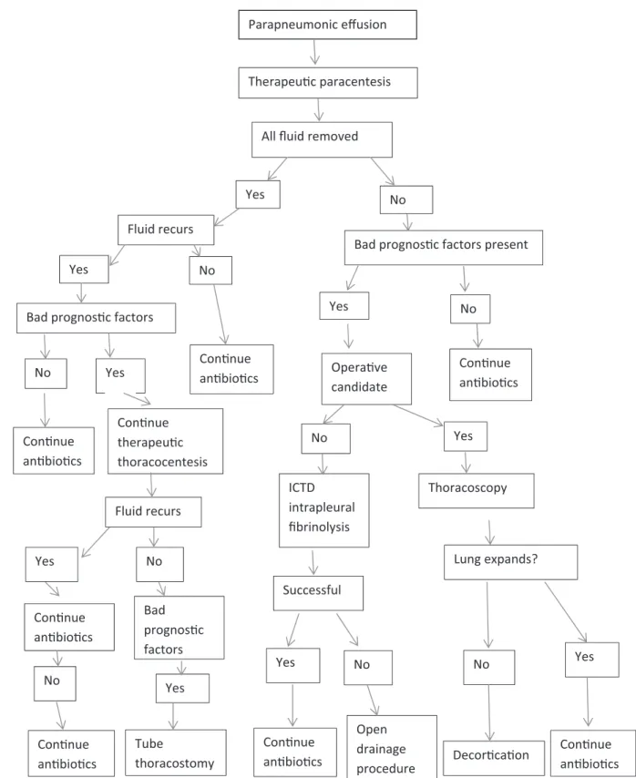
Video-assisted thoracoscopic surgery (VATS) is often used to debride multiloculated empyemas or Parapneumonic effusion
Therapeuc paracentesis
All fluid removed
Yes No
Yes No
Connue anbiocs No
Bad prognosc factors present
Bad prognosc factors
Yes Fluid recurs Connue therapeuc thoracocentesis Connue anbiocs No Yes Bad prognosc factors Connue anbiocs Connue anbiocs Yes No Tube thoracostomy Yes Connue anbiocs ICTD intrapleural fibrinolysis Successful Yes No Thoracoscopy Operave candidate No Fluid recurs Lung expands? Yes Connue anbiocs Open drainage procedure No No Yes Decorcaon Connue anbiocs
uniloculated empyemas that fail to resolve with antibiotics and chest tube drainage.[37] VATS allows for minimally invasive debridement and drainage. It can be followed by or converted to a thoracotomy if adequate pleural fluid drainage is achieved or lung expansion is not satisfactory. VATS can be used as a procedure to assist initial insertion of chest tube under vision. At the time chest tube is inserted, VATS can be used to irrigate the pleural space and break down all the fibrous strands.[38]One randomized study of 70 patients compared the results when the chest tube was inserted in the standard manner and when it was inserted in conjunction with VATS. In this study, patients with chest tubes inserted through VATS had a shorter hospital stay (8.3 vs. 12.8 days) and required less decortication (17 vs. 37%).[37]
Decortication
Decortication involves removal of all the fibrous tissue from the visceral and parietal pleura along with pus from the pleural space. It eliminates the pleural sepsis and thus assists expansion of underlying lung. In acute stage of infection, decortication helps in control of pleural infection. Decortication should not be performed just to remove thickened pleura because such thickening usually resolves spontaneously over several months.[39] Decortication is considered after 6 months if the pleura remains thickened and the patient’s pulmonary function is sufficiently reduced to limit activities. Decortication can be performed with VATS or with a full thoracotomy.
Open Drainage Procedures
Chronic drainage of the pleural space can be achieved with open drainage procedures. Two different types of procedures can be performed. The simplest procedure involves resecting segments of one to three ribs overlying the lower part of the empyema cavity and inserting a short/large-bore tubes into the empyema cavity. The tubes are irrigated daily with a mild antiseptic solution. The drainage from the tubes can be collected in a colostomy bag placed over the tubes. The advantage of this method over closed-tube drainage is that drainage is more complete and the patient is freed from attachment to the chest tube bottles. A similar but more complicated procedure is open drainage, in which a skin and muscle flap is positioned so that it lines the tract between the pleural space and the surface of the chest after two or more overlying ribs are resected. The advantage of this open flap (Eloesser flap) is that it creates a skin-lined fistula that provides drainage without tubes. Therefore, it can be more easily managed by the patient at home and permits gradual obliteration of the empyema space.
Algorithm for managing patients with parapneumonic effusions is shown in Figure 1.
Financial support and sponsorship
Nil.
Conflicts of interest
There are no conflicts of interest.
R
EFERENCES1. Light RW, Girard WM, Jenkinson SG, George RB. Parapneumonic effusions. Am J Med 1980;69:507.
2. Adams F. The Genuine Works of Hippocrates. New York, NY: William Wood; 1948. p. 266.
3. Eggers C. Radical operation for empyema. Ann Surg 1923;77:327. 4. Tillett WS, Sherry S, Read CT. The use of streptokinase-streptodornase in the treatment of post-pneumonic empyema. Thoracic Surg 1951;21:275-97.
5. Glenert J. Sugar levels in pleural effusions of different etiologies. Acta Tuberc Scand 1962;42:222-7.
6. Light RW, Mac Gregor MI, Ball WC Jr, Luchsinger PC. Diagnostic significance of pleural fluid pH and PCO2. Chest 1973;64:591-6.
7. Ferguson MK. Thoracoscopy for empyema, bronchopleural fistula, and chylothorax. Ann Thorac Surg 1993;56:644-5.
8. Sherman MM, Subramanian V, Berger RL. Management of thoracic empyema. Am J Surg 1977;133:474-9.
9. Bartlett JG, Finegold SM. Anaerobic infections of the lung and pleural space. Am Rev Respir Dis 1974;110:56-77.
10. Maeds RHA. History of Thoracic Surgery. Springfield, IL: CC Thomas; 1961.
11. Ehler AA. Nontuberculous thoracic empyema: A collective review of the literature from 1934–1939. Int Abstr Surg 1941;72:17. 12. Benfield CF. Recent trends in empyema thoracis. Br J Dis Chest
1981;75:358.
13. Ravitch MH, Fein R. The changing picture of pneumonia and empyema in infants and children. JAMA 1961;175:1039. 14. Freij BJ, Kusmiesz H, Nelson JD, McCracken GH. Parapneumonic
effusions in hospitalized children: A retrospective review of 227 cases. Pediatr Infect Dis J 1984;3:578.
15. Mangenet EDO, Kombo BB, Legg-Jack TE. Thoracic empyema: A study in 56 patients. Arch Dis Child 1993;69:587.
16. Kawanami T, Fukuda K, Yatera K, Kido M, Mukae H, Taniguchi H. A higher significance of anaerobes: the clone library analysis of bacterial pleurisy. Chest 2011;139:600.
17. Menzies SM, Rahman NM, Wrightson JM, Davies HE, Shorten R, Gillespie SH,et al.Blood culture bottle culture of pleural fluid in pleural infection. Thorax 2011;66:658.
18. Bartlett JG, Gorbach SL, Thadepalli H, Finegold SM. Bacteriology of empyema. Lancet 1974;1:338.
19. Civen R, Jousimies-Somer H, Marina M, Borenstein L, Shah H, Finegold SM. A retrospective review of cases of anaerobic empyema and update of bacteriology. Clin Infect Dis 1995;20:S224. 20. Gupta D, Hemachander SS. A study on parapneumonic effusions/
lower respiratory infections and their burden on rural population of Khammam district,Andhra Pradesh, South India. Ann Trop Med Public Health 2012;5:214-8.
21. Brixey AG, Luo Y, Skouras V, Awdankiewicz A, Light RW. The efficacy of chest radiographs to detect parapneumonic effusions. Respirology 2011;16:1000-4.
22. Colice GL, Curtis A, Deslauriers J, Hefner J, Light RW, Littenberg
B, et al. Medical and surgical treatment of parapneumonic
23. Maskell NA, Batt S, Hedley EL, Davies CW, Gillespie SH, Davies RJ. The bacteriology of pleural infection by genetic and standard methods and its mortality significance. Am J Respir Crit Care Med 2006; 174:817-23.
24. Sullivan KM, O’Toole RD, Fisher RH, Sullivan KN. Anaerobic empyema thoracis. Arch Intern Med 1973;131:521-7.
25. Porcel JM, Vives M, Cao G, Bielsa S, Ruiz-González A, Martínez-Iribarren A, et al. Biomarkers of infection for the differential diagnosis of pleural effusions. Eur Respir J 2009;34:1383. 26. Athanassiadi K, Gerazounis M, Kalantzi N. Treatment of
post-pneumonic empyema thoracis. Thorac Cardiovasc Surg 2003;5l:338–41.
27. Wrightson JM, Davies RJ. The approach to the patient with a parapneumonic effusion. Semin Respir Crit Care Med 2010;31:706-15. 28. Mandell LA, Barltett JG, Dowell SF, File TM Jr, Musher DM, Whitney C,et al.Update of practice guidelines for the management of community-acquired pneumonia in immunocompetent adults. Clin Infect Dis 2003;37:1405-33.
29. Davies HE, Davies RJ, Davies CWH. Management of pleural infection in adults: British Thoracic Society pleural disease guideline2010. Thorax 2010;65:ii41-53.
30. Clagett OT, Geraci JE. A procedure for the management of postpneumonectomy empyema. J Thorac Cardiovasc Surg 1963;45:141–5.
31. Bowditch HI. Paracentesis thoracic: An analysis of 25 cases of pleuritic effusion. Am Med Monthly 1853;3-45.
32. Rahman NM. Intrapleural agents for pleural infection: Fibrinolytics and beyond. Curr Opin Pulm Med 2012;18:326-32. 33. Maskell NA, Davies CWH, Nunn AJ, Hedley EL, Gleeson FV, Miller R,et al.U.K. controlled trial of intrapleural streptokinase for pleural infection. N Engl J Med 2005;352:865-74.
34. Sharma VP, Guleria R, Gupta R, Sharma SK, Pande JN. Intrapleural streptokinase in multiloculated empyema thoracis. J Assoc Physicians India 1998;46:227-9.
35. Barthwal MS, Deoskar RB, Rajan KE, Chatterjee RS. Intrapleural streptokinase in complicated parapneumonic effusions and empyema. Indian J Chest Dis Allied Sci 2004;46:257-61. 36. Rahman NM, Maskell NA, West A, Teoh R, Arnold A, Mackinlay
C,et al.Intrapleural use of tissue plasminogen activator and DNase
in pleural infection. N Engl J Med 2011;365:518.
37. Potaris K, Mihos P, Gakidis I, Chatziantoniou C. Video-thoracoscopic and open surgical management of thoracic empyema. Surg Infect (Larchmt) 2007;8:511.
38. Bilgin M, Akcali Y, Oguzkaya F. Benefits of early aggressive management of empyema thoracis. ANZ J Surg 2006;76:120-2. 39. Neff CC, van Sonnenberg E, Lawson DW, Patton AS. CT
follow-up of empyemas: Pleural peels resolve after percutaneous catheter drainage. Radiology 1990;176:195-7.