O R I G I N A L I N V E S T I G A T I O N
Open Access
Heparanase induced by advanced glycation end
products (AGEs) promotes macrophage migration
involving RAGE and PI3K/AKT pathway
Qiaojing Qin
1, Jianying Niu
1, Zhaoxia Wang
2, Wangjie Xu
2, Zhongdong Qiao
2and Yong Gu
1,3*Abstract
Background:Advanced glycation end products (AGEs), inflammatory-associated macrophage migration and accumulation are crucial for initiation and progression of diabetic vascular complication. Enzymatic activity of heparanase (HPA) is implicated strongly in dissemination of metastatic tumor cells and cells of the immune system. In addition, HPA enhances the phosphorylation of selected signaling molecules including AKT pathway
independent of enzymatic activity. However, virtually nothing is presently known the role of HPA during macrophage migration exposed to AGEs involving signal pathway.
Methods:These studies were carried out in Ana-1 macrophages. Macrophage viability was measured by MTT (3-(4,5-dimethylthiazol-2-yl)-2,5-diphenyltetrazolium bromide) assays. HPA and AKT protein expression in macrophages are analysed by Western blotting and HPA mRNA expression by real time quantitative RT-PCR. Release of HPA was determined by ELISA. Macrophage migration was assessed by Transwell assays.
Results:HPA protein and mRNA were found to be increased significantly in AGEs-treated macrophages.
Pretreatment with anti-HPA antibody which recognizes the nonenzymatic terminal of HPA prevented AGEs-induced AKT phosphorylation and macrophage migration. LY294002 (PI3k/AKT inhibitor) inhibited AGEs-induced
macrophage migration. Furthermore, pretreatment with anti-receptor for advanced glycation end products (RAGE) antibody attenuated AGEs-induced HPA expression, AKT phosphorylation and macrophage migration.
Conclusions:These data indicate that AGEs-induced macrophage migration is dependent on HPA involving RAGE-HPA-PI3K/AKT pathway. The nonenzymatic activity of HPA may play a key role in AGEs-induced macrophage migration associated with inflammation in diabetic vascular complication.
Keywords:Advanced glycation end products, Macrophage migration, Diabetes, RAGE, Heparanase, PI3K/AKT
Introduction
Advanced glycation end products (AGEs), final products of the non-enzymatic reaction between reducing sugars and amino groups in proteins, lipids and nucleic acids, promotes inflammation to accelerate the progression of vascular disease in patients with diabetes as well as other mechanisms [1]. Inflammatory-associated macrophage migration and accumulation in inflamed tissue sites are implicated in the major pathogenic process of vascular
complications in diabetes [2-4]. Although the accumula-tion of advanced glycaaccumula-tion end products (AGEs), chronic inflammation-associated macrophage migration and ac-cumulation play critical roles in vascular complication development of diabetes [5-7], knowledge regarding the relationship between AGEs and macrophage migration through extracellular matrix is still unclear.
Heparanase (HPA), an endo-β-glucuronidase, is strongly implicated in cell dissemination associated with tumor metastasis and inflammation. It can cleave heparan sulfate side chains of heparan sulfate proteoglycans to participate in extracellular matrix remodeling and regulate the release of many heparan sulfate-bonded molecules include inflammatory cytokines [8-10]. Moreover, HPA has non-* Correspondence:yonggu@vip.163.com
1
Department of Nephrology, Shanghai Fifth People’s Hospital, Fudan University, Shanghai 200240, China
3
Department of Nephrology, Huashan Hospital, Fudan University, Shanghai 200240, China
Full list of author information is available at the end of the article
enzymatic activities which play a part in different signaling cascades and selected protein kinase activation associated with cell migration [11,12]. Evidences have shown that over-expressed HPA in most human cancers allow them to penetrate the endothelial cell layer and basement mem-brane to invade target organs [13,14]. Increased expression of HPA is essential for the development of microvascular complication such as diabetic nephropathy in mice and associated with inflammation in human atherosclerosis [15-17].
Recently, several reports have indicated that AGEs increased HPA expression to facilitate migration of cell associated with inflammation in adult tubular and endo-thelial cells [18-20]. However, it is unknown whether macrophage migration is induced by AGEs in HPA-dependent manner.
Given the crucial role of AGEs and macrophage mi-gration in the progression of diabetic complications, we thoroughly investigated the effect of AGEs on macro-phage migration via HPA independent of enzyme acti-vity. In particular, we analyzed: the effects of AGEs on the mRNA, protein and secretion of HPA; the signaling pathways involved; the effect of an altered HPA expres-sion on macrophage migration and the mechanisms.
Materials and methods Materials
RPMI 1640 and fetal bovine serum (FBS) were from GibcoTM Invitrogen Corporation (Grand Island, NY). Advanced glycation end products (Glycated bovine serum albumin) was from Shanghai Yixin Bio-Technology Co. Ltd (Shanghai, China). RevertAid First Strand cDNA Synthesis Kit was from Fermentas International Inc (Graiciuno, Vilnius, Lithuania). Real time PCR Master Mix was from Delaware Biotechnology Institute (Newark,DE). SuperECL Plus and LY 294002 were from Beytime Insti-tute of Biotechnology (Haimeng, China). ELISA kit for mouse HPA was from Glory Science Co,Ltd (Hangzhou, China). Rabbit anti-mouse HPA, RAGE, AKT antibody and peroxidase-labeled goat anti-rabbit second antibody were from Wuhan Boster Bio-engineering Limited Com-pany (Wuhan, China). Rabbit anti-mouse GAPDH antibody was from Santa Cruz Biotechnology Inc (Santa Cruz, CA); Rabbit anti-mouse phospho-AKT antibody was from Cell Signaling Technology (Boston, MA). MTT was from Sigma-Aldrich (Shanghai, China). Falcon™ cell culture insert system was from Becton Dickinson and Company (Franklin Lakes, NJ).
Cell culture
Ana-1 mouse macrophage cell line was obtained from the cell bank of Shanghai Institutes for Biological Sciences, Chinese Academy of Sciences (Shanghai, China). Cells were maintained in RPMI 1640 medium supplemented
with 10% fetal bovine serum and 100 units/ml penicillin and 100 μg/ml streptomycin and were incubated at 37°C in 5% CO2 humidified air. Spent medium was replaced every 2–3 days. Cells were grown to 80% confluence and then serum-starved for 16 hours before use.
MTT assay
In order to determine the effects and mechanism of AGEs on HPA in macrophages, we performed assays to determine the concentrate of AGEs, LY294002 (PI3k/ Akt inhibitor), anti-HPA and RAGE antibody which didn’t change the viability of macrophages significantly. 100 μl macrophages were seeded at a density of 5 × 10⁴ cells/ml and incubated with AGEs, LY294002, anti-HPA and RAGE antibody at the indicated concentration in 96-well plates. After 24 h incubation, 3-(4,5-dimethyl-thiazol-2-yl)-2,5-diphenyltetrazolium bromide (MTT) solution was added to each well for 4 hours. Finally, the blue salt in each well was dissolved and the plates were read by using a microplate reader with RPMI 1640 as blank and cell culture medium as control. The results of cell viability determined 100 mg/L AGEs, 15 μM LY294002, 10μg/ml anti-RAGE and 10μg/ml anti-HPA antibody in the following experiments (The choice of dosage is showed in results and discussion).
Treatments with cells
First, we evaluate the role of RAGE, HPA, AKT in AGEs-induced macrophage migration. Cells were treated with AGEs (100 mg/L), and LY294002 (pre-treated with 15 μM LY294002), and anti-RAGE or HPA antibody (pre-treated with 10μg/ml anti-RAGE or HPA antibody) for 24 h. Subsequently, cells were treated with AGEs (100 mg/L), and anti-RAGE antibody (pre-treated with 10μg/ml anti-RAGE antibody) for 24 h and analyzed by RT-PCR, Western blot and ELISA assay to examine effects of AGEs on HPA expression and the role of re-ceptor for advanced glycaiton end products (RAGE) in AGEs-induced HPA expression. Finally, we treated cells with AGEs (100 mg/L), and anti-RAGE or HPA antibody (pre-treated with 10μg/ml anti-RAGE or HPA antibody) for 24 h, and harvested for Western blot analysis to assess of the role of HPA and RAGE in AGEs-induced activation of AKT.
Migration assay
for 15 min to fix cells adherent to the underside of the membrane. Migrated cells were stained with hematoxylin and counted (6 random fields per slide) in ten 40× fields.
ELISA for HPA
The Ana-1 macrophages were plated at 5 × 10⁵cells/well in 24 well plates overnight. Macrophages were cultured separately with 100 mg/L AGEs, and RAGE anti-body (pre-treated with 10 μg/ml anti-RAGE antibody) for 24 h. The levels of HPA in culture supernatants were determined using commercially available enzyme linked immunosorbent assay (ELISA) kits according to the manufacturer’s instructions. Briefly, 10 μl supernatant and 40 μl sample diluent was added to sample well, followed by the addition of 100 μl HRP-conjugate re-agent to each well and incubated for 60 minutes at 37°C. The reaction was visualized by the addition of 50 μl chromogen solution A and 50 μl chromogen solution B for 15 min at 37°C. The reaction was stopped with 50μl stop solution and absorbance at 450 nm was measured using ELISA plate reader within 15 min. The level of HPA was quantified by a standard curve established by a serial dilution of standard concentration.
Detection of mRNAs by real time quantitative RT-PCR RNA from Ana-1 macrophages was isolated using TRIZOL reagent following the manufacturer’s instruc-tions. Isolated RNA was reversed transcribed into comple-mentary DNA using RevertAid First Strand cDNA Synthesis Kit according to the protocol by the manufac-turer. To determine the fold-changes in the expression of HPA gene, real-time PCR was performed using the first strand cDNA, the forward and reverse primers (forward: GAACCTCCATAATGTCACCAAGC, resverse: GTCTG CTCATCCACCATCTTCAG), and Bestar™Taqman Real time PCR master mix. Thermocycling conditions for SYBR Green consisted of a denaturation step for 2 min at 95°C followed by 35 cycles of 95°C for 15 s, 59°C for 30 s and 72°C 15 s. Data analysis was performed using standard curve method andΔΔCt method. The mount of HPA was determined and normalized by the amount of GAPDH cDNA.
Western blot analysis
The cells were lysed with dissociation solution containing phosphatase, protease inhibitors and PMSF. Protein extracted from macrophages was electrophoresed on SDS-polyacrylamide gels (10%) and Western blotted. Proteins were transblotted onto nitrocellulose membrane. Membranes with the transferred proteins were blocked for 1 hour with 5% (w/v) skim milk powder diluted in PBS and then incubated with anti-HPA, AKT or phosphorylated (p)-AKT antibodies (1:1000 dilution)
respectively at 4°C overnight. The membranes were incubated with a peroxidase-conjugated secondary anti-body respectively and visualized by a supper enhanced chemiluminesence detection system. Densitometric ana-lysis was performed by normalizing band density to that for GAPDH.
Statistical analysis
The data were expressed as mean ± SEM. Statistical ana-lysis was performed by SPSS software using one-way analysis of variance (ANOVA). A value of P < 0.05 was considered statistically significant.
Results Cell viability
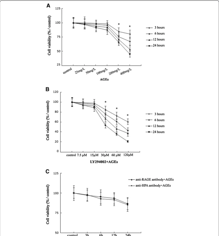
Incubation of Ana-1 macrophages with AGEs (25– 400 mg/L) for 3,6,12 and 24 h led to the significant dose-dependent reduction in the viability of cells compared with controls. AGEs didn’t change significantly the viabi-lity of Ana-1 macrophages within 25-100 mg/L AGEs con-centration range for 3,6,12 and 24 h incubation (Figure 1A). Therefore, we chose the max dosage of AGEs at 100 mg/L, which didn’t change the viability significantly in Ana-1 macrophages within 24 h incubation, to finish the following experiments involving the mechanism.
Subsequently, macrophages were pre-treated with LY294002 (7.5-120 μM) for 1 h before culture with 100 mg/L AGEs except control group. The treatment of macrophages with LY294002 (7.5-15μM) and 100 mg/L AGEs within 24 h culture didn’t affect cell viability sig-nificantly. Exposure to LY294002 (30–120 μM) and 100 mg/L AGEs for 3, 6, 12, 24 h decreased macrophage viability significantly (Figure 1B). We chose the max dosage of LY294002 at15 μM and 100 mg/L AGEs for 24 h incubation, which didn’t change the viability signifi-cantly in Ana-1 macrophages, to finish the following experiments.
In addition, the pre-treatment with 10 μg/ml anti-RAGE or 10 μg/ml anti-HPA antibody in 100 mg/L AGEs-induced macrophages for 24 h culture didn’t change the cell viability significantly (Figure 1C).
HPA play a key role in the AGEs-induced macrophage migration involving RAGE and PI3K/AKT pathway
Figure 1Viability analysis of Ana-1 macrophages after treatment with AGEs, LY294002, anti-RAGE or HPA antibody.Cell viability assay is performed using MTT assay.A, Cells (5 × 10⁴) were treated with AGEs (0, 25, 50, 100, 200 and 400 mg/L) for 3, 6, 12, 24 h.B, Cells (5 × 10⁴) were
pretreated with LY294002 (7.5-120μM) for 1 h before culture with 100 mg/L AGEs for 3, 6, 12, 24 h.C, Cells (5 × 10⁴) were pretreated with
infiltration. Our data showed that AGEs-induced macro-phage migration was reduced after pretreatment with anti-HPA antibody for 1 h. In addition, pretreatment with anti-RAGE antibody or LY294002 for 1 h decreased the AGEs-induced macrophages migration significantly (Figure 2). The results explore that HPA mediates AGEs-induced macrophage migration and may involve RAGE and AKT pathway.
AGEs changes HPA mRNA, protein expression and secretion via RAGE in macrophages
To test the possible effect and mechanism of AGEs treat-ment on HPA expression, Ana-1 macrophages were incubated for 24 h in the presence of 100 mg/L AGEs with or without the pretreatment of 10μg/ml RAGE anti-body for 1 h. Using RT-PCR and ELISA assays, we show that AGEs promoted the levels of mRNA and secretion significantly in macrophages. Pretreatment with anti-RAGE antibody significantly inhibited HPA secretion and mRNA production in AGEs-induced macrophages (Figure 3A and 3B).
Furthermore, the HPA latent form (65 kDa) and active form (50 kDa) were identified using an antibody against the C-terminus domain of HPA protein. We show that untreated cells primarily expressed the 65 kDa latent form. Both of 65 kDa and 50 kDa forms were increased signifi-cantly and the 50 kDa actived form was more strongly induced as compared with the 65 kDa latent form in AGEs-induced macrophages. Pretreatment with anti-RAGE antibody attenuated AGEs-induced 50 kDa actived and 65 kDa latent forms in macrophages (Figure 3C).
Phosphorylation of AKT are increased in AGEs-stimulated macrophages via HPA involving RAGE
We examine the expression of AKT protein in macrophages to explain the role of PI3K/AKT signaling pathway in AGEs-induced macrophage migration via HPA. We found that the phosphorylation of AKT was increased significantly at 24 h of stimulation with AGEs in macrophages. The expression of AKT phosphoryl-ation could be inhibited by the pretreatment of an antibody recognizing the COOH-terminal domain of HPA protein or anti-RAGE antibody in AGEs-induced macrophages. Furthermore, we didn’t find the changes of total AKT protein in macrophages with different treatment (Figure 4).
Discussion
Cardiovascular complications are the leading cause of death in patients in diabetic patients [21,22]. The current study provides evidence to support that excess accumulation of advanced glycation end products (AGEs) and inflammation is emerging as important mechanism for micro- and macrovascular complication of diabetes [23,24]. Monocytes transmigrate into the subendothelial space and differentiate into macrophages, which migrate, infiltrate and accumulate in the vascular tissues, involving in diabetic vascular complications. Clinical study has also observed significant increase of AGEs accumulation in diabetic vascular tissues [25], which may induce macrophage migration across an endothelial cell monolayer [26].
Heparanase (HPA), a mammalian endo-beta-D-glu-curoxnidase, has previously been shown to be a key
enzyme in the metastatic potential of tumor-derived cells and cells of the immune system [27-29]. Recently, evidences show that HPA enhances the phosphorylation of selected signaling molecules, in a manner that is mediated by its C-terminal domain but not enzymatic
activity [12,30]. In the study, we have characterized the role and mechanism of HPA in AGEs-induced macro-phage migration independent of enzymatic activity.
With the MTT assay we observed the dose-dependent reduction of viability in macrophages treated by AGEs.
Figure 3AGEs up-regulates HPA mRNA, protein expression and secretion in macrophages via RAGE.Cells were cultured with AGEs for 24 h with or without pre-treatment with antibody against RAGE for 1 h.A, The levels of HPA mRNA were assessed with real time quantitative RT-PCR.B, The secretion of HPA in supernatant was measured by enzyme-linked immunosorbent assay (ELISA).C, The expression of HPA protein in macrophages was determined by Western blotting. The results represent the mean of six culture wells (mean ± SEM). *p < 0.05 compared to control and #p <0.05 compared to AGEs. All of the experiments were performed independently in triplicate.
Figure 4The expression of AKT protein in AGEs-induced macrophages.Cells were cultured with AGEs or pretreated with antibody against RAGE or HPA for 1 h before exposed to AGEs for 24 h. AKT and p-AKT protein expression is determined by Western blot analysis using anti-AKT and p-AKT antibody. The results represent the mean of six culture wells (mean ± SEM).*P < 0.05 compared to control and#P <0.05 compared to
The viability revealed a nonlinear dose response to AGEs in macrophages. AGEs treatment at 25, 50 and 100 mg/L for 3,6,12 and 24 h incubation didn’t result in a significant loss of viability in macrophages compared with controls (0 mg/L) (Figure 1A). We then used 100 mg/L AGEs, a max dosage which didn’t changed the viability significantly within studied AGEs concentration range at 24 h culture, for further studies on the role of HPA in macrophages.
Moreover, pretreatment with LY294002 (7.5-15 μM), anti-RAGE or HPA antibody (10 μg/ml) for 1 h before culture with 100 mg/L AGEs for 3,6,12 and 24 h incuba-tion didn’t result in a significant loss of viability in macrophages compared with controls (Figure 1B). We then used preincubation of 15μM LY294002, 10 μg/ml anti-RAGE or HPA antibody, the dosage which didn’t changed the viability significantly within the studied concentration range at 24 h culture, for further studies in 100 mg/L AGEs-induced macrophages.
Evidences have shown that HPA play a role in signaling pathway [31]. C-terminus domain mediates nonenzymatic functions of HPA, facilitating the phosphorylation of phosphatidylinositol 3-kinase/protein kinase B (PI3K/ AKT), in an enzymatic activity-independent manner in pi-tuitary tumor and proximal tubular [32,33]. Recent studies show that AKT (protein kinase B), a serine/threonine pro-tein kinase, regulates monocyte/macrophage migration. Inhibition of AKT pathways decreased macrophage migra-tion while mammalian cell migramigra-tion can be promoted by enhancing AKT signaling [34-36].
In agreement with previous study [26,37], we have shown that AGEs induced macrophage migration significantly. However, more importantly, we have shown that pretreat-ment of anti-HPA antibody, which bind to the C-terminus domain of HPA specially, inhibited the macrophage migration significantly (Figure 2). This demonstrates that C-terminus domain of HPA mediates AGEs-induced macrophage migration. We speculate that the C-terminus domain of HPA may mediate AGEs-induced macrophage migration via AKT signaling pathway.
Subsequently, we detected the role of PI3K/AKT sig-naling pathway in AGEs-induced macrophage migration via HPA. AKT is a target of PI3K activation and its phosphorylation is prevented by PI3K/AKT inhibitors. Although AKT phosphorylation is increased by external HPA in a short time, the AKT phosphorylation induced by changes of HPA protein after AGEs treatment may need longer time. So we chose to determine the levels of AKT at 24 h culture. The results show that the levels of AKT phosphorylation was increased at 24 h in macro-phage cultured with AGEs. Pretreatment with an anti-body recognizing the C-terminus domain of HPA protein inhibited AKT phosphorylation significantly (Figure 4). The results suggest that AGEs could activate
PI3K/AKT signaling pathway via C-terminus domain of HPA protein. Furthermore, we observed that AGEs-induced cell migration was attenuated by using LY294002, a PI3K/AKT inhibitor, in macrophages (Figure 2). These data indicate that the AGEs-induced macrophage migra-tion is partially mediated by the activamigra-tion of PI3K/AKT signaling pathway in HPA-dependent manner. HPA-PI3K/ AKT signal pathway may be important in macrophage mi-gration induced by AGEs.
Recently, it was reported that AGEs could induce HPA expression through receptor for advanced glycation end products (RAGE) in human microvascular endothelial cells [19]. Substantial evidence has demonstrated that RAGE plays a central role in the etiology of diabetes complications and inflammation [38-40]. RAGE ligand, such as HMGB-1, could stimulate phosphorylation of AKT and cell proliferation/migration through RAGE/ PI3K/AKT signal transduction pathway [41,42]. Here we assess the role of RAGE on HPA expression and AKT pathway which associated with macrophage migration.
We observed that treatment with AGEs for 24 h cul-ture resulted into significant increase of HPA mRNA, HPA latent form (65 kDa) and enzyme form (50 kDa) compared with untreated cells. 50 kDa enzyme form is more strongly induced than 65 kDa latent form. All the changes could be attenuated by pretreatment with anti-RAGE antibody (Figure 3A and 3C). These results show that increased expression of HPA mRNA and protein are associated with AGEs stimulation via RAGE and HPA protein expression may partly dependent of post-transcriptional regulation in macrophages.
Furthermore, ELISA analysis was employed to determine the levels of HPA in supernatant. We discovered that pre-treatment with anti-RAGE antibody inhibited the increased secretion of HPA in AGEs-stimulated macrophages signifi-cantly (Figure 3B). The result from ELISA suggests that AGEs could induce HPA protein secretion via RAGE as well as the results from Western blot. These data provide the first evidence for AGEs-induced macrophage HPA mRNA, protein expression and secretion in a RAGE-dependent manner. RAGE-HPA pathway may play a key role in AGEs-induce macrophage migration.
Conclusions
Our results show for the first time that AGEs-induced macrophage migration may be mediated by RAGE-HPA-PI3K/AKT signal pathway. It supports the notion that HPA could mediate macrophage migration involving in RAGE-HPA-PI3K/AKT pathway independent of enzym-atic activity, providing new insights into the role of HPA in AGEs-induced macrophage migration associated with vascular complication of diabetes. The mechanism such as regulation of HPA expression and AKT phosphorylation mediated by HPA might be required for further studies.
Abbreviations
AGEs: Advanced glycation end products; RAGE: Receptor for advanced glycation end products; HPA: Heparanase; PI3K/AKT: Phosphatidylinositol 3-kinase/protein kinase B.
Competing interests
The authors declare that they have no competing interests.
Authors’contributions
QQ carried out the design, analysis and writing of the manuscript. YG participated in its design and coordination and helped to draft the manuscript. ZW, JN, WX and ZQ participated in conduct and analysis. All authors read and approved the final manuscript.
Acknowledgments
This work was supported by a grant from the Major State Basic Research Development Program of China (973 Program) (2012CB517700), the Key Basic Research Project of the Science and Technology Commission of Shanghai Municipality (10JC1413000) & the National Natural Science Foundation of China (30871175).
Author details
1Department of Nephrology, Shanghai Fifth People’s Hospital, Fudan University, Shanghai 200240, China.2School of Life Science and Biotechnology, Shanghai Jiaotong University, Shanghai 200240, China. 3Department of Nephrology, Huashan Hospital, Fudan University, Shanghai
200240, China.
Received: 15 January 2013 Accepted: 18 February 2013 Published: 26 February 2013
References
1. Watson AM, Gray SP, Jiaze L, Soro-Paavonen A, Wong B, Cooper ME, Bierhaus A, Pickering R, Tikellis C, Tsorotes D, Thomas MC, Jandeleit-Dahm KA:Alagebrium reduces glomerular fibrogenesis and inflammation beyond preventing RAGE activation in diabetic apolipoprotein E knockout mice.Diabetes2012,61(8):2105–2113.
2. Soetikno V, Sari FR, Veeraveedu PT, Thandavarayan RA, Harima M, Sukumaran V, Lakshmanan AP, Suzuki K, Kawachi H, Watanabe K:Curcumin ameliorates macrophage infiltration by inhibiting NF-κB activation and proinflammatory cytokines in streptozotocin induced-diabetic nephropathy.Nutr Metab (Lond)2011,8(1):35.
3. Rosenson RS, Fioretto P, Dodson PM:Does microvascular disease predict macrovascular events in type 2 diabetes?Atherosclerosis2011,
218(1):13–18.
4. Marino F, Maresca AM, Cosentino M, Castiglioni L, Rasini E, Mongiardi C, Maio RC, Legnaro M, Schembri L, Dentali F, Grandi AM, Guasti L:
Angiotensin II type 1 and type 2 receptor expression in circulating monocytes of diabetic and hypercholesterolemic patients over 3-month rosuvastatin treatment.Cardiovasc Diabetol2012,11:153.
5. Coughlan MT, Yap FY, Tong DC, Andrikopoulos S, Gasser A, Thallas-Bonke V, Webster DE, Miyazaki J, Kay TW, Slattery RM, Kaye DM, Drew BG, Kingwell BA, Fourlanos S, Groop PH, Harrison LC, Knip M, Forbes JM:Advanced glycation end products are direct modulators ofβ-cell function.
Diabetes2011,60(10):2523–2532.
6. Zhong Y, Li J, Chen Y, Wang JJ, Ratan R, Zhang SX:Activation of endoplasmic reticulum stress by hyperglycemia is essential for Muller cell-derived inflammatory cytokine production in diabetes.Diabetes2012,
61(2):492–504.
7. Raposeiras-Roubín S, Rodiño-Janeiro BK, Paradela-Dobarro B, Grigorian-Shamagian L, García-Acuña JM, Aguiar-Souto P, Jacquet-Hervet M, Reino-Maceiras MV, Alvarez E, González-Juanatey JR:Predictive value of advanced glycation end products for the development of post-infarction heart failure: a preliminary report.Cardiovasc Diabetol2012,11:102. 8. Peterson S, Liu J:Deciphering mode of action of heparanase using
structurally defined oligosaccharides.J Biol Chem2012,
287(41):34836–34843.
9. Shafat I, Agbaria A, Boaz M, Schwartz D, Baruch R, Nakash R, Ilan N, Vlodavsky I, Weinstein T:Elevated urine heparanase levels are associated with proteinuria and decreased renal allograft function.PLoS One2012,
7(9):e44076.
10. Bhattacharjee PS, Huq TS, Potter V, Young A, Davenport IR, Graves R, Mandal TK, Clement C, McFerrin HE, Muniruzzaman S, Ireland SK, Hill JM: High-glucose-induced endothelial cell injury is inhibited by a Peptide derived from human apolipoprotein e.PLoS One2012,7(12):e52152.
11. Purushothaman A, Babitz SK, Sanderson RD:Heparanase enhances the insulin receptor signaling pathway to activate extracellular signal-regulated kinase in multiple myeloma.J Biol Chem2012,
287(49):41288–41296.
12. Cohen-Kaplan V, Jrbashyan J, Yanir Y, Naroditsky I, Ben-Izhak O, Ilan N, Doweck I, Vlodavsky I:Heparanase induces signal transducer and activator of transcription (STAT) protein phosphorylation: preclinical and clinical significance in head and neck cancer.J Biol Chem2012,
287(9):6668–66678.
13. Purushothaman A, Uyama T, Kobayashi F, Yamada S, Sugahara K, Rapraeger AC, Sanderson RD:Heparanase-enhanced shedding of syndecan-1 by myeloma cells promotes endothelial invasion and angiogenesis.
Blood2010,115(12):2449–2457.
14. Xu X, Rao G, Quiros RM, Kim AW, Miao HQ, Brunn GJ, Platt JL, Gattuso P, Prinz RA:In vivo and in vitro degradation of heparan sulfate (HS) proteoglycans by HPR1 in pancreatic adenocarcinomas. Loss of cell surface HS suppresses fibroblast growth factor 2-mediated cell signaling and proliferation.J Biol Chem2007,282(4):2363–2373.
15. Osterholm C, Folkersen L, Lengquist M, Pontén F, Renné T, Li J, Hedin U:
Increased expression of heparanase in symptomatic carotid atherosclerosis.Atherosclerosis2013,226(1):67–73.
16. Gil N, Goldberg R, Neuman T, Garsen M, Zcharia E, Rubinstein AM, van Kuppevelt T, Meirovitz A, Pisano C, Li JP, van der Vlag J, Vlodavsky I, Elkin M:
Heparanase is essential for the development of diabetic nephropathy in mice.Diabetes2012,61(1):208–216.
17. Baker AB, Chatzizisis YS, Beigel R, Jonas M, Stone BV, Coskun AU, Maynard C, Rogers C, Koskinas KC, Feldman CL, Stone PH, Edelman ER:Regulation of heparanase expression in coronary artery disease in diabetic, hyperlipidemic swine.Atherosclerosis2010,213(2):436–442. 18. Masola V, Gambaro G, Tibaldi E, Onisto M, Abaterusso C, Lupo A:
Regulation of heparanase by albumin and advanced glycation end products in proximal tubular cells.Biochim Biophys Acta2011,
1813(8):1475–1482.
19. An XF, Zhou L, Jiang PJ, Yan M, Huang YJ, Zhang SN, Niu YF, Ten SC, Yu JY:
Advanced glycation end-products induce heparanase expression in endothelial cells by the receptor for advanced glycation end products and through activation of the FOXO4 transcription factor.Mol Cell Biochem2011,354(1–2):47–55.
20. Rao G, Ding HG, Huang W, Le D, Maxhimer JB, Oosterhof A, van Kuppevelt T, Lum H, Lewis EJ, Reddy V, Prinz RA, Xu X:Reactive oxygen species mediate high glucose-induced heparanase-1 production and heparan sulphate proteoglycan degradation in human and rat endothelial cells: a potential role in the pathogenesis of atherosclerosis.Diabetologia2011,
54(6):1527–1538.
21. Li H, Peng W, Jian W, Li Y, Li Q, Li W, Xu Y:ROCK inhibitor fasudil attenuated high glucose-induced MCP-1 and VCAM-1 expression and monocyte-endothelial cell adhesion.Cardiovasc Diabetol2012,11:65. 22. Teixeira-Lemos E, Nunes S, Teixeira F, Reis F:Regular physical exercise
23. Wen Y, Gu J, Li SL, Reddy MA, Natarajan R, Nadler JL:Elevated glucose and diabetes promote interleukin-12 cytokine gene expression in mouse macrophages.Endocrinology2006,147(5):2518–2525.
24. Li Y, Liu S, Zhang Z, Xu Q, Xie F, Wang J, Ping S, Li C, Wang Z, Zhang M, Huang J, Chen D, Hu L, Li C:RAGE mediates accelerated diabetic vein graft atherosclerosis induced by combined mechanical stress and AGEs via synergistic ERK activation.PLoS One2012,7(4):e35016.
25. Soulis V, Thallas S, Youssef RE, Gilbert BG, McWilliam RP, Murray-McIntosh RP, Cooper ME:Advanced glycation end products and their receptors co-localise in rat organs susceptible to diabetic microvascular injury.
Diabetologia1997,40(6):619–628.
26. Xu Y, Feng L, Wang S, Zhu Q, Lin J, Lou C, Xiang P, He B, Zheng Z, Tang D, Zuo G:Phytoestrogen calycosin-7-O-β-D-glucopyranoside ameliorates advanced glycation end products-induced HUVEC damage.J Cell Biochem2011,112(10):2953–2965.
27. Cohen E, Doweck I, Naroditsky I, Ben-Izhak O, Kremer R, Best LA, Vlodavsky I, Ilan N:Heparanase is overexpressed in lung cancer and correlates inversely with patient survival.Cancer2008,113(5):1004–1011. 28. Li JP, Vlodavsky I:Heparin, heparan sulfate and heparanase in inflammatory reactions.Thromb Haemost2009,102(5):823–828. 29. Blich M, Golan A, Arvatz G, Sebbag A, Shafat I, Sabo E, Cohen-Kaplan V,
Petcherski S, Avniel-Polak S, Eitan A, Hammerman H, Aronson D, Axelman E, Ilan N, Nussbaum G, Vlodavsky I:Macrophage Activation by Heparanase Is Mediated by TLR-2 and TLR-4 and Associates With Plaque Progression.
Arterioscler Thromb Vasc Biol2013,33(2):e56–e65.
30. Fux L, Feibish N, Cohen-Kaplan V, Gingis-Velitski S, Feld S, Geffen C, Vlodavsky I, Ilan N:Structure-function approach identifies a COOH-terminal domain that mediates heparanase signaling.Cancer Res2009,
69(5):1758–1767.
31. Ben-Zaken O, Gingis-Velitski S, Vlodavsky I, Ilan N:Heparanase induces Akt phosphorylation via a lipid raft receptor.Biochem Biophys Res Commun 2007,361(4):829–834.
32. Rubinfeld H, Cohen-Kaplan V, Nass D, Ilan N, Meisel S, Cohen ZR, Hadani M, Vlodavsky I, Shimon I:Heparanase is highly expressed and regulates proliferation in GH-secreting pituitary tumor cells.Endocrinology2011,
152(12):4562–4570.
33. Masola V, Gambaro G, Tibaldi E, Brunati AM, Gastaldello A, D’Angelo A, Onisto M, Lupo A:Heparanase and Syndecan-1 Interplay Orchestrates Fibroblast Growth Factor-2-induced Epithelial-Mesenchymal Transition in Renal Tubular Cells.J Biol Chem2012,287(2):1478–1488.
34. Boudot C, Saidak Z, Boulanouar AK, Petit L, Gouilleux F, Massy Z, Brazier M, Mentaverri R, Kamel S:Implication of the calcium sensing receptor and the Phosphoinositide 3-kinase/Akt pathway in the extracellular calcium-mediated migration of RAW 264.7 osteoclast precursor cells.Bone2010,
46(5):1416–1423.
35. Granado MH, Gangoiti P, Ouro A, Arana L, González M, Trueba M, Gómez-Muñoz A:Ceramide 1-phosphate (C1P) promotes cell migration Involvement of a specific C1P receptor.Cell Signal2009,21(3):405–412. 36. Ghosh P, Garcia-Marcos M, Bornheimer SJ, Farquhar MG:Activation of
Galphai3 triggers cell migration via regulation of GIV.J Cell Biol2008,
182(2):381–393.
37. Xu Y, Feng L, Wang S, Zhu Q, Zheng Z, Xiang P, He B, Tang D:Calycosin protects HUVECs from advanced glycation end products-induced macrophage infiltration.J Ethnopharmacol2011,137(1):359–370. 38. Monden M, Koyama H, Otsuka Y, Morioka T, Mori K, Shoji T, Mima Y,
Motoyama K, Fukumoto S, Shioi A, Emoto M, Yamamoto Y, Yamamoto H, Nishizawa Y, Kurajoh M, Yamamoto T, Inaba M:Receptor for advanced glycation End products regulates adipocyte hypertrophy and insulin sensitivity in mice: involvement of toll-like receptor 2.Diabetes2013,
62(2):478–489.
39. Reiniger N, Lau K, McCalla D, Eby B, Cheng B, Lu Y, Qu W, Quadri N, Ananthakrishnan R, Furmansky M, Rosario R, Song F, Rai V, Weinberg A, Friedman R, Ramasamy R, D’Agati V, Schmidt AM:Deletion of the receptor for advanced glycation end products reduces glomerulosclerosis and preserves renal function in the diabetic OVE26 mouse.Diabetes2010,
59(8):2043–2054.
40. Milutinovic PS, Alcorn JF, Englert JM, Crum LT, Oury TD:The receptor for advanced glycation end products is a central mediator of asthma pathogenesis.Am J Pathol2012,181(4):1215–1225.
41. Tang CH, Keng YT, Liu JF:HMGB-1 induces cell motility andα5β1 integrin expression in human chondrosarcoma cells.Cancer Lett2012,
322(1):98–106.
42. Rai V, Maldonado AY, Burz DS, Reverdatto S, Schmidt AM, Shekhtman A:
Signal transduction in receptor for advanced glycation end products (RAGE): solution structure of C-terminal rage (ctRAGE) and its binding to mDia1.J Biol Chem2012,287(7):5133–5144.
doi:10.1186/1475-2840-12-37
Cite this article as:Qinet al.:Heparanase induced by advanced glycation end products (AGEs) promotes macrophage migration involving RAGE and PI3K/AKT pathway.Cardiovascular Diabetology2013
12:37.
Submit your next manuscript to BioMed Central and take full advantage of:
• Convenient online submission
• Thorough peer review
• No space constraints or color figure charges
• Immediate publication on acceptance
• Inclusion in PubMed, CAS, Scopus and Google Scholar
• Research which is freely available for redistribution