DENISE L. MATTHEWS. Studies in Chemical Toxicology:
Synthesis of a Protected N -Deoxyguanosine Adduct of a
Cyclopenta-Polycyclic Aromatic Hydrocarbon. (Under the
Direction of Dr. AVRAM GOLD)
A pilot synthesis was undertaken to develop synthetic routes
for synthesis of N^-deoxyguanosine adducts of a
cyclopenta-polycyclic aromatic hydrocarbon (CP-PAH), which have
potential application for use as chromatographic standards
in the -^^P-postlabeling assay. A straightforward synthetic
route was developed for the synthesis of an
0°-p-nitrophenylethyl(NPE) and 3',5'-p-toluoyl group protected
derivative of 2-bromo-2'-deoxyinosine(2-BrdI) (compound 6B).
The successful strategy involved sodium salt gylcosylation
of 0^-NPE-protected 2-bromohypoxanthine (4) with
2-deoxy-3.5-di-O-p-toluoyl- g-D-erythro pentofuranosyl chloride (5B)
to give desired pure N-9 6 glycosylation product (6B) in 39%
yield. The direct glycosylation product (6B) was coupled
with a CP-PAH amino alcohol, trans acenaphthene l-amino-2-ol
(7), to generate the first example of a protected
N^-deoxyguanosine adduct of a CP-PAH (compound 8) in'*'5% yield.
This successful condensation reaction proved the usefulness
of the protected 2-BrdI derivative (6B) as a precursor to
target N^-modified-2'-deoxyguanosine. The p-toluoyl groups
can be removed by ammonolysis of compound 6B to give
0°-NPE-protected-2-BrdI in 63% yield. The structural identity and
purity of reaction products were confirmed by -'
ͣ
H NMR and
ACKNOWLEDGMENTS
I would like to deeply thank Dr. Avram Gold for his
personal interest, support, and guidance during my research
studies in the Department of Environmental Sciences and
Engineering. I am also very grateful to fellow laboratory
workers and friends including Rick, Rachel, Kirstin, Dr.
Sangaiah, and Dr. Jayaraj for all of their never-ending
support and encouragement throughout my research project. I
especially appreciate Dr. Sangaiah for his help and guidance
in synthetic organic chemistry. He was a true inspiration
during this investigation. I am also grateful to Dr. Ball
and Dr. Rappaport for their suggestions and support during
the preparation of this technical report.
The United States Coast Guard sponsored my education at
TABLE OF CONTENTS
Page
ACKNOWLEDGMENTS ...iii
LIST OF TABLES...vi
LIST OF FIGURES ...vii
Chapter I. INTRODUCTION...1
II. LITERATURE REVIEW...6
General Mechanism of Chemical Carcinogenesis ...6
Metabolism of Xenobiotics in Relation to DNA Adduct Formation ...10
Metabolic Activation of Alternant PAH...13
Metabolic Activation of Cyclopenta-PAH...18
Molecular Sites on DNA Susceptible to Covalent Reactions ...25
Methods of Identification and Quantitation of DNA Adducts ...26
ͣ
^^P-Postlabeling Techniques ...28
Application to DNA Adducts of Cyclopenta-PAH ...31
Use of DNA Adducts in Human Biomonitoring...34
Detection of Aromatic DNA Adducts in Human
Tissues by ^^P-Postlabeling...38
III. EXPERIMENTAL MATERIALS AND METHODS... 41
Basis for Synthetic Approach ...41
Instrumentation and Laboratory Materials ...45
Synthesis of 0°-NPE-Protected
2-Bromohypoxanthine (4) ...47
2-Bromohypoxanthine (1) ...47
Tritylated 2-Bromohypoxanthine (2) ...47
Tritylated 2-Bromo-0°-p-nitrophenylethyl purine {3) ...48
2-Bromo-0°-p-nitrophenylethylpurine (4) ...49
Lewis Acid Catalyzed Glycosylation of
0°-NPE-Protected 2-Bromohypoxanthine (4) ...52
1,3,5-Tri-0-acetyl-2-deoxy-a^
B-D-erythro-pentofuranose (5A) ...522-Bromo-0°-p-nitrophenylethyl-9-(2-deoxy-3,
5-di-O-acetyl-B-D-erythro-pentofuranosyl) purine (6A) and Its Isomers ...52Sodium Salt Glycosylation of 0 -NPE-Protected
2-Bromohypoxanthine (4) ...55
and Its 7-6 Isomer ...55
Condensation of Glycosylation Product (6B) with
Cyclopenta-PAH amino alcohol (7) ...61 Trans acenaphthene l-amino-2-ol (7) ...61
N^-[1-(trans-2-hydroxy-acenaphthenyl)]-0°-
p-nitrophenylethyl-9-(2-deoxy-3,5-di-O-p-toluoyl-5-D-erythro-pentofuranosyl)
purine (8) ...61
Ammonolysis of Glycosylation Product (6B) ...62
2-Bromo-0^-p-nitrophenylethyl-2'-deoxyinosine (9) ...65
Condensation of 0°-NPE-Protected 2-Bromo-2'-deoxyinosine (9) with Cyclopenta-PAH
amino alcohol (7) ...68
N^-[1-(trans-2-hydroxy-acenaphthenyl)]-0°-p-nitrophenylethyl-2'-deoxyguanosine (10).... 68 IV. RESULTS AND DISCUSSION ...75
Synthesis of 0^-NPE-Protected
2-Bromohypoxanthine ...75 Lewis Acid Catalyzed Glycosylation of
0°-NPE-Protected 2-Bromohypoxanthine ...78
Sodium Salt Glycosylation of
0^-NPE-Protected 2-Bromohypoxanthine ...82
Condensation of Glycosylation Product with
Cyclopenta-PAH amino alcohol ...85 Ammonolysis Reactions on Glycosylation Product ...88
Condensation of 0 -NPE-Protected
2-Bromo-2'-deoxyinosine with
Cyclopenta-PAH amino alcohol...97
V. CONCLUSIONS AND RECOMMENDATIONS ...100
Summary and Conclusions...100"
LIST OF TABLES
Table Page 1. Environmental Occurrence of Cyclopenta-PAH...21 2. PMO Delocalization Energies of Benzylic Carbonium
Ions Derived from Epoxides of PAH ...23 3. Physical and Immunological Methods to Identify
LIST OF FIGURES
Figure Page
1. The Multistage Process of Chemical Carcinogenesis ....7
2. Metabolic Activation of a Polycyclic Aromatic
Hydrocarbon ...15
3. Structures of Some Cyclopenta-PAH Studied for
Genotoxic Activity ...20
4. -^^P-Postlabeling of DNA Adducts ...29
5. Possible Structures of Aceanthrylene-Deoxyguanosine
Adducts ...336. Use of DNA Adducts in Hximan Biomonitoring ...37
7. Reaction Scheme I ...43
8. Reaction Scheme II ...44
9.
ͣ
'•H NMR Spectrum (400 MHz, CDCI3) of Tritylated
2-Bromo-0°-p-nitrophenylethylpurine (3),
N^ isomer ...50
10. •'
ͣ
H NMR Spectrum (400 MHz, DMSO-dg) of
2-Bromo-O^-p-nitrophenylethylpurine (4) ...51
^H NMR Spectrum (400 MHz, CDCI3) of N-9 a and 6
anomeric mixture of
2-Bromo-0°-p-nitrophenylethyl-11. " -p-nitrophenylethyl-9-(2-deoxy-3,5-di-O-acetvl-B-D-erythro-pentofuranosyl)purine (6A) ...54
12. ^H NMR Spectrum (400 MHz, CDCI3) of
2-Deoxy-3,5-di-O-p-toluoyl-g-D-erythro-pentofuranosylchloride (SB) ...56
13. ^H NMR Spectrum (400 MHz, CDCI3) of
2-Bromo-O^-p-
nitrophenylethyl-9-(2-deoxy-3,5-di-0-p-toluoyl-6-D-erythro-pentofuranosyl)purine (6B) ...58
14. FAB Mass Spectrum of
2-Bromo-O^-p-nitrophenylethyl-9-(2-deoxv-3.5-di-0-p-toluoyl-B-D-erythro-pentofuranosy1)purine (6B) ...59
16. UV-Visible Spectrum (methanol) of Fraction
Containing N'^-[l-(trans-2-hydroxy-acenaphthenyl)
]-
0°-p-nitrophenylethyl-9-(2-deoxy-3,5-di-0-p-toluoyl-B-D-erythro-pentofuranosyl)purine (8) ...63
17. FAB Mass Spectrum of
N^-[l-(trans-2-hydroxy-
acenaphthenyl)]-0°-p-nitrophenylethyl-9-(2-deoxy-3,5-di-O-p-toluoyl-fi-D-erythro-pentofuranosvl)
purine (8) ...64
18. ^H NMR Spectrum (400 MHz, DMSO-dg) of
2-Bromo-O^-p-nitrophenylethyl-2'-deoxyinosine (9) ...66
19. FAB Mass Spectrum of
2-Bromo-O^-p-nitrophenylethyl-2'-deoxyinosine (9) ...67 20. UV-Visible Spectrum (methanol) of Fraction Possibly
Containing N
-[l-(trans-2-hydroxy-acenaphthenyl)]-0 -p-nitrophenylethyl-2'-deoxyguanosine (1-[l-(trans-2-hydroxy-acenaphthenyl)]-0),
after 1st HPLC ...70
21. -^H NMR Spectrum (4 00 MHz, CD3OD) of Fraction
Possibly Containing
N^-[l-(trans-2-hydroxy-
acenaphthenyl)]-0°-p-nitrophenylethyl-2'-deoxyguanosine (10), after 1st HPLC ...71 22. UV-Visible Spectrum (methanol) of Fraction
Possibly Containing
N^-[l-(trans-2-hydroxy-
acenaphtheny1)]-0°-p-nitrophenylethyl-2'-deoxyguanosine (10), after 2nd HPLC ...73
23. Partial ^H NMR Spectrum (500 MHz, DMSO-dg) of
Fraction Possibly Containing
N^-[l-(trans-2-
hydroxy-acenaphthenyl)]-0^-p-nitrophenylethyl-2'-deoxyguanosine (10), after 2nd HPLC ...74 24. Reaction Mechanism of Lewis Acid Catalyzed
Glycosylation ...80
25. ^H NMR Spectrum (400 MHz, DMSO-de) of Side-Product
in Ammonolysis Rxn - Possibly
2-Bromo-2'-deoxyadenosine ...90
26. ^H NMR Spectrum (400 MHz, DMSO-dg) of Side-Product
in Ammonolysis Rxn - Possibly a
2-Bromo-2'-deoxyadenosine Derivative ...92
27. Mass Spectrum (EI Mode) of Side-Product in
Ammonolysis Rxn - p-Nitrophenylethanol ...94
28. ^H NMR Spectrum (400 MHz, DMSO-dg) of Side-Product
Covalent interaction of chemical carcinogens with
critical cellular macromolecules, especially DNA, is a key event in the overall mechanism of chemical carcinogenesis,
and irreversible chemical lesions formed can initiate the
carcinogenic process (Holbrook 1980; Miller and Miller 1981;
Harris 1985; Williams and Weisburger 1986; Hermo 1987).
Ultimate reactive and carcinogenic forms of chemicals
contain an electrophilic atom capable of covalently reacting with an available nucleophilic atom of a cellular
macromolecule, such as a DNA base, to form a DNA adduct (Miller and Miller 1981; Dipple, Michejda, and Weisburger
1985). Most organic genotoxic chemicals, including
polycyclic aromatic hydrocarbons (PAH) and cyclopenta-PAH
(CP-PAH), must undergo metabolic activation (also termed
bioactivation) by cytochrome P-450 dependent mixed-function oxidases to form the electrophilic reactive intermediates
(Holbrook 1980; Williams and Weisburger 1986).
Numerous studies conducted on alternant PAH, especially with benzo[a]pyrene, have determined that epoxides and
dihydrodiol epoxides (especially the bay region diol-epoxide) are the reactive metabolites responsible for
for the exocyclic amino groups of guanine (the N-2 position) and adenine (the N-6 position) (Holbrook 1980; Jeffrey 1985;
Swenberg et al. 1990).
Cyclopenta-PAH, nonalternant PAH that contain a
cyclopenta ring fused to the periphery of an alternant
aromatic nucleus, possess unique structures that can provide important insight into structure-activity relationships, especially the effects of molecular geometry and electronic
structure on mutagenic/carcinogenic activity. Many CP-PAH are environmental contaminants formed by incomplete
combustion of fossil fuels (Nesnow et al. 1986).
Cyclopenta-PAH have been the focus of many recent
investigations (see review by Gold, Sangaiah, and Nesnow
1988), and it has been determined that cyclopenta-ring
oxidation and (if applicable) bay region diol-epoxide
formation represent major pathways of metabolic activation for CP-PAH. A derivative of acenaphthylene, the smallest CP-PAH, will be utilized in this study to synthesize a
deoxyguanosine adduct of a cyclopenta-PAH.
Identification and quantitation of DNA adducts in
animal and human tissues, in vitro and in vivo, has become an important expanding area of research. Detection and
analysis of DNA adducts can aid in formulation of the
following: stucture-activity relationships for
in human populations; and relationships between levels of
DNA adducts and environmental chemical exposures - hence,
usefulness of DNA adducts in human biomonitoring as markers
of "biologically effective dose" and as "internal
dosimeters" (Wogan and Gorelick 1985; Harris 1985) of
exposure to genotoxic chemicals.
Of the various immunological and physicochemical
methods available to detect DNA adducts, the highly
sensitive ^^P-postlabeling assay is well suited for aromatic
adducts, and it generates autoradiograms useful as
fingerprint maps for identification of unknown chemicals and
mixtures (see review by Watson 1987).
Using -^^P-postlabeling techniques, partial structural
elucidation of DNA adducts (base/metabolite composition) has been accomplished by comparing autoradiograms from DNA of
C3H10T1/2 cells exposed to CP-PAH with autoradiograms from
homo oligodeoxynucleotides treated with chemically
synthesized ultimate active metabolites of CP-PAH (Nesnow et
al. 1989; Lasley et al. 1990). For detailed structural
characterization of adducts, co-chromatography of chemical
synthetic standards of known structure (modified
deoxynucleosides/deoxynucleotides) with cellular adducts is necessary. This type of identification procedure has been reported in the literature for styrene oxide-DNA adducts
molecular level about the initial interactions between
op
chemical mutagens/carcinogens and DNA. Based on -'ͣ
'p-postlabeling results for aceanthrylene (Nesnow et al. 1989)
and benz(j)aceanthrylene (Lasley et al. 1990) and the
documented regioselectivity of PAH towards the exocyclic amino group of guanine, initial synthetic targets should be
cyclopenta ring and bay region diol-epoxide adducts at N^ of
deoxyguanosine.
Cyclopenta-PAH amino alcohols of acenaphthylene and
aceanthrylene have been successfully synthesized and used to generate N-6 adenosine adducts (RNA adducts) expected from attack of the corresponding ring-opened cyclopenta epoxides
(Bartczak et al. 1989). Later these amino alcohol
derivatives were successfully condensed with
6-chloropurine-9-(2-deoxy-fi-D-ribose) to produce N°-deoxyadenosine adducts
(DNA adducts). Although N^-aryl and N^-alkyl
2'-deoxyguanosine derivatives have been reported in the
literature (Wright and Dudycz 1984; Casale and McLaughlins
1990; Lee et al. 1990; Hildebrand et al. 1990), no
N^-deoxyguanosine adduct of a cyclopenta-PAH has been
synthesized.
Thus, this pilot study was undertaken to develop
synthetic routes for synthesis of N^-modified deoxyguanosine
using the CP-PAH amino alcohol derivatives previously
derivatives of this type are not commercially available. Specific objectives of this study included the following: 1) Develop a straightforward synthetic route for generation of 2-bromo-2'-deoxyinosine (2-BrdI) or a suitably protected
derivative. This was to be accomplished by:
A) Synthesis of a protected 2-bromohypoxanthine
derivative.
B) Glycosylation of above compound with some derivative
of 2'-deoxyribose with the objective of obtaining
the N-9 ^-substituted isomer as predominant,
isolable glycosylation product.
2) Test applicability of 2-bromo-2'-deoxyinosine or a
derivative as a precursor to N -deoxyguanosine adducts of a
cyclopenta-PAH by using it to synthesize a (protected)
N^-modified deoxyguanosine:
A) Carry out condensation reaction(s) between 2-BrdI or its derivative and trans acenaphthene l-amino-2-ol, 3) Evaluate results of the synthetic strategy employed in this pilot study and make recommendations that will
facilitate the future synthesis of biologically significant
CP-PAH - deoxyguanosine adducts in quantity for eventual
application as chromatographic standards in the -^^p.
General Mechanism of Chemical Carcinogenesis
Environmental factors, including certain radiations,
specific viruses, some chemicals, and combinations of the
above, play a strong role in determining the occurrence of
many human cancers (Miller and Miller 1981). Of the six
million or so chemical compounds which have been identified
and catalogued, about 35 have been recognized by the
International Agency for Reseach on Cancer (lARC) to be causally associated with increased risk of human cancer
(lARC 1987). Despite the limited number of compounds which are recognized as human carcinogens, the list can be
expanded if animal test data is included. Also, many more
chemical compounds have been found to transform or mutate
cells in culture.
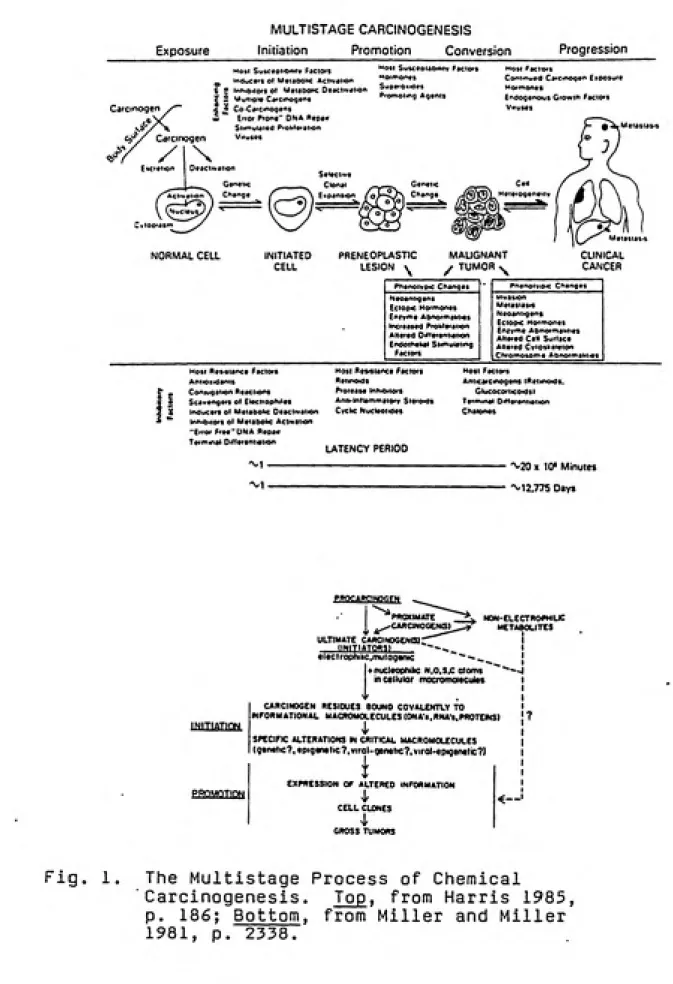
Currently, it is well accepted that chemical
carcinogenesis is a complex multistage process involving
initiation, promotion (including tumor conversion), and
progression, as shown in figure 1 (Holbrook 1980; Miller and Miller 1981; Harris 1985; Williams and Weisburger 1986;
Hermo 1987). Chemicals play key roles as initiating
carcinogens and promoting agents in this scheme.
E»ctetioo
c 2 NrtuU'D'e Ca'Cinogeris
£ • CoCatcinogens
ku EfiOf Prone ONA Repair
Stimutaied Ptoi>*eiatiQn
inogen Vnuses
P'omoiing Agents Endogenous G'OwiH Facto'S
Vifuses Genetic Change Selective Clonal Expansion
/ ^^^ \ expansion (oK^^^
NORMAL CELL INITIATED
CELL
fi
ueneiic Change eie'oge Metastasis MALIGNANT CLINICAL CANCER TUMORPhenoivpic Changes Plenor PRENEOPLASTIC
LESION \ Neoantigens Ectopic Hormones £niv">e Abnormalities Increased Proltteration Altered Oiftetsniiaiion Endothetiai Stimul4i)n9 Factors Invasion Metastasis Neoantigens EciODtc Moffnones Eniv"ie Abnormalities
Altered Celt Surface Altered Cytoskeleion Chfomosome Abnormaliiies Host Resistance factors
Antioiidants
Coniugation Reactions
Scavengers ot Electroo^iles Inducers ot Metabolic Deactivation Inhibitors Ot Metabolic Activation
ͣ
Error Free ' DMA ftepaii Terminal Oittereniiaiion
Most Resistance F»ctors
Retinoids Protease innibitors Anti-tntlammaiorv Steroids Cyclic Nucleotides LATENCY PERIOD Host Factors Aniicatcinogens iReimoids. Glucocorticoids) Terminal Oitterentiaiion C^atones
^'20x 10* Minutes
'^'12,775 Days PRQCARCIMOG^N PROXIMATE CARClNOGeNtSl non-ele:ctroph)lic metabolites INITIATIOM PROMOTION
ULTIMATE CARCINOOENOl-"-"^
(INITIATORS) ~"
el ttctrophi he, mutagenic
+ nucleophihc m,o,s,c atoms ^ "^-^--j
Incetlular mocromotecutfts
CARCINOGEN RESIDUES BOUND COVALEMTLY TO
INFORMATIONAL MACROMOLECULES (DNAt.HNA*«.PROTEINS)
SPECIFIC ALTERATIONS IN CRITICAL MACROMOLECULES
iqtm^KT, epigenetic 7. «irol-genetic?, virol-epiqenetic?)
EXPRESSION OF ALTERED INFORMATION
CELL CLONES
I
GROSS TUMORS
<—
Fig. 1. The Multistage Process of Chemical
Carcinogenesis. Top, from Harris 1985,
p. 186; Bottom, from Miller and Miller
chemical carcinogenesis, and irreversible chemical lesions formed can initiate the carcinogenic process. Such genetic events followed by cell proliferation may lead ultimately to
the induction of neoplasia, a process that can span twenty
years or longer in humans.
Hermo (1987) describes the series of events that are
generally referred to as "initiation". These include:
(1) absorption, distribution, and possible biotransformation (metabolic activation) of a chemical; (2) interaction of an electrophilic ultimate carcinogenic metabolite with cellular macromolecules, particularly DNA (but also RNA/proteins);
and (3) the fixation of carcinogen damage through cell
replication producing a permanent genetic change.
The ability of chemical carcinogens to covalently bind
to and damage DNA can lead to cellular mutational events, such as point mutations and frame-shift mutations, as well as other types of gross structural DNA alterations like gene
rearrangement and amplification (Hermo 1987). Codon
rearrangement may involve sequences known as oncogenes, and several mechanisms, including point mutation, have been identified as being responsible for the activation of cellular proto-oncogenes (Williams and Weisburger 1986).
Gene products of many oncogenes function in biochemical
processes that could be involved in malignant cell
latent preneoplastic cell) which may, in the future, undergo
cell divisions and changes in gene expression ultimately
leading to production of a neoplasm.
The "promotion" stage of carcinogenesis involves the
conversion of an initiated precancerous cell to the
transformed neoplastic state then capable of proliferation leading to clonal amplification and malignant tumor
formation (Hermo 1987). Cells that have undergone
neoplastic conversion may remain dormant, held in check by
tissue homeostatic factors, but their proliferation may be
facilitated by various chemical promoting agents, such as
phorbol esters (Williams and Weisburger 1986). Harris
(1985) mentions the concept of tumor conversion, whereby benign tumors can convert to malignant tumors, and he
suggests that DNA-damaging and mutagenic chemical agents may
cause an additional genetic event that hastens this
conversion.
Many of the specific steps in chemical carcinogenesis,
such as bioactivation and DNA repair, are controlled and
modified by endogenous and exogenous host factors including: species; sex; age; and immunological, hormonal, and
nutritional conditions (Williams and Weisburger 1986).
Besides the genetic mechanism described above, there is
an alternative, perhaps accompanying, mechanism for
effects on gene expression, possibly stemming from
carcinogenic interaction with proteins and/or RNA (Holbrook
1980; Williams and Weisburger 1986). As discussed by
Williams and Weisburger (1986), this has led to the attempted classification of chemical carcinogens as genotoxic if they interact with and alter DNA or as
epigenetic if a biologic effect other than reaction with DNA
could be the basis for their carcinogenicity.
Metabolism of Xenobiotics in Relation to DNA Adduct Formation
Many studies on a variety of chemical carcinogens have shown that their ultimate reactive and carcinogenic forms contain an electrophilic (relatively electron-deficient) atom capable of reacting nonenzymatically by covalent bond formation with an available nucleophilic (electron-rich) atom of a critical cellular macromolecule (DNA, RNA, and/or protein) (Miller and Miller 1981; Dipple, Michejda, and Weisburger 1985). Direct-acting or primary genotoxic
carcinogens contain intrinsically electrophilic centers and do not require metabolic activation. They include such chemicals as: alkyl imines, lactones, alkylene epoxides,
sulfate esters, mustards, halo ethers, and certain
nitrosamides (Holbrook 1980; Williams and Weisburger 1986).
In contrast, most organic genotoxic carcinogens are
indirect-acting or secondary carcinogens. They are not reactive as the parent compound (termed "pro-" or
activation (or bioactivation) to the reactive electrophilic "ultimate carcinogen" (Holbrook 1980; Hermo 1987). The term "proximate carcinogen" applies to any intermediate species
involved in the overall metabolic activation.
Indirect-acting chemicals include: polycyclic aromatic hydrocarbons, aromatic amines, quinolines, nitrofurans, nitrosamines, azo
compounds, and more (Williams and Weisburger 1986). The enzymes necessary for metabolic activation of
xenobiotics (chemicals entering an organism from the external environment) are concentrated primarily in the endoplasmic reticulum of hepatocytes. The primary role of these systems is the biotransformation and detoxication of
lipophilic compounds to more water-soluble hydrophilic
species which can be more readily excreted from the
organism. Sipes and Gandolfi (1986) and Singer and
Grunberger (1983) discuss the role of hepatic systems with
particular regard to activation of promutagens and
procarcinogens.
The enzyme-dependent reactions are divided into two
stages: Phase I, such as oxidation, reduction, and
hydrolysis reactions in which substrates are functionalized to increase hydrophilicity; and Phase II conjugation
reactions in which a highly polar endogenous group is added
to the hydrophilic site generated by Phase I action. Of most importance in metabolic activation are the Phase I
known as the mixed function oxygenase (MFO) system,
monooxygenases, or mixed-function oxidases.
For example, in the case of polycyclic aromatic
hydrocarbons or olefinic compounds, one oxidation pathway involves the cytochrome P-450-mediated addition of oxygen to
the carbon-carbon double bond to produce an epoxide.
Containing a highly strained 3-membered ring, epoxides are
very reactive intermediates capable of opening to an
electrophilic carbonium ion which can form covalent adducts with cellular nucleophiles including the DNA bases. Epoxide hydrolase (formerly epoxide hydrase/hydratase) is an
important hydrolytic enzyme that can deactivate epoxides by
catalyzing the hydration of arene oxides and aliphatic
epoxides to their corresponding trans-dihydrodiols, which
can be conjugated and excreted. Some aromatic
dihydrodiols, however, can undergo secondary metabolism by the MFO enzymes, giving diol-epoxides which may be potent mutagens and carcinogens. This pathway has been identified
as the major activation route of benzo[a]pyrene.
Thus, the same enzymes that biotransform and detoxify xenobiotics to protect the body can also produce reactive intermediates (including proximate and ultimate carcinogens) which are inherently capable of causing more harm than the
original parent chemical compound that entered the body.
The balance between rates of formation and rates of
determining whether adverse cellular events might later
occur.
Metabolic Activation of Alternant PAH
Polycyclic (or polynuclear) aromatic hydrocarbons (PAH)
are ubiquitous environmental pollutants formed from the incomplete combustion of fossil fuels and have been
identified in gasoline and diesel fuel exhaust, coke oven
emissions, coal soot, coal tar and its pitches, mineral
oils, tobacco smoke, and charred food. As potential
mutagens, carcinogens, and teratogens, PAH pose human health
hazards due to public exposure to these compounds through inhalation of contaminated air or consumption of certain
food products (Sawicki 1985; Boulos and von Smolinski 1986).
During the past two decades, the most commonly and
extensively studied subset of PAH has been the "alternant"
structures composed of fused aromatic 6-meinbered rings.
"Nonalternant" PAH such as cyclopenta-PAH are characterized by a cyclopenta ring fused to the periphery of an alternant aromatic nucleus. They possess unique structures that can provide important insight into structure-activity
relationships. In order to understand the metabolic
activation pathways of cyclopenta-PAH, it is important to consider the results of previous studies on alternant PAH, especially those supporting the role of epoxides and diol-epoxides as the reactive metabolites responsible for
As described by Dipple, Michejda, and Weisburger (1985)
and shown in figure 2(A), oxidation of a PAH double bond catalyzed by the cytochrome P-450-dependent arylhydrocarbon hydroxylases (AHH) can produce an arene oxide. There are
three known possible outcomes for this initially formed epoxide: nonenzymatic rearrangement to a phenol; hydration catalyzed by the epoxide hydrolase enzyme to a
trans-dihydrodiol; or glutathione conjugation catalyzed by the glutathione-S-transferases leading to mercapturic acid excretion products. One of these three routes can usually inactivate most primary epoxide metabolites of PAH, offering no chance for epoxide ring opening to a benzylic carbonium
ion capable of reacting with critical cellular
macromolecules.
For many years the simple "K-region" expoxide was
proposed as the metabolically formed DNA-reactive metabolite and the ultimate carcinogen of many PAH. Beginning in the 1970s, numerous studies on the metabolic activation of PAH focused on benzo[a]pyrene, abbreviated B[a]P (see review by Wislocki and Lu 1988). These investigations led to the
discovery of the vicinal dihydrodiol epoxide as the ultimate carcinogen, formed by further enzyme-dependent oxidation of the isolated double bond adjacent to a trans-dihydrodiol. Most of the biologically active diol-epoxides were formed on
a 3-sided peripheral indentation called the "bay region".
Figure 2(B) outlines the metabolic reaction scheme for a PAH
n
ph«nol
qlutathton«
ransierase
I tans -d ihydrodiot
Bay rtglon
glutathion* (R) conjugata
.OTOTQ
7 8 tS
K region
('fK7R.8S)-«poxida
OH
(-H7R.8R)-<Jiol
OTOIO
H0\>\-^ HO*^^/^
OH OH
(+ )(7R.8S,9S,10R) (- X7S.8R,9R,10S)
anti or trans dihydrodiol «poxidaa
br Ix
HO-^'
OH OH
(-K7R,8S.9R,10S) ( + X7S,8R.9S.iaR)
syn or cis dihydrodiol epoxidaa
___^ 0\^
(-X7S.8R)-apaxida
XX
HO*^ =
OH
9^
Fig. 2. Metabolic Activation of a Polycyclic Aromatic Hydrocarbon. (A), Disposition of an Epoxide (from Dipple, Michejda, and Weisburger 1985, p. 227); (B), Formation of a Bay Region Diol-Epoxide, (same ref.,p. 274); (C), Stereoisomers from Metabolism of 3 (a )P (same ref., p. 275);
proximate carcinogen to the bay region diol-epoxide as the
ultimate carcinogen.
As discussed by Grover (1982), the metabolic conversion
of dihydrodiols to diol-epoxides may depend on whether the
conformation of the dihydrodiol hydroxyl groups is diaxial
(approximately perpendicular to the plane of the molecule)
or diequatorial (approximately within the same plane as the plane of the molecule). When the diol groups are adjacent to a bay in the molecule or an alkyl substituent, they may be forced to adopt a quasi-diaxial conformation. In this conformation further metabolism to the diol-epoxide does not
readily occur possibly because of stereochemical effects
preventing the enzyme-substrate interaction. However, when
an unhindered dihyrodiol adopts the quasi-diequatorial
conformation, as it does for the precursor to the bay region diol-epoxide in which the epoxide is adjacent to the bay, further oxidative metabolism can and does take place.
According to in vitro studies, bay region diol-epoxides can be converted to glutathione conjugates but do not act as substrates for epoxide hydrolase, an important factor in determining their mutagenic and carcinogenic capabilities
(Grover 1982? Dipple, Michejda, and Weisburger 1985).
There are four possible stereochemical isomers of the
biological activity are highly stereoselective with
(+)-anti-7.8-dihydrodiol-9,10-epoxide determined to be the
most carcinogenic ultimate species (Jeffrey 1985; Dipple, Michejda, and Weisburger 1985; Wislocki and Lu 1988).
Figure 2(D) shows the structure of the trans B[a]P-DNA adduct expected upon epoxide ring opening of the ultimate
carcinogenic species to form an electrophilic carbonium ion
at C-10 that covalently binds to the exocyclic amino group
of deoxyguanosine. Because of the high stability of the C-10 carbonium ion, a mixture of cis and trans adducts would be predicted, but the trans isomer represents greater than
90% of the DNA adducts (Jeffrey 1985) .
In 1976, Jerina et al. proposed the "bay region theory"
to explain the high biological activity of bay region
diol-epoxides (see reviews by Wood et al. 1979; Lehr et al. 1982; Jeffrey 1985; and Wislocki and Lu 1988). The concept
postulates that epoxides situated on saturated, angular
benzo-rings located in the bay region of a PAH (bay region
diol-epoxides) should be highly reactive due to greater ease
of electrophilic carbonium ion formation. This prediction is based upon Dewar perturbational molecular orbital (PMO)
calculations which estimate resonance stabilization energies
( A E(jeloc /^ values) of benzylic carbonium ions formed upon
ring opening of the epoxides. Based upon studies on
alternant bay-region PAH, larger values of AEdeloc /^
activity of the metabolite with a value of A E(jeloc /^ ^ °''7
determined to be a possible threshold for carcinogenic activity (Jerina et al. 1976; Wood et al. 1979; Lehr et al.
1982) .
Besides B[a]P, the bay region diol-epoxides of other PAH including benz[a]anthracene, dibenz[a,h]anthracene, 7,12-dimethyIbenz[a]anthracene, and 3-methyIcholanthrene
have proven to be the most chemically reactive forms,
exhibiting higher mutagenic/carcinogenic activity than
metabolites at non-bay region molecular sites (Grover 1982;
Singer and Grunberger 1983).
The bay region theory predicts chemical reactivities of
the highly stabilized oxirane ring-opened benzylic carbonium
ions formed from bay region diol-epoxides solely on the
basis of perturbational delocalization energies. It does
not account for regioselectivity of metabolism,
stereochemical, conformational, and/or structural parameters
(like molecular size), which can all influence the interaction of the reactive intermediate species with
critical cellular nucleophiles.
Metabolic Activation of Cyclopenta-PAH
Cyclopenta-polycyclic aromatic hydrocarbons (CP-PAH)
are characterized by the fusion of an ethylene fragment to
an alternant PAH molecule to form a new PAH containing an
unsaturated five-membered ring (Nesnow et al. 1986). This
opportunities to investigate the roles of molecular
geometry, stereochemistry, and electronic structure in
mutagenic and carcinogenic activity, especially for
comparison with predictions based upon previous studies of
alternant PAH. In the past decade CP-PAH studies have
focused on the importance of cyclopenta ring and bay region
(if present) features in the mixed-function oxidase
activation of these compounds as well as the correlation of
PMO resonance stabilization energy ( AE^jeioc /B)
calculations with biological activity (see reviews by Nesnow
et al. 1986; Gold, Sangaiah, and Nesnow 1988).
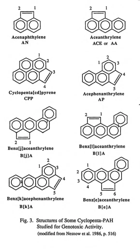
Figure 3 depicts the molecular structures, names, and
common abbreviations of eight cyclopenta-PAH which have been
well studied for genotoxic activity. Of these CP-PAH, at
least five have been identified as environmental
contaminants primarily from combustion processes, as
indicated in table 1. The smallest cyclopenta-PAH,
acenaphthylene, contains three rings and is also an
environmental contaminant but is not a bacterial mutagen
O
o
Acenaphthylene
AN
O
O O
Aceanthrylene
ACE or AA
o
o
o
o
Cyclopenta[cd]pyrene
CPP
o
o o
2 1
Benz[j]aceanthrylene
B[j]A
o
o o o
Benz[k]acephenanthrylene
B[k]A
o
o o
/
Acephenanthrylene
AP
o
o
o
o
Benz[l]aceanthrylene
B[1]A
o
o
o
o
5 6
Benz[e]aceanthrylene
B[e]A
Fig. 3. Structures of Some Cyclopenta-PAH
Studied for Genotoxic Activity,
Table 1. — Environmental Occurrence of Cyclopenta-PAH
Parent PAH CP-PAH Sources
Pyrene
Phenanthrene
Benz[a]anthracene
CPP
AP
B[e]A
B[j]A
B[1]A
Automobile exhausts
Coal combustion Wood combustion
Tobacco smoke Carbon black
Pitch
Grilled foods
Coal combustion Kerosene combustion
Wood smoke
Tobacco smoke
Carbon black
Coal combustion Coal combustion
Wood smoke
Source: modified from Nesnow et al. 1986, p. 517; Krishnan
and Kites 1981, p. 342.In 1980 Fu, Beland, and Yang presented PMO calculations
on the presumed ultimate carcinogenic metabolites, the
cyclopenta-PAH epoxides, for over fifty CP-PAH compounds.
They reported that the olefinic double bond of the fused
five-membered ring in most CP-PAH has the highest bond order
making it highly susceptible to epoxidation and that the
benzylic carbonium ion resulting from ring opening of the
cyclopenta epoxide may be effectively stabilized by the
aromatic system. In fact, 33 of the CP-PAH epoxides can
yield carbonium ion intermediates having higher resonance
stabilization/delocalization energies (AEdeloc /^) than
that of the mutagenic and carcinogenic bay region
diol-epoxide of B[a]P (7,8-dihYdrodiol-9,10-diol-epoxide; AEdeloc /^
cyclopenta-PAHs may present a mutagenic and/or carcinogenic
hazard similar to the classic alternant PAH since the
five-membered ring of CP-PAH provides a site for metabolic
activation by mixed-function oxidases.
Around the same time period, Eisenstadt and Gold (1978) and Gold et al. (1980) reported their studies on the
metabolic activation, mutagenesis, and morphological
transforming capability of the non-bay-region
cyclopenta[cd]pyrene (CPP) and its cyclopenta epoxide, the
3,4-oxide. They found CPP to be highly mutagenic with rat
liver S9 activation in the Ames assay, CPP 3,4-oxide to be a direct-acting mutagen in bacterial and mammalian assays, and
both compounds able to transform mammalian cells in culture.
Gold and co-workers suggested that CPP 3,4-oxide might be
the ultimate mutagenic form of the parent CP-PAH, and this conclusion was further supported by identification of the dihydrodiol, 3,4-dihyroxy-3,4-dihydro-cyclopenta[cd]pyrene, as the major metabolite in bacterial and C3H10T1/2 mouse cells (Gold, Schultz, and Eisenstadt 1979; Nesnow et al.
1981) .
Of six other CP-PAH studied, including aceanthrylene, acephenanthrylene, B[e]A, B[j]A, B[1]A, and B[k]A, liver
microsomes from Aroclor-1254 induced rats metabolized all
compounds to cyclopenta-ring dihydrodiols, implicating
typhimurium Ames assay (Nesnow et al. 1984; Sangaiah et al.
1983, 1986). The four cyclopenta-fused isomers of
benz[a]anthracene were also mutagenic in mammalian cells in
culture (Nesnow et al. 1984).
In S. typhimurium the peak mutagenic activity of B[j]A, B[e]A, and B[1]A occurred at considerably lower S9
concentrations than B[a]P and B[k]A (Nesnow et al. 1984).
Low optimum S9 concentration has been reported previously
for CPP and was interpreted as indicating activation via a
one-step cyclopenta-ring epoxidation as opposed to the multistep diol-epoxide activation pathway of B[a]P
(Eisenstadt and Gold 1978).
As shown in table 2, the delocalization energies for
oxirane ring-derived carbocations on the cyclopenta rings of
B[j]A, B[e]A, and B[1]A exceed that of the mutagenic and
carcinogenic bay region C-10 carbonium ion of
B[a]P-7,8-dihydrodiol-9,10-epoxide.
Table 2. — PMO Delocalization Energies of Benzylic Carbonium Ions Derived from Epoxides of PAH
Epoxide Carbonivim ion AEdgioc /^
ACE-l,2-oxide
B[j]A-l,2-oxide
B[e]A-5,6-oxide B[l]A-l,2-oxide
B[a]P-7,8-diol-9,10-oxide
CPP-3,4-oxide
B[k]A-4,5-oxide
AP-4,5-oxide
Source: modified from Bartczak 1988, p. 80.
C-1 0.931
C-1 0.879
C-5 0.879
C-1 0.833
C-10 0.794
C-3 0.794
C-5 0.722
The cyclopenta-ring arene oxides of B[j]A, B[e]A,
B[1]A, and B[k]A were synthesized and found to be
direct-acting mutagens in the Ames assay with B[k]A oxide the least
potent in agreement with its lower A E^^eioc /^ value
(Bartczak at al. 1987, 1988). The results of this study
support the hypothesis that the cyclopenta epoxides of the
three benzaceanthrylene isomers B[e]A, B[j]A, and B[1]A are
major contributors to mutagenicity in the Ames assay, but
other metabolic activation pathways may play a role in the
mutagenicity of B[k]A. They also reaffirm the utility of
the AEjjeloc /^ parameter as a predictor of mutagenic
activity.
Three of the four cyclopenta-benz[a]anthracene isomers
(B[e]A, B[j]A, and B[1]A) were active in morphologically
transforming C3H10T1/2 cells (Mohapatra et al. 1987). In
this assay B[j]A and B[1]A were metabolized to
cyclopenta-ring dihydrodiols as well as K-region dihydrodiols and the
dihydrodiol precursor to the bay region diol-epoxide as
major metabolite for B[j]A. These findings suggest that
alternative routes of metabolic activation besides
cyclopenta-ring oxidation, such as K-region or bay region
activation, may be operative in C3H10T1/2 cells.
Recently, the cell transforming activities of B[j]A,
its cyclopenta epoxide, cylcopenta dihydrodiol,
9,10-dihydrodiol, and bay region diol-epoxide were studied.
formation of the bay region diol-epoxide represent major independent pathways of metabolic activation (Lasley et al.
1990).
Gold, Sangaiah, and Nesnow (1988) provide a detailed
account of many CP-PAH studies including bioassay results,
metabolic characterization, and structure-activity
considerations.
Molecular Sites on DNA Susceptible to Covalent Reactions
The covalent reactions of elctrophilic ultimate
mutagens/carcinogens with susceptible nucleophilic centers
in DNA can lead to a wide variety of DNA adducts. Which
nucleophilic molecular sites on DNA are attacked depends
upon several factors including the reaction mechanism (as
unimolecular Sjjl or bimolecular Sjj2) , the strength of the
nucleophile, characteristics of the electrophile such as
hardness and stability, and steric constraints (Jeffrey
1985; Swenberg et al. 1990). The most strongly nucleophilic
sites in DNA involve the ring-nitrogens, particularly N-7 of guanine, and the more weakly nucleophilic sites include the
oxo and amino groups of the four DNA bases (Holbrook 1980;
Jeffrey 1985; Swenberg et al. 1990).
Alkylating agents, such as methyl methanesulfonate,
react by the Sfjl mechanism tend to be less selective and
react efficiently at oxygens and other nitrogens in DNA. This can lead to alkylation on positions such as: N-1, N-3,
and N-7 of adenine; N-3 and O^ of guanine; N-3 of cytosine;
O^ of thymine; and the hydroxyl of phosphates (Holbrook
1980; Jeffrey 1985; Swenberg et al. 1990).
Proceeding by the Sjjl mechanism, metabolites of
aromatic amines and amides react preferentially with C-8 of
guanine. N^ of guanine and N^ of adenine are frequent
positions of attack by the stable carbonium ions of
polycyclic aromatic hydrocarbons (Holbrook 1980; Jeffrey
1985; Swenberg et al. 1990).
Adducts formed at several sites that normally
participate in the hydrogen bonding of complementary bases
(such as N-3 of cytosine, O^ of guanine, or O of thymine)
are critical with respect to mutagenesis and carcinogenesis
because they have the potential to induce base mispairing
when DNA is replicated (Holbrook 1980; Hermo 1987; Swenberg
et al. 1990).
Methods of Identification and Quantitation
of DNA Adducts
Detection and characterization of DNA adducts can aid
in the formulation of structure-activity relationships for
mutagens/carcinogens (including stereo- and regioselectivity factors) and can also help establish relationships between
DNA adducts and exposure to environmental chemicals as well
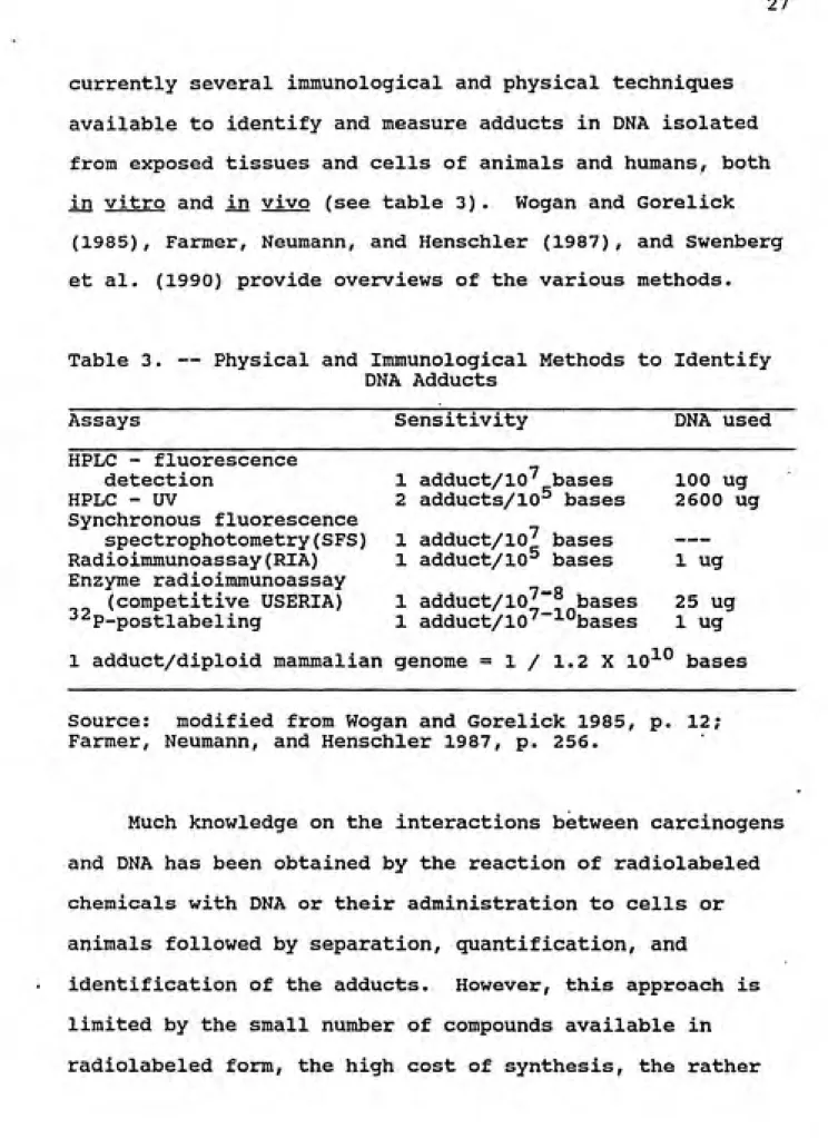
currently several immunological and physical techniques available to identify and measure adducts in DNA isolated
from exposed tissues and cells of animals and humans, both in vitro and in vivo (see table 3). Wogan and Gorelick
(1985), Farmer, Neumann, and Henschler (1987), and Swenberg
et al. (1990) provide overviews of the various methods.
Table 3. — Physical and Immunological Methods to Identify
DNA Adducts
Assays Sensitivity DNA used
HPLC - fluorescence
detection 1 adduct/10' bases 100 ug
HPLC - UV 2 adducts/10^ bases 2600 ug
Synchronous fluorescence
spectrophotometry(SFS) 1 adduct/10^ bases
---Radioimmunoassay(RIA) 1 adduct/10^ bases 1 ug
Enzyme radioimmunoassay
(competitive USERIA) 1 adduct/10^~^ bases 25 ug
ͣ
^^P-postlabeling 1 adduct/10^ -'
ͣ
'^bases 1 ug
1 adduct/diploid mammalian genome = 1 / 1.2 X 10 bases
Source: modified from Wogan and Gorelick 1985, p. 12;
Farmer, Neumann, and Henschler 1987, p. 256.
Much knowledge on the interactions between carcinogens
and DNA has been obtained by the reaction of radiolabeled
chemicals with DNA or their administration to cells or
animals followed by separation, quantification, and
identification of the adducts. However, this approach is
limited by the small number of compounds available in
"large" amounts (up to mg) of DNA required for analysis, and
inapplicability to studies in humans.
High pressure liquid chromatography (HPLC) in
conjunction with UV or fluorescence detection has been employed for detection of B[a]P-DNA adducts. Synchronous
fluorescence spectrophotometry (SFS) can be useful for
carcinogens that fluoresce, like PAH and aflatoxins, and may
be suitable for complex mixtures as contour maps of spectra
become available.
Monoclonal and polyclonal antibodies, which have been
prepared against a variety of specific carcinogen-DNA
adducts, can be used in specific and sensitive immunological
techniques which have applications to human tissue samples.
These competitive or noncompetitive immunoassays include the radioimmunoassay (RIA), enzyme-linked immunosorbent assay
(ELISA), and ultrasensitive enzymatic radioimmunoassay
(USERIA).
The •^^P-postlabeling techniques can successfully detect
adducts of aromatic amines and amides, PAH, and methylating agents. Sensitivity of this assay is, in theory, thehighest available to date for aromatic or bulky adducts.
•^^P-Postlabeling Techniques
In 1981 Randerath, Reddy, and Gupta reported a -^^p.
postlabeling method for the detection and quantitation of
covalent adducts formed by the reaction of DNA with chemical
A G C T a-iBPBP^
Carcinogan Modified ONA
1! Micrococcal nucteasa
2) Spleen phosphoamsisrase
HO I HO
\
HO 1 HO
J MP ATP
T4 Poiynuclaotida kinass
Oeoxyribonucleoside 3'-monophosphates
nBuQH
Extraction
IBul.N^CI-8-(BP)
Nuclaasa P1
Penicittum Citrinumt
DeoxvnbonucMOSides
OH OH OH
i "P ATPISp.Ac. - 7000 Ci/m moll
T4 Polynuctftotida kinasa
!( ^2P ATPISo.Ac. - 7000 Cl/m moll T4 Polyhuclaotide kinasa
^xfej^xN^,
B-(BP)
DeoxYribonucteoside 3'.5'-bisphosphatas
TLC Autoradiography
Fig. 4. ^2p_pQs|-ia|-|g;Ling of DNA Adducts (from Watson
1987, p. 320). (A), Standard Method; (B), Butanol Extraction Enhancement; (C), Nuclease
basic procedure involves enzymatic digestion of isolated DNA
to deoxynucleoside 3'-monophosphates which are then
converted to their corresponding S'-'^^P-labeled
3',5'-biphosphates by T4 polynucleotide kmase-catalyzed Ptransfer from adenosine CY--^^P]triphosphate. The mixture of
labeled adducts is then purified and resolved by four-directional thin layer chromatography, usually anion
exchange TLC on polyethyleneimine (PEI)-cellulose plates. Autoradiography is used to determine the presence of
chemically modified nucleoside 3',5'-biphosphates. Adduct spots are then cut from the plates and measured using
Cerenkov or liquid scintillation counting.
With the aim of increasing the sensitivity and accuracy of quantitative measurements, the original techniques were later modified with adduct enrichment procedures in a
butanol extraction modification (Gupta 1985) and nuclease PI
modification (Reddy and Randerath 1986). The butanol adduct
enrichment procedure, which involves extraction with 1-butanol in the presence of the phase-transfer agent tetrabutylammonium chloride prior to the labeling, is
particularly suited to analysis of aromatic carcinogen-DNA
adducts (especially PAH). It has been shown to enhance
assay sensitivity to a level of 1 adduct per lo^"-^^
nucleotides with 1-10 ug of DNA used (Gupta 1985; Gupta and
Earley 1988).
Randerath et al. (1985) used the butanol extraction
3?
individual PAH to mouse skin DNA in vivo, and they observed
a good correlation between carcinogenic potency and binding.
They failed to detect DNA binding of the noncarcinogens
anthracene, pyrene, and perylene while the strong
carcinogens B[a]P, 7,12-dimethylbenz[a]anthracene, and
3-methylcholanthrene exhibited highest levels of binding.
Watson (1987) and Randerath et al. (1985) review the
significant advantages of the -^^P-postlabeling assay which
include the following features: it is applicable to individual chemical compounds and mixtures; chemical
identity of adducts doesn't have to be known to detect DNA
binding; fingerprint patterns obtained after autoradiography
of known chemicals can be used for identification of unknown
chemicals and mixtures; it is highly sensitive for aromatics
enabling detection of a few adducts per mammalian genome; it
requires a minimal amount (1-10 ug) of DNA; it allows accurate quantitation of adducts; and it is potentially
useful for studying repair and removal of adducts from cell
or tissue DNA.
Application to DNA Adducts of Cyclopenta-PAH. Recently Nesnow et al. (1989) analyzed and identified
aceanthrylene-DNA adducts in C3H10T1/2 cells by using the
butanol extraction modification of the -^^P-postlabeling
technique. C3H10T1/2 cells treated with aceanthrylene for
adducts). Identities of this ACE metabolite and DNA base
portion of the adducts were confirmed by co-chromatography
of adduct mixtures from ACE-treated C3H10T1/2 cells, calf
thymus DNA incubated with ACE-1,2-oxide, and the
homopolymers of 2'-deoxyguanosine modified by ACE with
Aroclor-1254-induced rat liver S9 activation.
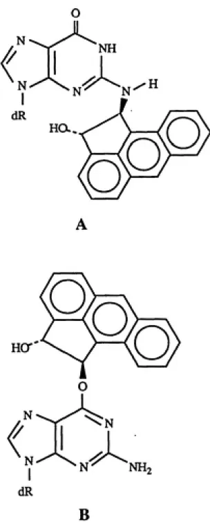
Because of the large delocalization energy ( A E^^gioc
/6) associated with the C-1 carbonium ion formed from the
opening of ACE-1,2-oxide (Sangaiah et al. 1986), possible
structures of the ACE-DNA adducts might be isomers of
N^-[l-(trans-2-hydroxy-aceanthrylenyl)]-2'-deoxyguanosine and
0°-[l-(trans-2-hydroxy-aceanthrylenyl)]-2'-deoxyguanosine, as
shown in figure 5 (Nesnow et al. 1989).
In order to accomplish the detailed structural
characterization of these adducts, co-chromatography with
known chemical synthetic standards is needed. Results from
this study support the choice of
ACE-1,2-oxide-deoxyguanosine adducts as targets for synthetic standards,
with exocyclic amino group modification given top priority
based on high susceptibility of this nucleophilic position
to covalent reaction with arene oxides.
Benz[j]aceanthrylene adducts in C3H10T1/2 cells have
been studied in a similar manner using the same -^2p_
postlabeling techniques (Lasley et al. 1990). DNA adduct
autoradiograms were generated after treatment of the cells
with B[j]A-l,2-oxide, B[j]A-9,lO-dihydro-9,10-diol, the
B[j]A-l,2-dihydro-O
O
o
Q
O
O
i
B
Fig. 5. Possible Structures of Aceanthrylene-Deoxyguanosine Adducts. (modified from
Nesnow et al. 1989, p. 232).
(A), N -[l-(trans-2-hydroxyaceanthryIenyI)]-2'-deoxyguanosine;
1,2-diol. These autoradiograms were compared with those
produced by treatment of homo polydeoxyguanylic acid with B[j]A-1,2-oxide and the B[j]A bay region anti diol-epoxide. Major adducts were determined to be formed by reaction of the bay region diol-epoxide with deoxyguanosine with minor contributions from reaction of B[j]A-1,2-oxide (the
cyclopenta epoxide) with deoxyguanosine. Synthetic
standards of deoxyguanosine modified at N-2 by the B[j]A diol-epoxide and B[j]A-1,2-oxide are needed in order to
confirm the structures of these DNA adducts to a higher
degree of specificity than base/metabolite composition.
This type of identification procedure, involving the
use of chromatographic standards in the ^P-postlabeling
assay, has been reported in the literature. Pongracz et al.
(1989) utilized chemical synthetic standards in the -^^p.
postlabeling assay to identify two of the six DNA adducts
formed by styrene oxide, which corresponded to aralkylation
at 0-6 of guanine.
Use of DNA Adducts in Human Biomonitorinq
Traditionally, human exposure to occupational and
environmental chemicals has been assessed by measuring
concentrations of a substance in ambient environmental media
- air, food, and water. However, biological monitoring,
measurements that can be made on cells, tissues, or body
fluids of exposed people, is presumably more directly
environmental monitoring method (Miller 1984; Wogan and
Gorelick 1985; Farmer, Neumann, and Henschler 1987; Wogan
1989).
Biological monitoring indicates the "internal dose" or
"biologically effective dose" on an individual basis and
takes into account variation in absorption, distribution,
and metabolism of xenobiotics (Wogan and Gorelick 1985;
Wogan 1989; Farmer, Neumann, and Henschler 1987).
Measurements of levels of the parent compound or its
metabolite(s) in body tissues/fluids or excreta (usually
blood, urine, and expired air) provides information on the
"internal dose" of a chemical substance, which reflects
environmental/occupational exposure (Miller 1984; Perera
1987). "Biologically effective dose" (also known as "target
dose" or "molecular dose") refers to the amount of a
chemical agent (mutagen/carcinogen) or its metabolite
(ultimate carcinogen) that has reacted with critical
cellular macromolecules (DNA, RNA, and/or proteins) of the
target tissue or its surrogate (Perera 1987; Farmer,
Neumann, and Henschler 1987).
For human biological monitoring (or biomonitoring),
markers of biologically effective dose include the
following: covalently bound DNA adducts, excreted DNA adducts, protein (i.e. hemoglobin or albumin) adducts,
somatic cell mutations, micronuclei in lymphocytes,
chromosomal aberrations, sister chromatid exchanges,
(Perera 1987). Figures 6(A) and 6(B) portray the role of
DNA adduct determination as a biomonitoring technique in the overall assessment of human exposure to environmental
mutagens/carcinogens.
Wogan and Gorelick (1985) and Harris (1985) discuss the application of carcinogen-DNA adducts as "internal
dosimeters" of exposure to genotoxic chemicals. Qualitative
and quantitative identification of DNA adducts in accessible
human tissue (such as white blood cells or biopsy samples) can provide indication of an individual's exposure history as well as one's ability to metabolize a mutagen/carcinogen to its reactive form. As shown in figure 6(C), many factors
affect the levels of carcinogen-DNA adducts detected in
cells at any given time including the exposure-sampling interval, carcinogen exposure concentration, the metabolic
balance between carcinogen activation and detoxication, and
DNA repair rates (Harris 1985).
Although information from some experimental animal models suggests that levels of DNA adducts in tissues are
linearly related to carcinogen dose (Wogan and Gorelick
1985), meaningful interpretation of DNA adduct levels as
quantitative measures of exposure in humans is much more complex. This is due to the factors previously discussed as
well as diverse human exposures to chemicals from a variety
of sources beyond the workplace, all of which may influence adduct levels in a manner not easily determined. The
Environmental \ Biomonitoring monitoring ^
I
Xenobiotic Xenobiotic Absorption in vivo Metabolites Excreted xenobiotic or metabolite Protein adducts, Nucleic acid adducts,Sulptiydryl (e.g.
glutathione) adducts,
Ottier detoxification products Excreted adduct or repair product Determination of
xenobiotic or metabolites,
in blood or excreta
Haemoglobin alkylation. DNA adduct determination
(chemical or immunoassay). Post-labelling of modified DNA
Thioethers. DNA repair products
DETERMINANTS OF CARCINOGEN-ONA ADDUCT LEVELS
Carcinogen Expotur* Activation Deactivation ONA Repair high high B Adduct Laval Excretion Chemical] in — Air — Water — Food Absorption
}
Ambient Monitoring Distribution -Biotransformation Biological» monitoring of
exposure
Binding to critical sites
Adverse effects t
Preclinical lesions
t
Clinical lesions
• Binding to
noncritlcal sites Non adverse effects
1
Health Surveillance (Biomonitoring of effects)Fig. 6 Use of DNA Adducts in,Human Biomonitoring.
(A), Methods Used for Biomonitoring (from
Farmer, Neumann, and Henschler 1987, p. 252);
(B), Biomonitoring in Assessment of Human
Exposure (from Miller 1984, p. 189A); (C),
Determinants of Adduct Levels (from Harris
reflect past or recent chemical exposures (adduct
persistence) and acute or chronic exposures needs to be extensively investigated. According to Wogan and Gorelick
(1985) and Harris (1985), DNA adducts can currently serve as
useful qualitative markers of exposure.
Regarding the use of carcinogen-DNA adduct levels as
indicators of long-term cancer risk, further complications
arise including the validity of surrogate cell measurements
in lieu of actual target tissue at risk and the necessary
simplifying assumptions made about the multistage
carcinogenic process (see Harris 1985; Perera 1987; Farmer,
Neumann, and Henschler 1987 for information on DNA adducts
in cancer risk assessment). Perera (1987) thoroughly
reviews the growing field of molecular cancer epidemiology
which involves conducting studies that establish
relationships between biologic markers of dose and cancer
risk in human populations.
Detection of Aromatic DNA Adducts in Human Tissues bv
-^^P-Postlabeling
Wogan (1989) thoroughly reviews the numerous human
biomonitoring studies that have involved detection of DNA
adducts of genotoxic agents in the cells and tissues of
exposed persons. In the past, many have focused on
detection of adducts arising from the bay region
diol-epoxide (BPDE) metabolite of B[a]P through immunoassays
,such as ELISA or USERIA, or physicochemical methods like
blood cells of cigarette smokers, foundry workers, aluminum
plant workers, and coke oven workers (see review by Wogan
1989).
Recently, numerous studies, many involving smokers,
have employed the -^^P-postlabeling techniques for the
detection of aromatic DNA adducts in human cells or tissues.
Everson et al. (1986) investigated the presence of DNA
adducts formed in placentas of women who smoked during
pregnancy, and they found a major aromatic adduct strongly
related to maternal smoking.
Through co-chromatography in the postlabeling assay, Randerath et al. (1986) compared DNA adducts detected in
mouse skin treated with cigarette tar with DNA adducts
detected in tissues from the bronchus and larynx of smokers. They observed several identical adducts present in the DNA
samples from exposed mice and smokers.
DNA from normal human bone marrow cells was analyzed by
•^^P-postlabeling, and ten out of ten individuals showed the
presence of aromatic adducts that were not detected in human fetal bone marrow (Phillips, Hewer, and Grover 1986). Their findings suggest that adducts reisulted from environmental exposure to unidentified genotoxic chemicals.
Chacko and Gupta (1987) analyzed DNA from human oral
mucosal cells of smokers and nonsmokers. Two
chromatographically distinct major adducts were detected in
most smokers but not in nonsmokers. In addition, they
chromatographic standards to aid in the chemical
identification of the mucosal DNA adducts. These
co-chromatography experiments eliminated the possibility that any lesions were formed from reaction of B[a]P with N'' of guanine or reaction of 4-aminobiphenyl with C-8 of guanine.
Phillips et al. (1987) detected aromatic adducts in DNA from white blood cells of foundry workers classified
according to their exposure to airborne BCa^P in the
workplace. Interestingly, none of the detected adducts in the individuals' DNA showed the chromatographic behavior
expected of B[a]P-DNA adducts, and adduct spots were
detected in a few unexposed workers. These results indicate that there may be significant interindividual variations in
DNA adduct levels among similarly exposed workers.
DNA from white blood cells and placentas of non-smoking women exposed to residential wood combustion smoke during
pregnancy was analyzed by the postlabeling assay for PAH-DNA
adducts (Reddy, Kenny, and Randerath 1987). All placental
DNA postlabeling maps (from exposed and nonexposed women)
Basis for Synthetic Approach
Trans B-amino alcohol derivatives (enantiomeric
mixtures) of acenaphthylene and aceanthrylene were
previously synthesized and successfully used in condensation
reactions with 6-chloropurine-9-6-D-ribofuranose to give
N°-adenosine adducts, RNA adducts expected from the
corresponding 1,2-oxides (Bartczak et al. 1989). The same
CP-PAH derivatives were later successfully condensed with
6-chloropurine-9-(2-deoxy-B-D-ribose) to produce
N^-deoxyadenosine adducts (Gold: unpublished results).
For synthesis of N^-modified deoxyguanosine (DNA
adducts), the appropriate halopurine precursor is a
2-halo-2'-deoxyinosine. Generation of the 2-chloro derivative had
been attempted previously in this laboratory and was
achieved by Lewis acid catalyzed glycosylation of
2-chlorohypoxanthine (from partial hydrolysis of
2,6-dichloropurine) with
2-deoxy-3.5-di-O-p-toluoyl-a-D-erythro-pentofuranosyl chloride (unpublished results). However, due
to high cost of 2,6-dichloropurine and presumed increased
efficiency of the condensation reaction expected by the more
reactive bromoinosine derivative, synthesis of
Recently, Hildebrand et al. (1990) indicated use of
2-broino-2'-deoxyinosine (2-BrdI) in generation of
N^-n-hexyl-2'-deoxyguanosine, but no details of the synthesis of
2-BrdI were provided in the literature. In addition,
although N^-aryl and N^-alkyl 2'-deoxyguanosine derivatives
have been reported in the literature (Wright and Dudycz
1984; Casale and McLaughlin 1990; Lee et al. 1990;
Hildebrand et al. 1990) , no synthesis of
N^-itiodified-2'-deoxyguanosine involving a cyclopenta-PAH has been
published.
Efficient synthesis of 2-bromohypoxanthine (1) has been
reported in the literature (Beaman, Gerster, and Robins
1962). The Lewis acid catalyzed glycosylation of
2-bromohypoxanthine with the anomeric mixture
1,3,5-tri-O-acetyl-2-deoxy-<x. S-D-erythro-pentofuranose (synthesized
according to Gold and Sangaiah 1990) yielded a mixture of N^
a plus 6 and N a plus B isomers which couldn't be readily
separated (Gold: unpublished results).
To avoid problems previously encountered in separating
isomeric glycosylation products and to presumably enhance
yields of N^ isomers over N^ isomers, synthesis of 0°-NPE
(p-nitrophenylethyl group)-protected 2-bromohypoxanthine (4)
by the method of Raju, Robins, and Vaghefi (1989) was
undertaken in this study, as shown in reaction scheme I of
figure 7. The synthetic strategy was originally based on
Lewis acid catalyzed glycosylation of the NPE-protected
1,3,5-tri-0-acetyl-2-^
Br' "^N-^^
TrCl
-^^
dryEtjN
lOO^C, 2 hrs.
(Tr = CPhg)
N
Br-^N
O —NPE
•N,
PPhj
diethyl azodicarboxylate
4-nitrophenylethanol
dry dioxane
8(fC, 6 hrs.
t
>
80% HOAc
H
--- N^
80 C, 45 min. I
BrA^N
(NPE group)
(85% yield from 3)
(24% yield from 1)
(28% yield from 1)
O—NPE
dryMeCN
OAc---ͨ
BSA
TMSOTf ^^'
AcO
(Ac = C - CH3)
II
o 5A
^
AcO 6A
(56% yield for N-9«x' + ^ mixture)
A.
\N
Br' ^N N
I
H
>
O—NPE
A
HO 9
(56% yield from 6B)
2-methoxy-ethanol
T
Et-,N
O —NPE
p-Tol-O 5 B
(p-Tol= C-^-CH3)
O dry MeCN NaH
Y
O —NPE
MeOH
anhydrous NH3
p-Tol-O
H NH2
HO-I---l-H
o
Q
p-Toi-o 6B
(39% yield)
2-methoxy-ethanol
EtaN
O —NPE
D
p-ToI-O
10
P-Tol-O
8 (~ 5% yield)
deoxy-g.,B-D-erythro-pentofuranose (5A), as shown in figure
7. However, the synthetic scheme was later modified due to
difficulties in efficiently separating the N^ a and 6
isomeric glycosylation products (6A) .
Thus, reaction scheme II (shown in figure 8) was
developed. This involved a sodium hydride catalyzed
glycosylation of 0^-NPE-protected 2-bromohypoxanthine (4)
with 2-deoxy-3,5-di-O-p-toluoyl-a-D-erythro-pentofuranosyl chloride (5B). The direct glycosylation product (6B) was then coupled with trans acenaphthene l-amino-2-ol (7) to
generate protected N^-deoxyguanosine adducts (8).
In hopes of improving yields of the final adduct
products, ammonolysis reactions were carried out on the glycosylation product (6B) to remove the bulky p-toluoyl
groups (giving 0^-NPE-protected 2-BrdI, 9) prior to
condensation with the cyclopenta-PAH derivative.
Condensation reactions were conducted with the NPE group still intact on the 0-6 position of the purine because the resulting aromatic 6-membered ring is expected to enhance
the nucleophilic substitution reaction at C-2.
Instrumentation and Laboratory Materials
^H nuclear magnetic resonance (NMR) spectra were
acquired at the UNC Department of Chemistry on a Varian XL-400 at 400 MHz. Chemical shifts are given in parts per million (ppm) relative to tetramethylsilane (TMS). Mass