ACC/AHA PRACTICE GUIDELINES
1999 Update:
ACC/AHA Guidelines for the Management
of Patients With Acute Myocardial Infarction
A Report of the American College of Cardiology/
American Heart Association Task Force on Practice
Guidelines (Committee on Management of Acute Myocardial Infarction)
COMMITTEE MEMBERS
THOMAS J. RYAN, MD, FACC,Chair
ELLIOTT M. ANTMAN, MD, FACC NEIL H. BROOKS, MD, FAAFP ROBERT M. CALIFF, MD, FACC L. DAVID HILLIS, MD, FACC LOREN F. HIRATZKA, MD, FACC
ELLIOT RAPAPORT, MD, FACC BARBARA RIEGEL, DNSC, FAAN RICHARD O. RUSSELL, MD, FACC EARL E. SMITH III, MD, FACEP W. DOUGLAS WEAVER, MD, FACC
TASK FORCE MEMBERS
RAYMOND J. GIBBONS, MD, FACC,Vice Chair
JOSEPH S. ALPERT, MD, FACC KIM A. EAGLE, MD, FACC
TIMOTHY J. GARDNER, MD, FACC ARTHUR GARSON, JR, MD, MPH, FACC
GABRIEL GREGORATOS, MD, FACC RICHARD O. RUSSELL, MD, FACC THOMAS J. RYAN, MD, FACC SIDNEY C. SMITH, JR, MD, FACC
The American College of Cardiology/American Heart As-sociation (ACC/AHA) Guidelines for the Management of Patients With Acute Myocardial Infarction have been
reviewed over the past 21⁄2years since their initial publica-tion (J Am Coll Cardiol 1996;28:1328 – 428) to ensure their continued relevancy. The guidelines have been updated to include the most significant advances that have occurred in the management of patients with acute myocardial infarc-tion (AMI) during that time frame. This Update was developed to keep the guidelines current without republish-ing them in their entirety. The Update represents a new procedure of the ACC/AHA Task Force on Practice Guidelines. These guidelines will be reviewed and updated as necessary until it is deemed appropriate to revise and republish the entire document.
This Update, as printed here in the Journal of the American College of Cardiology, appears without the full text guidelines. The full text guidelines, incorporating the Update, are available on the Web sites of both the American College of Cardiology (www.acc.org) and the American Heart Association (www.americanheart.org). In the Web site version, deleted text is indicated by s
兾兾t兾r兾ık兾e兾o兾u兾兾t, and new/revised text is presented in a high-lighted typeface. Reprints of the original 1996 document with the revised sections appended are available from both organizations (see footnote).
The “1999 Update: ACC/AHA Guidelines for the Management of Patients With Acute Myocardial Infarction: A Report of the American College of Cardiology/ American Heart Association Task Force on Practice Guidelines (Committee on Management of Acute Myocardial Infarction)” was approved by the American College of Cardiology Board of Trustees and the American Heart Association Science Advisory and Coordinating Committee in June 1999.
The American College of Cardiology and the American Heart Association request that the following format be used when citing this document: Ryan TJ, Antman EM, Brooks NH, Califf RM, Hillis LD, Hiratzka LF, Rapaport E, Riegel B, Russell RO, Smith EE III, Weaver WD. 1999 update: ACC/AHA guidelines for the management of patients with acute myocardial infarction: a report of the American College of Cardiology/ American Heart Association Task Force on Practice Guidelines (Committee on Man-agement of Acute Myocardial Infarction). J Am Coll Cardiol 1999;34:890–911. Available at http://www.acc.org/clinical/guidelines.
This Update is presented in 4 sections as follows:
1. Changes/additions to text
2. New and revised figures and tables
3. Changes in Class I, II, and III recommendations 4. Changes in references
This Update to the guidelines represents a new proce-dure. Comments about this approach and format are en-couraged and should be sent to Chair, ACC/AHA Task Force on Practice Guidelines, American College of Cardi-ology, 9111 Old Georgetown Road, Bethesda, MD 20814.
New section, page 1340, column 2: new text replaces paragraphs beginning “An ideal serum marker. . .” through page 1342, column 1, “Assays for biochemical. . .”
Serum cardiac markers. When myocytes become necrotic, they lose membrane integrity, and intracellular macromol-ecules diffuse into the cardiac interstitium and ultimately into the cardiac microvasculature and lymphatics (55). Eventually, these macromolecules are detectable in the peripheral circulation. The term currently used to collec-tively describe these macromolecules is serum cardiac mark-ers. An ideal serum cardiac marker of MI should be present early and in high concentration in the myocardium and should be absent from nonmyocardial tissue and serum (55–57). It should be rapidly released into the blood at the time of the myocardial injury, and there should be a stoichiometric relation between the plasma level and the extent of myocardial injury. The marker should persist in blood for a sufficient length of time to provide a convenient diagnostic time window. Finally, measurement of the marker should be easy, inexpensive, and rapid.
The nomenclature of the acute coronary syndromes (ACS) is illustrated in revised Figure 2. The central position of the 12-lead electrocardiogram (ECG) and initial triage of patients are emphasized. Listed at the bottom of the figure is the information sought by clinicians when measuring serum cardiac marker levels in patients at different ends of the ACS spectrum. Serum cardiac markers are useful for confirming the diagnosis of MI when patients present without ST-segment elevation, when the diagnosis may be unclear, and when clinicians must distinguish patients with unstable angina from those with a non–Q-wave MI. Serum cardiac markers also provide valuable prognostic informa-tion. For patients with ST-segment elevation, the diagnosis of MI is secure; clinicians are interested in prognostic information as well as a noninvasive assessment of the likelihood that the patient has undergone successful reper-fusion when thrombolytic therapy is administered.
Because the conventional serum cardiac marker, creatine kinase (CK) and its MB isoenzyme (CK-MB) lack sufficient sensitivity and specificity, there is a need for more sensitive and cardiac-specific markers of myocardial necrosis (792– 794). The troponin complex consists of 3 subunits: troponin T, troponin I, and troponin C (795). The ternary troponin complex is a calcium-sensitive molecular apparatus that
regulates the interaction of actin and myosin. Troponin T binds the troponin complex to tropomyosin, and troponin I binds to actin and inhibits interactions between actin and myosin. Troponin C is responsive to changes in intracellular calcium concentration. Amino acid sequences of the skeletal and cardiac isoforms of troponin I and troponin T have sufficient dissimilarity that monoclonal antibody-based im-munoassays have been developed to detect cardiac-specific troponin T (cTnT) and cardiac-specific troponin I (cTnI). Because the amino acid sequence of troponin C is the same in cardiac and skeletal muscle, no immunoassays of troponin C have been developed for clinical purposes.
Because CK-MB is found in the skeletal muscle and blood of healthy subjects, the cutoff value for an elevated CK-MB level is typically set a few units above the upper end of the reference (normal) range. In contrast, because cardiac troponin I and cardiac troponin T are not normally detected in the blood of healthy people, the cutoff value for elevated cTnI and cTnT levels may be set only slightly above the noise level of the assay, permitting clinicians to diagnose lesser degrees of myocardial necrosis (ie, increased sensitiv-ity) (796). Because CK and CK-MB are characteristically used as the gold standard for diagnosing MI, investigators may face a dilemma when a new diagnostic test is more sensitive than the gold standard, particularly for identifying episodes of minor myocardial cell necrosis. Case reports confirm histologic evidence of focal myocyte necrosis in patients with elevated cardiac troponin levels and normal Figure 2. Patients with ischemic discomfort may present with or without ST-segment elevation on the electrocardiogram. The majority (large arrow) of patients with ST-segment elevation ultimately develop a Q-wave AMI, whereas a minority (small arrow) develop a non–Q-wave AMI. Of patients who present without ST-segment elevation, the majority (large arrows) are ultimately diagnosed as having either unstable angina or non–Q-wave AMI based on the presence or absence of a cardiac marker such as CK-MB detected in the serum; a minority of such patients ultimately develop a Q-wave AMI. The spectrum of clinical conditions ranging from unstable angina to non–Q-wave AMI and Q-wave AMI is referred to as acute coronary syndromes. AMI⫽ acute myocardial infarction.
CK values (796). It is estimated that ⬇30% of patients presenting without ST-segment elevation who would oth-erwise be diagnosed with unstable angina are actually experiencing a non–Q-wave MI when assessed with cardiac-specific troponin assays (797). Furthermore, numer-ous investigators have now reported that elevated levels of cTnI or cTnT provide more prognostic information than that supplied by the patient’s demographic characteristics or the ECG at presentation (798,799). Elevated cTnI or cTnT levels, even in the presence of normal CK-MB levels, identify patients without ST-segment elevation who are at an increased risk of death. Finally, patients presenting without ST-segment elevation who are characterized as high risk because of elevated cardiac-specific troponin levels demonstrate a greater benefit from treatment with new therapies such as glycoprotein (GP) IIb/IIIa inhibitors than patients without elevated cardiac-specific troponin levels who receive such new pharmacotherapeutic interventions (800).
CK-MB isoforms are another new serum cardiac marker that may be useful for evaluating patients with an acute coronary syndrome. CK-MB exists in only 1 form in myocardial tissue but in different isoforms (or subforms) in the plasma. An absolute level of CK-MB2⬎1 U/L or a ratio of CK-MB2to CK-MB1of 1.5 has improved sensitivity and specificity for diagnosis of MI within the first 6 hours compared with conventional assays for CK-MB (59). Myo-globin, a low-molecular-weight heme protein found in cardiac and skeletal muscle, is not cardiac specific but is released more rapidly from infarcted myocardium than CK-MB and may be detected as early as 2 hours after MI. The diagnostic sensitivity and specificity for MI were compared for total CK-MB (activity and mass), CK-MB subforms, myoglobin, cTnI, and cTnT in the Diagnostic Marker Cooperative Study (DMCS) (801). The DMCS was a large, prospective, multicenter, double-blind study of patients presenting in the emergency department (ED) with chest pain. CK-MB subforms were most efficient for early diagnosis (within 6 hours) of MI, whereas cTnI and cTnT were highly cardiac specific and particularly efficient for late diagnosis of MI. The DMCS investigators concluded that either a single assay (CK-MB subforms) or a select combi-nation (CK-MB subform and a cardiac-specific troponin) reliably triages patients with chest pain and could potentially lead to improved therapy and reduced cost of care of ACS patients. It should be noted that serum levels of cTnT and cTnI may be present for several days after MI (up to 7 days for cTnI and up to 10 to 14 days for cTnT). Therefore, the ability to diagnose recurrent infarction is significantly com-promised if the clinician relies solely on cardiac-specific troponins and fails to obtain a concomitant CK or CK-MB measurement within the first 12 to 24 hours of admission of an MI patient. Thus, although CK and CK-MB are not as cardiac specific as the troponins, they will return to normal levels within the first 24 to 36 hours, making it more likely that a reelevation is associated with recurrent myocardial
necrosis. For patients presenting within the first 2 or 3 hours of symptom onset, the 2 markers most appropriate for the early diagnosis of AMI are myoglobin and CK-MB sub-forms.
In patients presenting with ST-segment elevation, clini-cians usually use peak CK as a rough estimate of the magnitude of the infarct and assessment of the patient’s prognosis. Release of cardiac-specific troponins is stoichio-metrically correlated with the amount of myocardial necro-sis, and the new serum cardiac markers can also be used to estimate infarct size and prognosis (58). Cardiac-specific troponins may not be detectable for up to 6 hours after onset of chest pain. Thus, when cTnI and cTnT levels are elevated early after onset of discomfort in patients with ST-segment elevation MI, clinicians should suspect that an antecedent episode of unstable angina was in fact MI and the patient is exhibiting a stuttering course of occlusion and release of the infarct-related artery. Data from the Global Utilization of Streptokinase and TPA for Occluded Arteries (GUSTO) III Study suggest that patients with elevated cardiac troponin T levels and who are ⬍6 hours from the onset of discomfort have an increased mortality risk (802). In addition to monitoring the patient for resolution of ischemic-type chest discomfort and regression of the mag-nitude of ST-segment elevation on the ECG, clinicians can obtain serial measurements of serum cardiac markers to buttress the noninvasive diagnosis of reperfusion of the infarct-related artery after thrombolytic therapy (65,803). Because of its rapid-release kinetics, myoglobin is a partic-ularly attractive marker for the early diagnosis of reperfu-sion.
New section: Follows “Serum Cardiac Markers” (see page 1340, column 2)
Text added to “Risk of Stroke,” page 1348, column 1: new text added after paragraph beginning “Thrombolytic therapy is associated with. . .”
More recent trials show that as use of thrombolysis has increased, a greater proportion of patients who are ⬎75 years old or female are now included. This change has been associated with a higher rate of intracranial hemorrhage (ICH) than that seen in earlier studies. For example, the rate of ICH after administration of alteplase was⬇0.7%; in more recent studies, it is 0.8 to 0.9% (new Table 2.1). It should be noted that the streptokinase without heparin administration regimen has the lowest rate of ICH.
Text added to “Primary Percutaneous Transluminal Coronary Angioplasty,” page 1351, column 1: new text replaces paragraph beginning “A meta-analysis suggests. . .”
In the GUSTO-IIb trial (805), 1138 patients with evolving ST-segment elevation MI within 12 hours of onset of chest pain were randomly assigned to receive primary percutane-ous transluminal coronary angioplasty (PTCA) (n⫽565) or accelerated tissue plasminogen activator (tPA) (n⫽ 573). Thirty days after enrollment, the incidence of death, recur-rent MI, or disabling stroke was 9.6% in those who underwent PTCA and 13.6% in those who received tPA (P ⫽ 0.033). However, 6 months after enrollment the difference between the 2 treatments did not reach statistical significance; the incidence of the composite adverse out-come was 13.3% in the PTCA group and 15.7% in the tPA group (P⫽NS).
Recently published data from the Second National Reg-istry of Myocardial Infarction (NRMI-2) (124) suggest that primary PTCA and thrombolytic therapy offer similar efficacy. Over 17 months, 4939 subjects with evolving ST-segment elevation MI received primary PTCA, and 24,705 received alteplase. For patients without cardiogenic shock, the in-hospital mortality rate was similar (5.4% for
the alteplase group, 5.2% for the PTCA group), and this was true even when the data from certain “high-risk” subgroups, such as those ⬎75 years old and those with anterior MI, were analyzed.
Among the most important contributions to these revised guidelines are the data in the preliminary report of the Should We Emergently Revascularize Occluded Coronaries for Cardiogenic Shock? (SHOCK) Trial, presented by Dr Judith Hochman on March 7, 1999, at the 48th Scientific Sessions of the American College of Cardiology, held in New Orleans, Louisiana and to be published in the August 26, 1999 issue of the New Engl J Med(805a). When this multicenter study was designed in 1992, it was postulated that emergency revascularization (ERV) of cardiogenic shock due to an ST-elevation/Q-wave or new left bundle-branch block (LBBB) MI would result in a 20% (absolute) reduction in the primary end point, all-cause 30-day mor-tality compared with initial medical stabilization (IMS), and delayed revascularization as clinically determined.
In this study, 152 patients were randomly assigned to the ERV strategy, and 150 patients were assigned to a strategy of IMS. The 30-day mortality rate for ERV patients was 46.7% versus 56.0% for IMS patients (95% confidence interval [CI],⫺20.5 to⫹1.9%,P⫽0.11), a nonsignificant trend. However, the mortality rate at 6 months (a secondary end point) was significantly lower in the ERV group (50.3% versus 63.1%,P⫽0.27). The prespecified subgroup analysis of patients⬍75 years old showed a 15.4% reduction in the primary end point (IMS group, 56.8%, versus ERV group, 41.4%,P⬍0.01), whereas outcome in patients⬎75 years old was worse for the ERV group. Intra-aortic balloon pump (IABP) support was used in 86% of both groups; 63% of the IMS group received thrombolytic agents, and 25% underwent delayed revascularization. Of the ERV group of patients who underwent emergency early revascularization,⬇60% received PTCA, and 40% had coronary artery bypass graft (CABG); the 30-day mortality rate was 45% and 42%, respectively.
New Table 2.1. Intracranial Hemorrhage in Recent Thrombolytic Trials
Patient Characteristics
GUSTO-I (497)
GUSTO-II (805)
COBALT (832)
GUSTO-III
(802,833) ASSENT-2* In Time-II*
Number 41,021 3473 7169 15,059 16,950 15,078
Average age (y) 62 62.5 62.4 63 — —
⬎75 y (%) 10.5 11.8 13.0 13.6 — —
Female (%) 25.2 22.4 23.4 27.4 — —
Intracranial Hemorrhage Rates
SK 0.51 0.37 — — — —
tPA 0.70 0.72 Double bolus 1.12
Accl infusion 0.81
0.87 0.93 0.62
rPA — — — 0.91 — —
TNK-tPA 0.7 0.72 — — 0.94 —
nPA — — — — — 1.13
An early meta-analysis of the randomized clinical trials that compared primary PTCA with thrombolytic therapy was reported in early 1995 (121) and included data on in-hospital or 6-week mortality and nonfatal MI for all 7 trials reported to that time. The combined data showed a mortality rate at 6 weeks of 3.7% in the PTCA group and 6.4% in the thrombolysis group (odds ratio [OR], 0.56; 95% CI, 0.33 to 0.94). In the combined outcome of short-term mortality and nonfatal reinfarction, the event rate was 6.1% for the PTCA group and 11.0% for the thrombolytic therapy group (OR, 0.53; 95% CI, 0.35 to 0.80). By 1 year, however, none of these end point differences were statisti-cally significant. The analyses showed that ⬇30% of the thrombolytic therapy patients underwent PTCA sometime during hospitalization or within the first 6 weeks of infarc-tion. Therefore, the contrast in the proportions of patients receiving any PTCA versus patients receiving no PTCA was substantial (64%). The authors conclude that the data on primary PTCA appear promising but should be interpreted with caution and viewed as a strong impetus for the conduction of larger trials in a more diverse range of hospitals, with clinical outcomes being the primary end points of interest.
A more recent meta-analysis by Weaver et al (806) provides a quantitative review of the treatment effects of primary coronary angioplasty versus intravenous thrombol-ysis for AMI from 10 randomized trials that involved 2606 patients. When the results of all studies were combined, the mortality rate atⱕ30 days was 4.4% for the 1290 patients treated with primary angioplasty, compared with 6.5% for the 1316 patients treated with thrombolysis (34% reduction; OR, 0.66%; 95% CI, 0.46 to 0.94;P⫽0.02). The pooled rate of death or nonfatal reinfarction was also lower in patients treated with primary PTCA than in those treated with thrombolytic therapy, from 11.9% to 7.2%, respectively (OR, 0.58; 95% CI, 0.44 to 0.76). Angioplasty was associ-ated with a significant reduction in total stroke (9/1290, 0.7%; versus 26/1390, 2%; P ⫽ 0.007) and hemorrhagic strokes as well (0.1% versus 1.1%;P⬍0.001). On the basis of outcomes at hospital discharge or 30 days, this analysis concluded that “primary PTCA appears to be superior to thrombolytic therapy for treatment of patients with AMI, with the proviso that success rates for PTCA are as good as those achieved in these trials. Data evaluating longer-term outcome, operator expertise, and time delays before treat-ment are needed before primary PTCA can be recom-mended universally as the preferred treatment.” Recently Brodie et al (788) pointed out that patients who underwent angioplasty within 2 hours of onset of symptoms showed a striking 53% relative reduction in 30-day mortality com-pared with those who underwent angioplasty⬎2 to 6 hours (4.3% versus 9.2%; P⬍ 0.04). Because their data failed to show an important time-dependent worsening of mortality beyond 2 hours, it has been suggested that the time delay in transferring patients with AMI to tertiary centers for primary PTCA may be permissible if the procedure cannot
be done within the first 2 hours of symptom onset. Clearly, it becomes critical to measure the outcomes of larger numbers of patients stratified by time to answer this important question. On the other hand, if a time-dependent worsening of mortality does exist for patients undergoing angioplasty (as seems likely since it does so for patients reperfused with fibrinolytic therapy), it seems reasonable to explore the theoretical advantage of combining the admin-istration of smaller doses of fibrinolytic agents on presenta-tion at the community hospital (for early patency) with prompt transfer to a tertiary center for percutaneous coro-nary intervention (PCI) (sustained patency). The safety of such an approach has been reported by Dr. Allan Ross for the PACT Trial at the 71st Scientific Sessions of the American Heart Association, in Dallas, Texas, on November 10, 1998.
Until more data have more reliably quantified a benefit of primary PTCA over thrombolytic therapy in the commu-nity setting, it seems prudent to suggest that institutions that do not have the capability of offering primary PTCA should not feel compelled to develop such services at this time (121,807).
Text added to “Primary PTCA,” page 1351, column 2: new paragraph added before “Recommendations for Early Coronary Angiography. . .”
The most recent developments in acute reperfusion by mechanical interventions in the management of patients with AMI are the emerging reports of randomized compar-isons between primary PTCA and routine deployment of stents (127,808). The Amsterdam group (127) published a randomized comparison of coronary stenting with balloon angioplasty in selected patients with AMI that showed that primary stenting can be used safely and effectively, resulting in a lower incidence of recurrent infarction and a significant reduction in the need for subsequent target-vessel revascu-larization compared with balloon angioplasty. These data support the concept that with improved stent technique and use of more effective antiplatelet regimens, including ticlo-pidine, the thrombus-laden lesion no longer represents a strict contraindication to stenting. An appropriate note of caution has been made about interpreting these data (809), pointing out the highly selective nature of the study popu-lation. Only 50% of patients with AMI who underwent primary PTCA were considered eligible for this study, raising serious questions about the generalizability of the results.
compared with PTCA alone (12.4% versus 20.1%, P ⬍ 0.01), this was determined solely by the incidence of target-vessel revascularization at 6 months (7.5% versus 17%, P⬍ 0.0001, respectively). Unfortunately, there were more deaths in the stent placement arm (4.2%) than in the PTCA arm (2.7%), although the difference was not statis-tically significant in this trial, which had a total of only 31 deaths.
Text added to “Patient Characteristics,” page 1352, column 1: new text replaces paragraphs beginning “Ischemic-type chest discomfort. . .” through “The early descriptions of MI. . .”
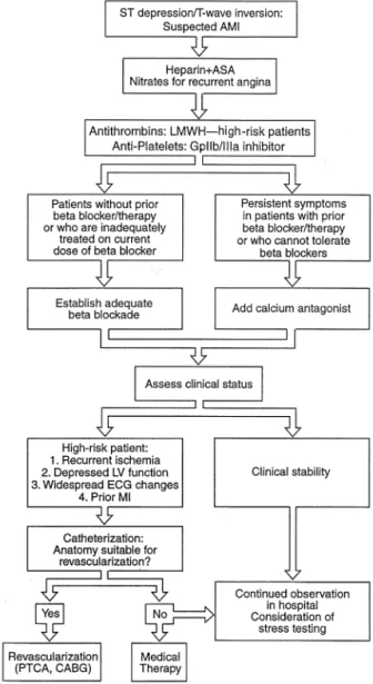
In the setting of nondiagnostic ECG findings (non-ST elevation), ACS represents a continuum between chronic stable angina and AMI with ST-segment elevation. Al-though the prognosis of the patient with chronic stable angina can be stratified and the emergency situation engen-dered by ST-elevation MI is readily evident, patients with acute symptoms but nondiagnostic ECG findings range from those with noncardiac chest pain to very high-risk MI with multivessel disease. Unstable angina and MI without ST elevation represent 2 of the most common cardiac emergencies requiring hospitalization and account for ⬎650,000 discharges per year in the United States. Al-though the optimal treatment regimen or strategies for such patients is under investigation, a proposed diagnostic schema is presented in Figure 2 and a therapeutic approach is depicted in revised Figure 4.
AMI accompanied by nondiagnostic ECG changes is believed to be related to acute disruption of an atheroscle-rotic plaque in the setting of chronic inflammatory infiltra-tion of its fibrous cap; this underlying pathophysiology is not thought to differ from AMI accompanied by ST-segment elevation. As more angiographic and clinical cor-relation studies are done, it is becoming clear that total occlusion of the culprit vessel is much less common in AMI without ST-segment elevation than in MI with ST eleva-tion (82,138 –140). Furthermore, patients without ST-segment elevation are more likely to have multivessel disease and prior MIs than are those with ST-elevation MI (810). In the clinical history, patients with MI without ST-segment elevation are more likely than those with ST elevation to have a history of diabetes, hypertension, heart failure, and peripheral vascular disease but less likely to be smokers or to have hyperlipidemia (810). Importantly, the elderly are less likely to have ST-segment elevation with MI, probably because of the more common presence of prior myocardial damage and multivessel disease (25,149).
Thus, during initial evaluation of the patient with acute ischemic-type chest discomfort, the clinician should classify patients as those with ST elevation or LBBB (acute reper-fusion indicated) and those with nondiagnostic ECGs. The nondiagnostic ECG group will include patients with non-cardiac symptoms, those with unstable angina and no myocardial necrosis, those with small MIs, those with direct
posterior infarctions caused by circumflex artery occlu-sion, and those at very high risk with multivessel coronary disease and significant left ventricular dysfunction. Stud-ies with different mixes of these subgroups have reported different morbidity and mortality rates for the population as a whole.
Figure 4. All patients without ST elevation should be treated with an antithrombin and aspirin (ASA). Nitrates should be adminis-tered for recurrent episodes of angina. Adequate-adrenoceptor blockade should then be established; when this is not possible or contraindications exist, a calcium antagonist can be considered. Current data indicate that either an invasive or noninvasive treatment strategy is suitable for non–ST-elevation AMI patients. AMI ⫽ acute myocardial infarction, CABG ⫽ coronary artery bypass graft; ECG⫽electrocardiographic, GpIIb/IIIa⫽ glyco-protein IIb/IIIa receptor for platelet aggregation, LMWH ⫽
low-molecular-weight heparin, LV⫽ left ventricular, PTCA ⫽
percutaneous transluminal coronary angioplasty.
The initial importance of classifying patients on the basis of the ECG should not be confused with the question of whether the patient has a Q-wave or a non–Q-wave MI. This classification can be made only after 24 hours, well beyond the point at which critical decisions about treatment must be made. Whether or not the patient initially has ST elevation, those with a normal QRS complex who do not develop Q waves with MI have a low in-hospital mortality rate, but recurrent ischemia, recurrent MI, and death in the weeks after discharge occur frequently. In contrast, patients with a significant QRS abnormality who do not develop new Q waves with a new MI are at high risk of both early and later death. The overall incidence of non–Q-wave MI may be increasing with the advancing age of the population and the greater use of thrombolytic therapy, aspirin, and -adrenoreceptor blockers.
New section, page 1353, column 2: new text added before “Interventional Therapy”
Antithrombotic therapy. Thousands of patients with ACS without ST-segment elevation have now been randomly assigned to treatment with various antithrombotic regimens. In these trials, approximately half the patients had enzymes positive for myocardial necrosis on the first measurement, indicating that they were having an MI without ST-segment elevation at the time of randomization. Patients with positive enzymes on the first draw not only had a higher mortality rate than patients without positive enzymes, but they also had a higher risk of repeat MI, hemodynamic complications, and arrhythmias. Fortunately, the response to newer antithrombotic agents has been homogeneous in pa-tients with ACS without ST elevation, whether or not they had positive enzymes at the time of admission.
New section, page 1353, column 2: new text added before “Interventional Therapy.” Follows “Antithrombotic Therapy.”
Glycoprotein IIb/IIIa inhibitors. The GP IIb/IIIa recep-tor is a member of the integrin family of receprecep-tors that is found in the membrane of platelets (811). When platelets are activated by a variety of stimuli, including thrombin, collagen, adenosine diphosphate (ADP), and epinephrine, the GP IIb/IIIa receptor changes conformation to be receptive to one end of a fibrinogen dimer. Occupancy of a GP IIb/IIIa receptor by the other end of the dimer provides the basis for platelet aggregation. Thus, the GP IIb/IIIa receptor is considered the final common pathway of platelet aggregation (812). Multiple therapeutic agents have now been developed to block the receptor.
More than 30,000 patients with ACS without ST-segment elevation have now been randomly assigned into trials comparing GP IIb/IIIa inhibitors with placebo in addi-tion to treatment with aspirin and unfracaddi-tionated heparin (UFH). A systematic overview has demonstrated a definite reduction in the composite end point of death and MI and in the composite end point of death, MI, and the need for revascularization procedures (813). A slight trend toward a
reduction in mortality may exist but does not reach statistical significance. The reduction in events is present while patients are treated with active drug, and the difference in event rates does not change after that point. When treatment is discon-tinued, no further effect, either beneficial or detrimental, is seen. Thus, intravenous GP IIb/IIIa inhibitors may be consid-ered as a method to reduce acute events and stabilize patients in the acute phase of MI without ST-segment elevation. Direct comparisons of the agents are not available, so the specific choice of which agent to use is speculative.
Three agents are available for clinical practice:
1. Abciximab is a chimeric Fab fragment of a monoclonal antibody to the GP IIb/IIIa receptor. Although multiple clinical trials have documented the reduction in the composite of death and nonfatal MI with abciximab in the setting of percutaneous intervention (814 – 817), only 1 trial (Chimeric 7E3 Antiplatelet in Unstable Angina Refractory to Standard Treatment [CAPTURE]) (814) has been completed in the setting of non–ST-elevation ACS.
2. Eptifibatide is a cyclical heptapeptide, which binds to the receptor with a short half-life (818). It has been evalu-ated in a trial of 11,000 patients with non–ST-elevation ACS, 45% of whom had enzymes positive for myocardial necrosis on admission.
3. Tirofiban is a small nonpeptide compound that also has a short half-life. It has been evaluated in 5147 patients in 2 randomized trials of non–ST-elevation ACS (819,820). In the PRISM-PLUS Study (Platelet Receptor Inhibition in Ischemic Syndrome Management in Patients Limited by Unstable Signs and Symptoms) (820), 45% of patients also had positive enzymes for myocardial necrosis.
New section, page 1353, column 2: new text added to end of paragraph 1, before “Interventional Therapy”
Low-molecular-weight heparin and direct antithrom-bins. Low-molecular-weight heparin (LMWH) is a sub-fraction of standard heparin with a greater degree of inhibition of factor Xa relative to thrombin when compared with standard UFH. In addition to its convenience—it can be administered subcutaneously with high bioavailability— LMWH has a number of theoretical benefits over UFH. These include the potential to prevent thrombin generation as well as inhibit thrombin, the lack of a need to monitor with coagulation testing, and a lower rate of heparin-associated thrombocytopenia. Four trials have compared the use of LMWH and UFH for non–ST-elevation ACS (507,838 – 840). In 2 trials, a clear benefit of LMWH was observed (839,840), whereas in another, LMWH was su-perior to placebo (507). The fourth trial did not show a clear difference in outcomes (838).
impor-tant component of leech saliva, has been studied in many thousands of patients; the results show a consistent reduc-tion in the composite of death and nonfatal MI. Hirulog, a synthetic direct thrombin inhibitor, has been studied in only limited populations.
Text added to “Interventional Therapy,” page 1353, column 2, new paragraph added before paragraph beginning “There is considerable variation. . .”
Recently, the Veterans Affairs Non–Q-Wave Infarction Strategies In Hospital Trial (VANQWISH) (821) has shed important light on the question of intervention in patients with non–Q-wave infarction. The VANQWISH Trial evaluated a somewhat different population than the Thrombolysis in Myocardial Infarction (TIMI-3B) Trial. VANQWISH investigators randomly assigned to an inva-sive or conservative strategy 920 patients who did not have a major complication within 24 to 72 hours of onset of symptoms. The ECG criteria required the absence of new Q waves; therefore, the trial included patients with and without ST-segment elevation on admission. The aggres-sive strategy called for routine cardiac catheterization with revascularization of significant lesions, whereas the conser-vative strategy used intensive medical therapy; angioplasty was used only in patients with recurrent ischemia or hemodynamic compromise. In this trial of management of non–Q-wave MI, there was a 28% rate of cardiac events during follow-up of 12 to 44 months but no early or late clinical benefit with routine invasive management. There was no difference in the primary end point of combined death or nonfatal MI during the average follow-up of 23 months (138 patients assigned to the invasive strategy versus 123 patients assigned to the conservative strategy, P ⫽ 0.35). There was a significantly higher rate of death among patients assigned to invasive treatment both at hospital discharge (21 versus 6,P⫽0.007) and at 1 year (58 versus 36,P⫽0.025). Concern has been raised about the operative mortality rate observed in the trial (7.7% for the composite group and 11.6% for those assigned to the invasive strategy); however, it has been demonstrated that the centers enrolling patients in the trial had operative mortality rates within the expected range for all centers in the United States.
Although the TIMI-3B and VANQWISH trials did not involve identical populations, both studies failed to support the notion that an aggressive approach to revascularization in non–ST-segment elevation ACS reduces the risk of death or nonfatal MI. A contrary view was expressed in the preliminary report of the Fragmin During Instability in Coronary Artery Disease (FRISC) Trial II presented on March 7, 1999, at the 48th Scientific Sessions of the American College of Cardiology, in New Orleans, Louisi-ana. The FRISC report indicated that, when combined with an early invasive strategy, the LMWH dalteparin may reduce early events in patients with unstable coronary artery disease. In the open acute phase of the trial, 2267 patients with unstable angina or non–Q-wave MI received
daltepa-rin, 120 IU/kg every 12 hours during the first 5 to 7 days. In the subsequent double-blind phase, 2015 of these patients were randomly assigned to receive subcutaneous dalteparin 5000 to 7500 IU/kg twice daily or placebo for 3 months.
Results at 90 days showed no significant difference between the dalteparin and placebo groups in terms of the primary end point (death or MI); however, during the first 45 days, there was a significant reduction in the primary end point among those receiving dalteparin compared with those receiving placebo (3.7% versus 6.5%, respectively;P⫽ 0.003). During the prolonged treatment phase, the inci-dence of bleeding events was 26% with dalteparin and 10% with placebo. In addition to being randomly assigned to receive dalteparin or placebo, patients enrolled in FRISC II were assigned within 48 hours to invasive or noninvasive early management. The invasive strategy consisted of early coronary angiography (within 2 to 7 days), whereas the noninvasive strategy consisted of exercise testing with refer-ral to coronary angiography if the test was positive or further events warranted it. At 6 months the rate of death or MI in the invasive group was 9.5% versus 12% in a noninvasive group (P ⫽ 0.045). According to subgroup analyses, men particularly benefited from an early invasive strategy, with the rate of death or MI among invasive versus noninvasive groups at 9.1% versus 13.9%;P⫽ 0.002.
It will be interesting to learn whether other antithrom-botic/antiplatelet therapies will produce an environment in which medical therapy alone will be sufficient or whether it will foster improved results with aggressive interventions, which is being addressed in ongoing clinical trials (822).
New section, page 1354, column 1: new text added before “Hospital Management”
was observed in patients treatedⱕ12 hours after symptom onset (RR, 0.43; 95% CI, 0.2 to 0.9;P⫽0.021). Because these results show that a metabolic modulating strategy is feasible in the early hours of an AMI with a GIK infusion in contemporary practice, it is hoped that an appropriately sized clinical trial will get under way soon. The results may have strong implications for incorporating this rather simple and inexpensive therapy for the routine care of AMI patients worldwide.
New text added to “Triage of Patients With Acute Myocardial Infarction and Other Coronary Syndromes,” page 1357, column 2: new paragraph added after line 37, “. . .and treatment success.”
Two large studies have been published that support these concerns (210,825). A survey of 7560 nurses from across the United States suggests that nurses are caring for increasing numbers of patients and are required to cross-train for more responsibilities; 74% report having less time to teach pa-tients and families, and 69% report less time to provide basic nursing care. Forty-nine percent reported that registered nurses working on a part-time or temporary basis have replaced full-time staff, and 36% reported an increase in nonlicensed assistive personnel. Staffing and perceived qual-ity of care were significantly lower in the Pacific and northeastern regions of the country, where managed care is prevalent (210).
Objective data on quality outcomes were obtained from the American Nurses’ Association (825) from a recent study of 502 hospitals in California, Massachusetts, and New York. These data demonstrated that adverse outcomes (ie, pressure ulcers, pneumonia [not community acquired], urinary-tract infections, and postoperative infections) and hospital lengths of stay were associated with RN staffing levels. Adverse events were higher in institutions with lower RN staffing levels. As RN staffing levels decreased, patient length of stay increased, presumably because of adverse events.
A recent report on the adequacy of staffing from the Institute of Medicine (826) concluded that there was sufficient evidence from several studies using different types of quality measures to conclude that there is a positive relationship between nursing-staff levels and the quality of care in nursing homes. The evidence is not sufficient, however, to conclude that such a relationship exists in hospitals. It has been suggested that patient variables (eg, severity of illness) contribute significantly to the variance in outcome and that adverse events may be a more sensitive marker of differences in organizational quality (ie, collabo-ration, leadership, organizational culture, job satisfaction) than staffing ratios (827,828). Taken together, the research in this area suggests that adverse events are not simply the result of changes in staffing levels but more a function of fundamental changes in institutions as a result of reorgani-zation and restructuring. If so, quality-monitoring activities
in hospitals will be essential as the current trend in managed care penetrates the rest of the country.
Text added to “Management of Mechanical Defects After AMI,” page 1370, column 2: new text replaces first 2 sentences of paragraph beginning “Coronary angiography can. . .”
Coronary arteriography can delineate the presence of surgi-cally correctable coronary artery disease, and cardiac cathe-terization may better delineate the presence of a mechanical defect if other studies are not clear. However, the evidence for concomitant CABG associated with surgical repair of an acute ventricular septal defect (VSD) is inconclusive (829). Although there is a need to minimize invasive angiographic procedures before early surgical correction of the ruptured septum, initial coronary arteriography to assess the coronary anatomy seems warranted in most cases.
Text added to “Postinfarction Ventricular Septal Defect,” page 1371, column 1: new text replaces paragraph
Increased frequency of acute rupture of the interventricular septum (VSD) as well as earlier presentation may be noted in patients who have undergone thrombolytic therapy (383). Although emergency surgical repair was formerly thought to be necessary only in patients with pulmonary edema or cardiogenic shock, it is now recognized as equally important in hemodynamically stable patients (384,385,830). Because all septal perforations are exposed to sheer forces and necrotic tissue removal processes by macrophages, the rup-ture site can abruptly expand, resulting in sudden hemody-namic collapse even in patients who appear to be clinically stable with normal left ventricular function (830). For this reason, prompt insertion of an intra-aortic balloon pump and referral for emergency operation are recommended for every patient with acute VSD as soon as the septal rupture is diagnosed. Simultaneous CABG, if feasible, seems war-ranted in patients with extensive coronary artery disease (386).
Section renamed and text added, page 1374, column 2: new text added as a second paragraph under new head
TICLOPIDINE AND CLOPIDOGREL
been reported, and in a review of 60 cases, 20% occurred after only 3 to 4 weeks of therapy, but only 3% of patients treated for ⱕ14 days developed TTP. Furthermore, mor-tality is high:⬇50% of untreated cases and 25% of treated cases (830a).
Ticlopidine and clopidogrel are ADP-receptor antago-nists and quite similar chemically. However, TTP has not been reported with use of clopidogrel, and in the large CAPRIE Trial (Clopidogrel versus Aspirin in Patients at Risk of Ischemic Events) (831), the incidence of a signifi-cant reduction in neutrophils was only 0.10% in the clopi-dogrel group and actually slightly higher, at 0.17%, in the aspirin group. In that trial, there was a statistically signifi-cant relative risk reduction in vascular death, MI, or stroke of 8.7% in favor of clopidogrel. For these reasons, in many catheterization laboratories, ticlopidine has been replaced with clopidogrel combined with aspirin for the prevention of adverse cardiac events after stent implantation. The effectiveness of this regimen, however, is unknown. Clopi-dogrel is also preferable to ticlopidine for patients who demonstrate aspirin resistance or for whom aspirin is con-traindicated because of hypersensitivity.
Text added to “Thrombolytic Agents: General Mechanisms of Action and Pharmacological Properties,” page 1375, column 2: text added between paragraph ending “. . .center of plasmin,” and paragraph beginning “Aside from this. . .”
Aside from this similarity, some comparative features of the Food and Drug Administration–approved thrombolytic agents for intravenous therapy (streptokinase, anistreplase, alteplase, and reteplase) are presented in the revised Table 8. Streptokinase and urokinase are approved for intracoronary use, but this route of administration for AMI is now virtually obsolete. In addition, newer agents have been developed (eg, TNK-tissue plasminogen activator [TNK-tPA] and lanetoplase). Recent trials with alteplase have used an accelerated regimen given over 90 minutes. The accel-erated regimen leads to the highest patency rate without an
increase in ICH and has become the preferred method of administration. The advantage of reteplase is that it can be given by bolus, which is convenient. A recent trial compared the effectiveness and safety of continuous infusion versus double-bolus administration of alteplase (832). The trial was stopped prematurely because of concern about the safety of the double-bolus injection. The rate of hemorrhagic stroke was 1.12% after double-bolus injection of alteplase compared with 0.81% after accelerated infusion of alteplase.
Text added to “Comparative Thrombolytic Efficacy,” page 1376, column 2: new paragraph added before
“Considerations in Selecting Thrombolytic Regimens”
Since the initial publication of these guidelines, the Food and Drug Administration has approved the fibrinolytic agent reteplase for use. Reteplase, a mutant of wild-type tPA, has a longer half-life than its parent molecule and has been compared with alteplase in a large clinical trial (833). An angiographic trial (834) found that 60- and 90-minute TIMI grade 3 flow and coronary patency rates were higher with reteplase than with the accelerated dose of alteplase. When compared with an accelerated infusion of alteplase, reteplase did not provide any additional survival benefit. The mortality rate at 30 days was 7.5% for reteplase and 7.2% for alteplase; and the rates of the combined end point, death or nonfatal MI– disabling stroke, were 7.98% and 7.91%, respectively.
Text added to “Considerations in Selecting Thrombolytic Regimens,” page 1376, column 2: text added as first paragraph
GUSTO-I (228), GUSTO-III (833), and other recent studies (467,468) suggest that accelerated alteplase and reteplase with intravenous heparin are currently the most effective therapies for achieving early coronary reperfusion, but both are substantially more expensive and carry a slightly greater risk of ICH than streptokinase. Thus, the cost-benefit ratio is greatest in patients presenting early after onset of chest pain or symptoms and in those with a large
Replacement Table 8. Comparison of Approved Thrombolytic Agents
Streptokinase Anistreplase Alteplase Reteplase
Dose 1.5 MU in 30–60 min 30 mg in 5 min 100 mg in 90 min 10 U⫻2 over 30 min
Bolus administration No Yes No Yes
Antigenic Yes Yes No No
Allergic reactions
(hypotension most common)
Yes Yes No No
Systemic fibrinogen depletion Marked Marked Mild Moderate
90-min patency rates (%) ⬇50 ⬇65 ⬇75 ⬇75
TIMI grade 3 flow (%) 32 43 54 60
Mortality rate in most recent comparative trials (%)
7.3 10.5 7.2 7.5
Cost per dose (US) $294 $2116 $2196 $2196
area of injury (eg, anterior infarction) and at low risk of ICH. Other promising thrombolytic agents under investi-gation are TNK-tPA and lanetoplase, both of which are mutant forms of wild-type tPA and can be given as a single bolus.
Two equivalence trials comparing these agents with the accelerated infusion of alteplase reported preliminary results in March 1999 at the 48th Scientific Sessions of the American College of Cardiology.
Data from the In TIME-II Study showed the single-bolus thrombolytic lanetoplase (nPA) was as effective in reducing the 30-day mortality rate as tPA in patients with AMI. The trial randomly assigned 15,078 patients within 6 hours of symptom onset to receive single-bolus lanetoplase (120,000 U/kg) or front-loaded alteplase (up to 100 mg). The 30-day mortality rate (primary end point) in the nPA and tPA groups was 6.7% and 6.6%, respectively. At 24 hours, mortality was slightly lower with nPA than with tPA (2.39% versus 2.49%). The nPA group had a significantly higher incidence of ICH than the tPA group (1.13% versus 0.62%;P⫽ 0.003).
The ASSENT-2 trial reported preliminary results from TNK-tPA, the other novel thrombolytic agent delivered by single bolus (790). Within 6 hours of symptom onset, 16,950 patients with AMI were randomly assigned to weight-adjusted TNK-tPA or accelerated tPA. The 30-day mortality rate was 6.17% in the TNK-tPA group and 6.18% in the accelerated tPA group. The incidence of total stroke was similar (1.78% versus 1.66%) as was hemorrhagic stroke (0.93% versus 0.94%), and mild to moderate bleeding was observed less often in the TNK-tPA group than in the tPA group (26% versus 28.1%;P⬍0.002). Although the efficacy of these agents appears to be equivalent to tPA, it will be important to carefully assess the adverse event rates when these studies are published.
There is considerable ongoing investigation of the effec-tiveness of thrombolytic therapy alone compared with the combination of either direct-acting antithrombins or the GP IIb/IIIa receptor antagonists as a means to improve effectiveness over the currently available regimen. In 2 studies that evaluated the combination of hirudin (desiru-din) with alteplase and streptokinase, there was no im-provement in mortality rate, and the therapeutic-to-severe bleeding profile appeared to be very close (TIMI-9 and GUSTO-IIb trials).
Over the past few years, there has been an increase in the number of patients who undergo primary angioplasty for treatment of AMI in hospitals with tertiary cardiac facilities. This has been driven to a large extent by the observed higher patency in TIMI-3 flow rates associated with coronary angioplasty as well as the desire of cardiologists to assess coronary anatomy and ventricular function early in patient management. Still, however, this represents only a small portion of patients with AMI, and thrombolytic therapy remains the major means of reperfusion.
New text added to “Current Use Rates for Thombolytic Therapy,” page 1377, column 1: new text replaces paragraph 1
The industry-sponsored NRMI tracks the use of thrombo-lytic therapy in the United States and has enrolled 330,928 patients treated at 1470 US hospitals during its second phase (NRMI-2) from June 1994 through July 1996. Barron et al (789) recently reported an analysis of this database, attempting to determine what proportion of patients with an MI who are eligible for reperfusion therapy do not receive this proven treatment. Barron used a conservative definition of thrombolytic eligibility (diagnostic changes on ECG or LBBBⱕ6 hours after onset of symptoms and no contraindication to thrombolytic therapy indicated); inves-tigators found that 31% of their cohort were eligible for reperfusion therapy; 25% had nondiagnostic initial ECGs; 41% presented⬎6 hours from onset of symptoms, and 3% had contraindications to thrombolytic therapy.
Of those who were eligible for thrombolytic therapy, 24% did not receive any form of reperfusion therapy (7.5% of all patients). Multivariate analysis revealed that the indepen-dent predictors for eligible patients not being given reper-fusion therapy were the presence of LBBB, the disappear-ance of chest pain at the time of presentation, age ⬎75 years, female gender, and various preexisting cardiovascular conditions. Perhaps most disconcerting was the finding that patients with the highest risk of death from AMI were the least likely to receive reperfusion therapy (eg, patients with a history of congestive heart failure or the presence of LBBB). Both groups had an in-hospital mortality rate of ⬇20%, well above the mortality rate of 7.9%, yet the presence of LBBB made it 78% less likely that a patient would receive reperfusion therapy than patients who pre-sented with ST-segment elevation.
New section, page 1380, column 1: new text added before paragraph beginning “Newer direct antithrombin. . .”
required for binding to antithrombin and inhibition of factor Xa. Thus, through the creation of a mixture of short-and long-chain heparin fragments, preparations of varying antiXa:antiIIa activity may be developed. Additional fea-tures of LMWHs of particular clinical relevance are a decreased sensitivity to platelet factor IV, a more stable, reliable anticoagulant effect, and lower rates of thrombocy-topenia and heparin-induced thrombocythrombocy-topenia syndrome. Thus, LMWHs are clinically attractive because of better bioavailability, ease of administration via the subcutaneous route, and enriched anti-Xa activity (837). Higher anti-Xa activity is important because of the multiplier effect in which 1 molecule of factor Xa leads to production of many molecules of thrombin.
Gurfinkel and colleagues (507) compared placebo treat-ment, UFH, and the LMWH nadroparin in 219 patients with unstable angina who were also treated with aspirin. Combination therapy with aspirin plus nadroparin signifi-cantly reduced the number of patients with an adverse end point event (combined death, MI, and recurrent angina) during the study period, from 59% in the aspirin group and 63% in the aspirin-plus-heparin group to 22% in the aspirin-plus-nadroparin group (P⬍0.0001 for comparisons of the nadroparin group with each of the other 2 groups).
The FRISC Trial (506) was designed to determine whether subcutaneous administration of the LMWH dalte-parin (Fragmin) would reduce ischemic events during the acute in-hospital period after an episode of unstable angina/ non–Q-wave MI. A secondary goal was to determine whether long-term anticoagulation therapy would provide additional benefit compared with anticoagulation restricted only to the acute phase (the first few days after hospitaliza-tion) of an acute coronary syndrome. Patients presenting ⱕ72 hours after onset of unstable angina/non–Q-wave MI were randomly assigned to receive either dalteparin (120 IU/kg subcutaneously twice daily for 6 days followed by daily subcutaneous injections of 7500 IU for an addi-tional 35 to 45 days; n ⫽ 746) or placebo (n⫽ 760). All patients received aspirin. Compared with the placebo group, dalteparin-treated patients experienced a 63% reduction in death and nonfatal MI at the 6-day evaluation (4.8% in the placebo group compared with 1.8% in the dalteparin group, P ⫽ 0.001). However, with longer-term follow-up, event rates for the 2 groups began to converge, and a nonsignif-icant trend toward improved outcome was observed in the dalteparin group (10.7% event rate for the placebo group, compared with 8.0% with dalteparin; RR, 0.75;P⫽ 0.07) by 40 days. By 150 days, there was no significant difference between the 2 groups.
The Fragmin in Unstable Coronary Heart Disease (FRIC) Study (838) compared dalteparin with IV heparin in patients with unstable angina/non–Q-wave MI present-ing ⱕ72 hours after an episode of ischemic chest pain. During the acute phase (the first 6 days after hospitaliza-tion), patients received either subcutaneous dalteparin twice daily or UFH infused intravenously during the first 48
hours; during the chronic phase, subcutaneous dalteparin or placebo was continued until day 45. All patients received aspirin throughout the course of the study. The occurrence of the composite outcome of death, MI, or recurrent angina was similar for the UFH and dalteparin groups during the 6-day acute period (7.6% versus 9.3% for the UFH and dalteparin groups, respectively). Similarly, after 45 days, the incidence of death, MI, or recurrent angina was 12.3% for both groups.
The Efficacy and Safety of Subcutaneous Enoxaparin in Non–Q-Wave Coronary Events (ESSENCE) Study (839) examined the effectiveness of enoxaparin in unstable angina/ non–Q-wave MI. In this large, multicenter, double-blind trial, 3171 patients were randomly assigned to receive either twice-daily subcutaneous injections of enoxaparin (1 mg/kg) or continuous intravenous infusion of UFH during the acute period (2 to 8 days) after hospitalization for unstable angina/non–Q-wave MI. The primary end point was a composite of death, MI, or recurrent anginaⱕ14 days after hospitalization. The median duration of treatment with the study drug was 2.6 days. The rate of end point events was significantly reduced in the enoxaparin group compared with UFH (16.6% versus 19.8% for the enoxaparin and UFH groups, respectively; P ⫽ 0.019). The enoxaparin group continued to have fewer events than the UFH group through 30 days, at which time a primary end point event had occurred in 19.8% of the enoxaparin group and 23.3% of the UFH group (P ⫽ 0.016). Patients treated with enoxaparin were also significantly less likely to require revascularization procedures within 30 days (27.0% versus 32.2%; P ⫽ 0.001). A cost-effectiveness analysis showed that despite a small increase in drug cost ($75 per patient), the lower rate of cardiac catheterization and revasculariza-tion procedures led to a savings of $1172 per patient if enoxaparin was used instead of UFH.
Although LMWHs share many pharmacological similar-ities, they also vary in important respects, and it is important to consider each drug individually rather than as members of a class of interchangeable compounds. The varying effec-tiveness of these drugs in clinical trials may reflect differing anti-Xa:anti-IIa ratios (835). For example, nadroparin and enoxaparin, both of which have been shown to reduce ischemic events after unstable angina or unstable angina/ non–Q-wave MI, have in vitro anti-Xa:anti-IIa ratios be-tween 3 and 4; dalteparin, which appeared to be less effective, has an anti-Xa:anti-IIa ratio of⬇2.2. It is not clear to what extent these pharmacological parameters influence the clinical usefulness of the various LMWHs. However, it is also possible that the lack of sustained effect of LMWH in the FRISC and FRIC trials was due to the long patient-enrollment period after the last episode of qualifying chest pain (72 hours in both studies), in contrast to a 24-hour enrollment period used in most other studies.
hemorrhage was 6.5% in patients who received 1.25 mg/kg enoxaparin subcutaneously every 12 hours for 2 to 8 days but decreased to 1.9% in patients receiving 1.0 mg/kg every 12 hours.
TIMI-11B enrolled 4020 patients with unstable angina/ non–Q-wave MI to compare 2 strategies of antithrombotic therapy: UFH during the acute phase followed by placebo subcutaneous injections during the chronic phase versus uninterrupted therapy with subcutaneous enoxaparin during both the acute and chronic phases (840a). The primary efficacy end point is the occurrence through day 43 of the sum of death/nonfatal MI not present at enrollment or severe recurrent ischemia requiring urgent revascularization. The primary safety end point is the development of major hemorrhage or serious adverse event(s) related to study drug.
Kaplan-Meier curves of the primary end point showed a lower rate of events beginning 8 hours after randomization in the enoxaparin-treated patients. At 48 hours there was a statistically significant 24% relative risk reduction from 7.3% in the UFH group to 5.5% in the enoxaparin group. The superiority of enoxaparin was seen in both patients who were treated with UFH and were outside the target aPTT range and patients who were in the target aPTT range. By 14 days, the rate of death/MI/urgent revascularization was 16.7% in the UFH group and 14.2% in the enoxaparin group, a relative risk reduction of 15% (P ⫽ 0.03). All individual elements of the composite end point were re-duced in the enoxaparin group.
After treatment in the acute phase, eligible patients entered the long-term phase. Kaplan-Meier curves contin-ued through day 43 showed maintenance of the initial benefit in favor of enoxaparin but no additional relative decrease in events during long-term treatment with enox-aparin compared with placebo.
New section, page 1380, column 1: text added before the paragraph beginning “Newer direct antithrombin. . .”
CONCLUSION
Enoxaparin for the acute management of patients with unstable angina/non–Q-wave MI has been shown to be superior to UFH for reducing death and serious cardiac ischemic events. This superiority is achieved without an increase in the rate of either spontaneous or instrumented major hemorrhage. The initial benefit observed with enox-aparin is sustained through day 43; however, no further relative decrease in events was observed in the chronic phase. There was an increase in the rate of major hemor-rhage (both spontaneous and instrumented) with long-term enoxaparin treatment.
New section, page 1380, column 1: new text before paragraph beginning “Newer direct antithrombin. . .” (text follows new Conclusion)
Low-molecular-weight heparins as an adjunct to throm-bolysis. Another phase II trial in progress, the Hyperten-sion Audit of Risk Factor Therapy (HART-II) Trial, is comparing enoxaparin with UFH as adjunctive antithrom-bin therapy for patients receiving a front-loaded tPA regi-men for ST-segregi-ment elevation MI. The primary end point is TIMI-3 flow 90 minutes after initiation of thrombolytic therapy.
Text added to “Secondary Prevention,” page 1396, column 1: new paragraph added before “These results firmly. . .”
Recently, the results of the large Long-Term Intervention With Pravastatin in Ischemic Disease (LIPID) Study have been reported. More than 9000 patients are randomly assigned to either placebo or 40 mg pravastatin daily. The trial was carried out in a group of patients with a prior history of MI or unstable angina. It was stopped prema-turely because of the efficacy of pravastatin in reducing major cardiovascular events, including a 24% decrease in coronary heart disease deaths, a 23% decrease in the total mortality rate, and a 20% decrease in stroke. Benefit has also been seen in patients with symptomatic coronary disease who were treated with fluvastatin. In the Lescol in Severe Atherosclerosis (LiSA) Study, patients with symptomatic coronary heart disease and hypercholesterolemia who were given fluvastatin had 71% fewer cardiac events than those in the placebo group. These results firmly establish the desir-ability of lowering atherogenic serum lipid levels among patients who have recovered from AMI.
Text added to “Smoking Cessation,” page 1397, column 1: new paragraph added before “Long-Term Use of Aspirin”
com-mon in smokers who quit. Bupropion appears to be another option for patients who need to quit smoking after AMI.
New section, page 1398, column 1: new text added before “Antioxidants”
Quality care alert. Indeed, the data supporting the bene-ficial effect of the long-term use of-blocker therapy after AMI is considered so compelling that the Department of Clinical Quality Improvement of the American Medical Association has circulated a document endorsed by the American College of Cardiology, the American Heart Association, the American College of Physicians, the American Academy of Family Physicians, and numerous other societies. The document provides a synthesis and consensus for the long-term use of -blockers after AMI. An expert review panel acknowledged that the data for use of -blockers after non–ST-segment elevated AMI are limited but generally agreed that the totality of evidence demonstrates the following: use of-blockers after AMI 1) decreases cardiovascular mortality, 2) decreases reinfarc-tions, and 3) increases the probability of long-term survival by up to 40%.
Although relative contraindications once may have been thought to preclude the use of-blockers in some patients, new evidence suggests that the benefits of -blockers in reducing reinfarctions and mortality may actually outweigh its risks, even in patients with 1) asthma; 2) insulin-dependent diabetes mellitus; 3) chronic obstructive pulmo-nary disease; 4) severe peripheral vascular disease; 5) PR interval ⬎0.24 second; and 6) moderate left ventricular failure. It is also emphasized that the use of-blockers in such patients requires careful monitoring of the patient to be certain that adverse events do not occur (842– 849).
Text added to “Estrogen Replacement Therapy and Myocardial Infarction,” page 1399, column 2: new text replaces paragraphs beginning “In 1993 the American Heart Association. . .” through “Given the overall. . .”
The first large-scale, randomized, double-blind, placebo-controlled trial that addresses the question of estrogen plus progestin for secondary prevention of coronary heart disease in postmenopausal women was recently published by Hulley et al (791) for the Heart and Estrogen-Progestin Replace-ment Study (HERS) Research Group. Contrary to con-ventional wisdom and several observational studies (761– 764), this trial of 3763 postmenopausal women with established coronary disease and an average age of 66.7 years found no reduction in overall risk for nonfatal MI or coronary death, nor any other cardiovascular outcome, during an average of 4.1 years of follow-up while taking either 0.625 mg conjugated equine estrogen plus 2.5 mg medroxyprogesterone acetate in 1 tablet daily (n⫽1380) or placebo (n⫽ 1383).
This lack of an overall effect occurred despite a net 11% lower low-density lipoprotein (LDL) cholesterol level and a 10% higher high-density lipoprotein (HDL) cholesterol in the group given hormone therapy compared with the placebo group (P⬍0.001). There was a statistically signif-icant time trend, however, with more primary coronary events in the hormone therapy group than in the placebo group in year 1 and fewer in years 4 and 5. More women in the hormone group than in the placebo group experienced venous thromboembolic events (34 versus 12; RR, 2.89; 95% CI, 1.50 to 5.58) and gallbladder disease (84 versus 62; RR, 1.38; 95% CI, 1.00 to 1.92). On the basis of the finding of no overall cardiovascular benefit and a pattern of early increase in risk of coronary events, it was concluded that starting estrogen plus progestin should not be recommended for the purpose of secondary prevention of coronary disease in postmenopausal women after an AMI. However, given the favorable pattern of coronary events after several years of therapy, it was considered appropriate for women already receiving treatment to continue.
This study did not evaluate the cardiovascular effect of treatment with unopposed estrogen, which is commonly used in women who have had a hysterectomy, or other estrogen plus progestin formulations. This study also did not investigate women without coronary disease. Other randomized trials of postmenopausal hormone therapy are likely to answer some of the questions raised by HERS. The Women’s Health Initiative Hormone Replacement Trial (HRT) includes a group of women who have had hysterec-tomies and received unopposed estrogen as well as women with intact uteruses who receive the same estrogen plus progestin used in HERS. Participants are not required to have coronary heart disease and are generally younger than those in the HERS cohort. The HRT has completed its enrollment of 27,348 women and plans to report the results of the trial in 2005 after 9 years of treatment.
The dose of estrogen for postmenopausal women who have had a hysterectomy is usually 0.625 mg oral conjugated estrogen or its equivalent once a day. In postmenopausal women with a uterus, 2 dosing schedules are commonly used: 0.625 mg conjugated estrogen or its equivalent once a day plus 10 mg progestin (medroxyprogesterone) orally per day for 10 to 14 days each month or 2.5 mg progestin orally every day. Screening procedures for women without a uterus who are taking estrogen are no different than for the nontreated population. Women who take cyclic progestins and develop bleeding other than at time of withdrawal or women who take continuous progestin and develop heavy, prolonged, frequent, or intermittent bleeding lasting ⬎10 months after the start of progestin should be evaluated (765).
ACC/AHA Guidelines for the Management of Patients With Acute Myocardial Infarction 1999 Update: Revisions and Additions to Recommendations
The following is a listing of the specific recommendations that have been revised:
JACC page no.
CIRC
page no. Original Recommendation (1996) Revised Recommendation (1999)
1344 2344 Aspirin Class IIb
1. Other antiplatelet agents such as dipyridamole or ticlopidine may be substituted if true aspirin allergy is present.
Aspirin Class IIb
1. Other antiplatelet agents such as dipyridamole, ticlopidine, or clopidogrel may be substituted if true aspirin allergy is present or if the patient is unresponsive to aspirin.
1348–49 2345 Primary PTCA
Class I
1. As an alternative to thrombolytic therapy only if performed in atimely fashion by individuals skilled in the procedure†andsupported by experienced personnel in high-volume centers.‡ Class IIa
1. As a reperfusion strategy in patients who are candidates for reperfusion but who have arisk of bleedingcontraindication to thrombolytic therapy (Table 3).
2. Patients in cardiogenic shock.
Class IIb
1. As a reperfusion strategy in patients who fail to qualify for thrombolytic therapy for reasons other than a risk of bleeding contraindication.
Class III
New.
Primary PTCA Class I
1. As an alternative to thrombolytic therapy in patients with AMI and ST-segment elevation or new or presumed new LBBB who can undergo angioplasty of the infarct-related artery within 12 hours of onset of symptoms or beyond 12 hours if ischemic symptoms persist, if performed in atimely fashion* by persons skilled in the procedure† and supported by experienced personnel in an appropriate laboratory environment.‡
*Performance standard: balloon inflation within 90 (⫾30) minutes of admission.
†Individuals who perform⬎75 PTCA procedures per year.
‡Centers that perform⬎200 PTCA procedures per year and have cardiac surgical capability (110). 2. In patients who are within 36 hours of an acute
ST-elevation/Q-wave or new LBBB MI who develop cardiogenic shock are⬍75 years old, and revascularization can be performed within 18 hours of onset of shock.
Class IIa
1. As a reperfusion strategy in candidates for reperfusion who have a contraindication to thrombolytic therapy (Table 3).
Class IIb
1. In patients with AMI who do not present with ST elevation but who have reduced (less than TIMI grade 2) flow of the infarct-related artery and when angioplasty can be performed within 12 hours of onset of symptoms.
Class III
This category applies to patients with AMI who 1. Undergo elective angioplasty of a non–infarct-related artery at the time of AMI
2. Are beyond 12 hours after onset of symptoms and have no evidence of myocardial ischemia 3. Have received fibrinolytic therapy and have no symptoms of myocardial ischemia