4th International Consultation
on Incontinence
Recommendations of the
International Scientific Committee:
Evaluation and Treatment
of Urinary Incontinence,
Pelvic Organ Prolapse and
Faecal Incontinence
◆◆◆
P. Abrams, K.E. Andersson, L. Birder, L. Brubaker,
L.Cardozo, C.Chapple, A. Cottenden, W. Davila,
D. de Ridder, R. Dmochowski, M.Drake, C. DuBeau,
C. Fry, P. Hanno, J. Hay Smith, S. Herschorn, G.Hosker,
C.Kelleher, H.Koelbl, S. Khoury, R. Madoff, I.Milsom,
K.Moore, D. Newman, V.Nitti, C. Norton, I.Nygaard,
C. Payne, A. Smith, D. Staskin, S.Tekgul, J. Thuroff,
A. Tubaro, D.B. Vodusek, A. Wein, JJ. Wyndaele,
and the Members of the Committees
Co-Sponsored by
I
NTERNATIONALC
ONSULTATION ONU
ROLOGICALD
ISEASES(ICUD)
I
NTERNATIONALS
OCIETY OFU
ROLOGY(SIU)
In collaboration
with
the Major
International
Associations of
Urology,
Gynecology and
Urodynamics
and other medical
associations
INTRODUCTION
The 4th International Consultation on Incontinence met from July 5th – 9th 2008 in Paris
and was organised by the International Consultation on Urological Diseases, in order
to develop recommendations for the diagnosis evaluation and treatment of urinary
incontinence, faecal incontinence, pelvic organ prolapse and bladder pain syndrome.
The recommendations are evidence based following a thorough review of the available
literature and the global subjective opinion of recognised experts serving on focused
committees. The individual committee reports were developed and peer reviewed by
open presentation and comment. The Scientific Committee, consisting of the Chairmen
of all the committees then refined the final recommendations.
These recommendations published in 2009 will be periodically re-evaluated in the
light of clinical experience, technological progress and research.
1. DEFINITIONS
2. EVALUATION
3. MANAGEMENT RECOMMENDATIONS
4. RECOMMENDATIONS FOR PROMOTION, EDUCATION,
AND PRIMARY PREVENTION
5. RECOMMENDATIONS FOR BASIC SCIENCE RESEARCH
6. RECOMMENDATIONS FOR EPIDEMIOLOGY
7. RECOMMENDATIONS FOR CLINICAL RESEARCH
International Consultation on Incontinence Modular Questionnaire
(ICIQ) - ICIQ UI SF(short-form)
Annex 1
: Bladder Charts and Diaries
X. FAECAL INCONTINENCE IN NEUROLOGICAL PATIENTS
IX. FAECAL INCONTINENCE
VIII. PAINFUL BLADDER SYNDROME, INCLUDING
IC (PBS/IC)
VII. URINARY INCONTINENCE IN FRAIL OLDER
MEN AND WOMEN
VI. NEUROGENIC URINARY INCONTINENCE
V. PELVIC ORGAN PROLAPSE
IV. VESICOVAGINAL FISTULA IN THE DEVELOPING
WORLD
III. URINARY INCONTINENCE IN WOMEN
II. URINARY INCONTINENCE IN MEN
I. URINARY INCONTINENCE IN CHILDREN
CONTENTS
The consultation agreed to use the current International Continence Society definitions (ICS) for lower urinary tract dysfunction (LUTD) including incontinence, except where stated. These definitions appeared in the journal Neurourology and Urodynamics (2002; 21:167-178 and 2006; 25: 293) or can be viewed on the ICS website:www.icsoffice.org
LUTS are divided intostorage symptoms and voiding
symptoms.
Urinary incontinenceis a storage symptom and
defined as the complaint of any involuntary loss of urine. This definition is suitable for epidemiological studies, but when the prevalence of bothersome incontinence is sought, the previous ICS definition of an “Involuntary loss of urine that is a social or hygienic problem " can be useful.
Urinary incontinence may be further defined according to the patient’s symptoms:
• Urgency Urinary Incontinenceis the complaint
of involuntary leakage accompanied by or immediately preceded by urgency.
• Stress Urinary Incontinenceis the complaint of
involuntary leakage on effort or exertion, or on sneezing or coughing.
• Mixed Urinary Incontinenceis the complaint of
involuntary leakage associated with urgency and also with effort, exertion, sneezing and coughing.
• Nocturnal Enuresisis any involuntary loss of
urine occurring during sleep.
• Post-micturition dribbleand continuous urinary
leakage denotes other symptomatic forms of
incontinence.
Overactive bladder is characterised by the storage
symptoms of urgency with or without urgency incontinence, usually with frequency and nocturia.
• Overactive Detrusor Function, is characterised
by involuntary detrusor contractions during the filling phase, which may be spontaneous or provoked.
The overactive detrusor is dividedinto:
- Idiopathic Detrusor Overactivity, defined as
overactivity when there is no clear cause.
- Neurogenic Detrusor Overactivityis defined
as overactivity due to a relevant neurological condition.
• Urodynamic stress incontinenceis noted during
filling cystometry, and is defined as the involuntary leakage of urine during increased abdominal pressure, in the absence of a detrusor contraction.
• Bladder pain syndrome is defined as an
unpleasant sensation (pain, pressure, discomfort) perceived to be related to the urinary bladder, associated with lower urinary tract symptom(s) of more than 6 weeks duration, in the absence of infection or other identifiable causes.
• Uro-genital prolapse is defined as the
symptomatic descent of one or more of : the anterior vaginal wall, the posterior vaginal wall, and the apex of the vagina (cervix/uterus) or vault (cuff) after hysterectomy. Uro-genital prolapse is measured using the POPQ system.
• Rectal prolapseis defined as circumferential full
thickness rectal protrusion beyond the anal margin.
• Anal incontinence, defined as “any involuntary
loss of faecal material and/or flatus” and may be divided into:
- Faecal incontinence, any involuntary loss of
faecal material
- Flatus incontinence, any involuntary loss of
gas (flatus)
* To date, these definitions are not included in the
current ICS terminology
5. Anal Incontinence *
4. Pelvic Organ Prolapse
3. Bladder Pain Syndrome *
2. Urodynamic Diagnosis
1. Lower Urinary Tract Symptoms
(LUTS)
The following ICS definitions
are relevant:
The following phrases are used to classify diagnos-tic tests and studies:
• A highly recommended test is a test that should
be done on every patient.
• A recommended test is a test of proven value in
the evaluation of most patients and its use is strongly encouraged during initial evaluation.
• An optional test is a testof proven value in the
evaluation of selected patients; its use is left to the clinical judgement of the physician.
• A not recommended testis a test of no proven
value.
The recommendations are intended to apply to chil-dren and adults, including healthy persons over the age of 65.
These conditions are highly prevalent but often not reported by patients. Therefore, the Consultation strongly recommends case finding, particularly in high risk groups.
The main recommendations for this consultation
have been abstracted from the extensive work of the 23 committees of the 4th International Consultation on Incontinence (ICI, 2008).
Each committee has written a report that reviews and evaluates the published scientific work in each
field of interest in order to give Evidence Based
recommendations. Each report ends with detailed recommendations and suggestions for a pro-gramme of research.
Theinitial evaluation should be undertaken, by a
clinician, in every patient presenting with symptoms/ signs suggestive of these conditions.
Management of a disease such as incontinence
requires caregivers to assessthe sufferer in a
holis-tic manner. Many factors may influence a parholis-ticular individual’s symptoms, some may cause inconti-nence, and may influence the choice and the
suc-cess of treatment. The following components of
the medical history are particularly emphasized:
• Presence, severity, duration and bother of any
urinary, bowel or prolapse symptoms. Identifying symptoms in the related organ systems is critical to effective treatment planning. It is useful to use validated questionnaires to assess symptoms.
• Effect of any symptoms on sexual function:
validated questionnaires including impact on quality of life are a useful part of a full assessment.
• Presence and severity of symptoms suggesting
neurological disease
• Previous conservative, medical and surgical
treatment, in particular, as they affect the
genitourinary tract and lower bowel. The effectiveness and side effects of treatments should be noted.
• Coexisting diseases may have a profound effect
on incontinence and prolapse sufferers, for example asthma patients with stress incontinence will suffer greatly during attacks. Diseases may also precipitate incontinence, particularly in frail older persons.
• Patient medication:it is always important to review
every patient’s medication and to make an assessment as to whether current treatment may be contributing to the patient’s condition.
• Obstetric and menstrual history.
• Physical impairment: individuals who have
compromised mobility, dexterity, or visual acuity may need to be managed differently
• Environmental issues: these may include the
social, cultural and physical environment.
c) Social History: b) Past Medical History: a) Review of Systems:
1. History and General Assessment
The main recommendations should be read in conjunction with the management algorithms for children, men, women, the frail older person, neu-rogenic patients, bladder pain, pelvic organ pro-lapse, and anal incontinence.
I.
HIGHLY RECOMMENDED TESTS
DURING INITIAL EVALUATION
This section primarily discusses the Evaluation
of Urinary Incontinence with or without Pelvic
Organ Prolapse (POP) and Anal
Incon-tinence.
• Lifestyle: including exercise, smoking and the amount and type of food/fluid intake.
• Desire for treatmentand the extent of treatment
that is acceptable.
• Patient goalsand expectations of treatment
• Patient supportsystems (including carers).
• Cognitive function: all individuals need to be
assessed for their ability to fully describe their symptoms, symptom bother and quality of life impact, and their preferences and goals for care, and to understand proposed management plans and to discuss, where appropriate, alternative treatment options. In some groups of patients formal testing is essential e.g. cognitive function testing for individuals for whom the clinician has concerns regarding memory deficits and/or inattention/confusion, and depression screening for individuals for whom the clinician has concerns about abnormal affect. Proxy respondents, such as family and carers, may be used to discuss history, goals of care, and treatment for individuals with dementia only if the individual is incapable of accurate reporting or weighing treatment decisions.
The more complicated the history and the more
extensive and/or invasive the proposed therapy, the
more complete the examination needs to be.
Depending on the patients symptoms and their severity, there are a number of components in the examination of patients with incontinence and/or pelvic organ prolapse.
• Mental status
• Obesity (BMI)
• Physical dexterity and mobility
• Examination of the perineum and external genitalia
including tissue quality and sensation.
• Vaginal (half-speculum) examination for prolapse
• Bimanual pelvic and anorectal examination for
pelvic mass, pelvic muscle function, etc.
• Stress test for urinary incontinence.
Neurological examination should be performed regardless of whether the patient is a child, a woman, a man, someone with neurological disease or a frail elderly person.
In patients with urinary symptoms a urinary tract
infectionis a readily detected, and easily treatable
cause of LUTS, and urine testing is highly recom-mended. Testing may range from dipstick testing, to urine microscopy and culture when indicated.
The tests below are recommended when the
appropriate indication(s) is present. Some
recom-mended tests become highly recomrecom-mended in specific situations.
This section should also be read in conjunction with the relevant committee reports.
In patients withurinary symptoms the use of a
simple frequency volume chartor bladder diary
(examples in Annex 1) is highly recommended to document the frequency of micturition, the volumes of urine voided, incontinence episodes and the use of incontinence pads.
1. Further Symptom and Health-Related
QoL Assessment
II. RECOMMENDED FURTHER
ASSESSMENT PRIOR TO, OR
DURING, SPECIALIST
ASSESSMENT
Conclusion
For simple treatments, particularly
non-invasive and inexpensive therapies,
man-agement may start without the need for
the further investigations listed below.
3. Urinalysis
d) Neurological testing (see chapter on assessment)
c) Pelvic examination:
b) Abdominal/flank examination: for masses, bladder distention, relevant sur-gical scars
a) General status:
2. Physical Examination
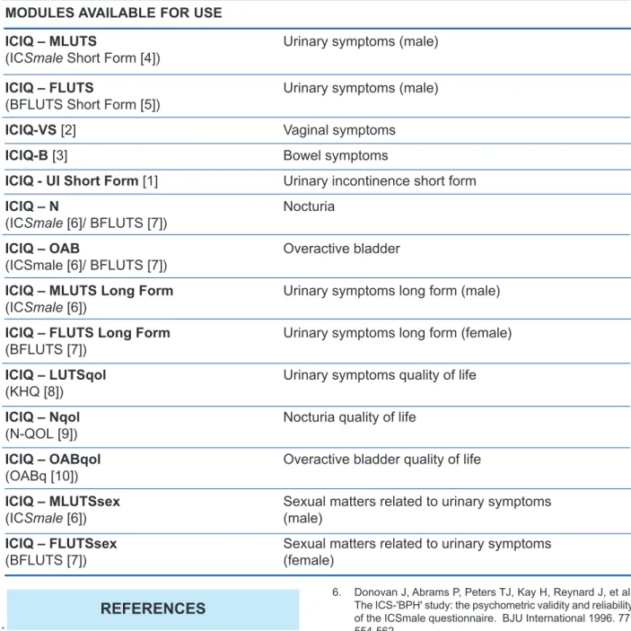
The use of the highest quality questionnaires
(Grade A, where available) is recommended for the assessment of the patient’s perspective of symptoms of incontinence and their impact on quality of life. The ICIQ is highly recommended (Grade A) for the basic evaluation of the patient’s perspective of urinary incontinence; with other Grade A questionnaires
recommended for more detailed assessment. Further
development is required in the areasof pelvic organ
prolapse, bladder pain syndrome and faecal incon-tinence, and for specific patient groups, as only Grade B questionnaires are currently available (see Appendix).
Standard biochemical tests for renal function are
recommended in patients with urinary incontinence and
a probability of renal impairment.
Uroflowmetry with the measurement of postvoid
residual urine is recommended as a screening test for symptoms suggestive of urinary voiding dysfunction or physical signs of POP or bladder distension.
In patients with suspected voiding dysfunction,
PVR should be part of the initial assessment if the result is likely to influence management, for example, in neurological patients.
Although routine imaging is not recommended,
imaging of the lower urinary tract and pelvis ishighly
recommended in those with urinary symptoms
whose initial evaluation indicates a possible co-existing lower tract or pelvic pathology. Initial imaging may be by ultrasound, or plain X ray.
Imaging of the upper urinary tract is highly recom-mended in specific situations. These include:
• Haematuria
• Neurogenic urinary incontinence e.g.
myelodys-plasia, spinal cord trauma,
• Incontinence associated with significant post-void
residual,
• Co-existing loin/kidney pain,
• Severe pelvic organ prolapse, not being treated
• Suspected extra-urethral urinary incontinence,
• Children with incontinence and UTIs, where
indicated
• Urodynamic studies which show evidence of poor
bladder compliance.
In anorectal conditionsanal US or MRI prior to anal
sphincter surgery is highly recommended, when obvious anatomic defects are not evident (cloacal formations). Defaecating proctography or dynamic MRI is recommended in suspected rectal prolapse which cannot be adequately confirmed by physical examination.
Although routine cystourethroscopy is not
recom-mended, LUT endoscopy is highly recommended:
• When initial testing suggest other pathologies, e.g.
haematuria
• When pain or discomfort features in the patient’s
LUTS : these may suggest an intravesical lesion
• When appropriate in the evaluation of vesicovaginal
fistula and extra-urethral urinary incontinence (in childbirth fistulae, endoscopy is often unnecessary).
In anorectal conditions, proctoscopy or flexible
sigmoidoscopy should routinely be performed in the evaluation of patients with faecal incontinence. Colonoscopy, air contrast barium enema or CT colography is highly recommended in the presence of unexplained change in bowel habit, rectal bleeding or other alarm symptoms or signs (see Basic Assessment chapter).
• When the results may change management, such
as prior to most invasive treatments for UI and POP
• After treatment failure, if more information is
needed in order to plan further therapy.
• As part of both initial and long-term surveillance
programmes in some types of neurogenic lower urinary tract dysfunction
• In “complicated incontinence” (for details please
see relevant subcommittee reports).
a) Urodynamic evaluation is recommended
7. Urodynamic Testing
6. Endoscopy
5. Imaging
4. Estimation of Post Void Residual
Urine (PVR)
3. Uroflowmetry
• To reproduce the patient’s symptoms and correlate these with urodynamic findings
• The assessment of bladder sensation
• The detection of detrusor overactivity
• The assessment of urethral competence during
filling
• The determination of detrusor function during
voiding
• The assessment of outlet function during voiding
• The measurement of residual urine
These tests are recommended in the presence of unexplained diarrhoea or when Crohn’s disease is suspected.
Video-urodynamics may be useful in the
management of UI in children, in patients who fail surgery and in some neurogenic patients, to obtain additional anatomical information. Both US and X-ray imaging can be used.
If a more detailed estimate of urethral functionis
required, then the following optional tests may give useful information:
• Urethral pressure profilometry
• Abdominal leak point pressures
• Video-urodynamics
• Electromyography
If initial urodynamics have failedto demonstrate the
cause for the patient’s incontinence then the following tests are optional:
• repeated routine urodynamics
• ambulatory urodynamics
Pad testing is an optional test for the routine evaluation of urinary incontinence and, if carried out, a 24 hr test is suggested.
Theinformationgained by clinical examination and
urodynamic testing may be enhanced by
neuro-physiological testingof striated muscle and nervous
pathways.
Appropriately trained personnel should perform these tests. The following neurophysiological tests can be considered in patients with peripheral lesions prior to treatment for lower urinary tract or anorectal dysfunction.
• Concentric needle EMG
• Sacral reflex responses to electrical stimulation of
penile or clitoral nerves.
Pudendal nerve latency testing is not
recom-mended.
Further imaging of the central nervous system,
including spine, by myelography, CT and MRI may prove useful if simple imaging, for example by spinal X-rays in patients with suspected neurological disease, proves normal.
Cysto-urethrography, US, CTand MRI may have an
indication in case of :
• Suspected pelvic floor dysfunction
• Failed surgery, such as recurrent posterior vaginal
wall prolapse or failed sling surgery
• Suspected fixed urethra
This is anoptional testin patients with complicated
or recurrent UI (e.g. after failed SUI surgery)
Anal manometryis useful to assess resting and
squeeze anal pressures.
6. Anorectal physiology testing
5. Cysto-urethroscopy
4. Further Imaging
3. Neurophysiological Testing and
Imaging
2. Pad Testing
1. Additional Urodynamic Testing
III. OPTIONAL DIAGNOSTIC TESTS
8. Small bowel follow-through, CT
entography or capsule endoscopy.
The management recommendations are derived from the detailed work in the committee reports
on the management of incontinence in children, men, women, the frail elderly and neurological patients, obstetric fistula, pelvic organ prolapse, bladder pain syndrome, and faecal incontinence. The management of incontinence is presented in
algorithm form with accompanying notes.
The Consultation recognised that no algorithm can be applied to every patient and each patient’s management must be individualised. There are algorithms for
• I. Urinary Incontinence in Children • II. Urinary Incontinence in Men • III. Urinary Incontinence in Women • IV. Obstetric Fistulae
• V Urinary Incontinence in Frail Older Men and Women
• VI. Urinary Incontinence in Neurological Patients
• VII Bladder Pain Syndrome • VIII. Pelvic Organ Prolapse • IX. Faecal Incontinence in
Non-Neurological Patients
• X. Faecal Incontinence in Neurological Patients
These algorithms are divided into two for groups I to III, IX, XI and X : the two parts, initial
management and specialised management
require a little further explanation.
Although the management algorithms are designed to be used for patients whose predominant problem is incontinence, there are many other patients in whom the algorithms may be useful such as those patients with urgency and frequency, so-called
“OAB dry”.
are intended for use by all clinicians including health care assistants, nurses, physiotherapists, generalist doctors and family doctors as well as by specialists such as urologists and gynaecologists. The consultation has attempted to phrase the
recommendations in the basic algorithms in such a way that they may be readily used by clinicians in all countries of the world, both in the developing and the developed world.
are intended for use byspecialists. The specialised algorithms, as well as the initial management algorithms arebased on evidence where possible
and on the expert opinion of the 700 healthcare professionals who took part in the Consultation. In this consultation, committees ascribed levels of evidence to the published work on the subject and devised grades of recommendation to inform patient management.
It should be noted that these algorithms, dated
March 2009, represent the Consultation
con-sensus at that time. Our knowledge, developing
from both a research base and because of evolving expert opinion, will inevitablychange with time.
The Consultation does not wish those using the algorithms to believe they are “carved in tablets of stone”: there will be changes both in the relatively short term and the long term.
Each algorithm contains a core of recommendations in addition to a number of essential components of basic assessment listed in sections I to III.
The patient’s desires and goals for treatment :
Treatment is a matter for discussion and joint decision making between the patient and his or her health care advisors. This process of
◆
Joint decision making
• General assessment
• Symptom assessment
• Assessment of quality of life impact
• Assessment of the desire for treatment
• Physical examination
• Urinalysis
◆
Essential components of basic
assessment
➦
The
specialised
algorithms
➦
The algorithms for
initial
management
3. Management
Recommendations
consultation includes the specific need to assess whether or not the sufferer of incontinence wishes to receive treatment and, if so, what treatments he or she would favour. Implicit in this statement is the assumption that the health care provider will give anappropriate explanation of the patient’s
problem and the alternative lines of
management,and the indications and the risks
of treatment.The assumption that patients almost
always wish to have treatment is flawed, and the need to consult the patient is paramount.
In each algorithm,treatments are listed in order
of simplicity, the least invasive being listed first.
This order does not imply a scale of efficacy or cost, two factors which need to be considered in choosing the sequence of therapy. The order is likewise not meant to imply a suggested sequence of therapy, which is determined jointly by the treating health care providers and the patient, considering all the relevant factors listed above.
In theinitial management algorithms, treatment
is empirically based, whilst, the specialized
managementalgorithms usually rely on precise
diagnosis from urodynamics and other testing. The assumption is made that patients will be reassessed at an appropriate time to evaluate their progress.
The possible role of continence productsshould be considered at each stage of patient assessment, treatment and, if treatment is not (fully) successful, subsequent management.
• Firstly, intermittent catheterisation or indwelling
catheter drainage often have a role to play in addressing urinary retention .
• Secondly, assisted toileting using such devices
as commodes, bedpans, and handheld urinals may help to achieve dependent continence*
where access, mobility and / or urgency problems under-mine a patient’s ability to maintain independent continence*, be it urinary and / or faecal.
• Finally, containment products (to achieve
contained incontinence*) for urine and / or
faeces find an essential role in enhancing the quality of life of those who:
• Elect not to pursue treatment options
• Are awaiting treatment
• Are waiting for treatment to take effect
• Are unable to be (fully) cured
Further guidance and care algorithms on which products might be suitable for a given patient are given in Committee 20.
* Useful terms suggested by Fonda & Abrams.(Cure sometimes, help always – a “continence paradigm” for all ages and conditions. Fonda D and Abrams P, Neurology & Urodynamics 25: 290-292, 2006).
A.
I
NITIAL
MANAGEMENT
I. CHILDREN
Children produce specific management problems for a variety of reasons:
assessment requires help from their parents and carers; consent to
treatment may be problematic; and cooperation in both assessment
and treatment may be dif
ficult.
➦
Referrals for specialist treatment are recommended for a group
of children who have complicated incontinence associated with:
•
recurrent urinary infection
•
voiding symptoms or evidence of poor bladder emptying
•
urinary tract anomalies,
•
previous pelvic surgery
•
neuropathy
➦
Initial treatment is recommended for the remaining patients who
have:
•
Nocturnal enuresis without other symptoms (mono-symptomatic
enuresis).
•
Daytime symptoms of frequency
, urgency
, urgency incontinence with
or without night-time wetting
➦
Initial treatment for
mono-symptomatic nocturnal
enuresis should
include:
-
parental counselling and motivation
- a choice between either alarm (Grade
A) and anti-diuretic hormone
analogues desmopressin (Grade
A)*. It may be a parental choice if
advantages and disadvantages are well explained.
➦
Daytime incontinence
should be managed holistically including:
-
counselling, bladder training (timed voiding), behaviour modification
and bowel management when necessary (Grade B).
-
antimuscarinics may be used if there are symptoms that suggest
detrusor overactivity (Grade C).
*
A
regulatory warning exists for the danger of overhydration in children
on desmopressin.
Should initial treatment be unsuccessful for either enuresis
or daytime symptoms, then after a reasonable period of time
(8-12 weeks), referral for a specialist’
s advice is highly
recommended.
2.
T
reatment
1. Initial assessement should involve a detailed
HIST
OR
Y/
SYMPT
OM
ASSESSMENT
CLINICAL
ASSESSMENT
TREA
TMENT
Initial Management of Urinary Incontinence in Children
PRESUMED
DIAGNOSIS
• Explanation/education
• Enuresis Diary
• Alarm
• Desmopressin
MONOSYMPT
OMA
TIC
NOCTURNAL
ENURESIS
“Complicated” Incontinence
associated with:
- Urinary tract anomaly
- Neuropathy
- Pelvic surgery
- V
oiding (emptying) symptoms
- Recurrent urinary injection
SPECIALIZED MANAGEMENT
Failure
Failure
Any other
abnormality
detected
e.g. Post
void
residual
General assessment (see relevant chapter)
• Physical examination: abdominal, perineal, ext. genitalia, back/spine, neurological
•
Assess bowel function -> if constipated, treat and reassess
• Urinalysis ± Urine culture -> if infected, treat and reassess
•
Assess post-void residual urine by abdominal examination
(optional : by ultrasound)
Nocturnal enuresis
(monosymptomatic)
Daytime ± Nighttime wetting
± Urgency / frequency
URGENCY
INCONTINENCE
RECURRENT
INFECTION
DYSFUNCTIONAL
VOIDING
B.
S
PECIALIZED
MANAGEMENT
I. CHILDREN
➦ T wo groups of children with “ complicated” incontinence should havespecialist management from the outset.
•
Children whose incontinence is due to, or associated with,
urinary tract anomalies and neuropathy . • Children
without urinary tract anomalies, but with
recurrent infection and, proven or suspected, voiding dysfunction. ➦ Children who
failed the basic treatment
, but who have neither neurogenic
nor anatomical problems, should also receive specialist management:
➦
As part of further assessment, the measurement of
urine flow
(in children
old enough), together with the
ultrasound estimate of residual urine
and
the
upper urinary tracts
is
highly recommended.
Those who
fail treatment
and have neither neurogenic nor anatomical problems
should be reassessed
using micturition charts, symptom scores, urinalysis,
Uroflowmetry and residual urine determination. If there are recurrent infections
, upper tract imaging and possibly a VCUG should
be considered. However
, endoscopy is rarely indicated.
➦
Urodynamics should be considered:
•
If the type and severity of lower tract dysfunction
cannot be explained by
clinical findings
•
If
invasive treatment
is under consideration, for example, stress incontinence
surgery or bladder augmentation, when there is sphincteric incompetence, or if there is detrusor overactivity
.
•
If
upper tract dilatation exists
and is thought to be due to bladder dysfunction.
Urodynamic studies are not recommended
if the child has normal upper tract
imaging and is to be treated by non invasive means. ➦
Spinal Imaging
(US/Xray/MRI) may be needed if a bony abnormality or
neurological condition is suspected.
The treatment of incontinence associated with
urinary tract anomalies
is
complex and cannot easily be dealt with in an algorithm. In many children, more than one pathophysiology
demands treatment. If there are
complex
congenital abnormalities present,
the treatment is mostly surgical and it
should be individualised according to the type and severity of the problem (please see Children’
s Committee Report).
Care should be given by specialist children’
s nurses and therapists.
➦
Initial treatment should be non-surgical.
•
For stress urinary incontinence
(SUI): pelvic floor muscle training (Grade
C)
•
For suspected detrusor overactivity
(DO): bladder training and
antimuscarinics (Grade C)
•
For voiding dysfunction
: timed voiding, biofeedback to teach pelvic floor
relaxation, intermittent catheterisation (when PVR > 30% of bladder capacity). (Grade B/C)
•
For bowel dysfunction:
as appropriate
The
child’
s progress
should be assessed and, if quality of life is still significantly
impaired, or if the upper urinary tracts are at risk,
surgical treatment
is likely
to be necessary
.
➦
If surgical treatment is required,
then urodynamics is recommended to
confirm the diagnosis.
•
For SUI,
sling surgery
, bulking agent injection and
AUS may be considered
•
For DO / poor compliance,
botulinum toxin
*
and bladder augmentation may
be performed.
•
If the child cannot do IC
then a Mitrofanof
f channel may be needed.
*
At the time of writing, botulinum toxin is being used “off label” for refractory DO and must be used with caution.
2. T
reatment
Specialized Management of Urinary Incontinence in Children
STRESS URINAR
Y
INCONTINENCE
VOIDING
DYSFUNCTION
DETRUSOR OVERACTIVITY
/
POOR COMPLIANCE
ANA
T
OMIC CAUSES
OF URINAR
Y
INCONTINENCE
• Pelvic floor and
muscle training
• AUS
• Sling
• Bulking agent
injection
• Bladder training
• Antimuscarincs
• Bowel management.
• Botulinum toxin
• Bladder augmentation
Mitrofanoff if IC fails
• Correct anomaly
(see : surgical
treatment in children)
EXPERT
HIST
OR
Y
&
PHYSI-CAL
EXAMINA
TION
CLINICAL
ASSESSMENT
TREA
TMENT
DIAGNOSIS
• Urinalysis: if UTI, treat and reassess
as appropriate
• T
reat bowel dysfunction and reassess
• Renal / bladder ultrasound
•
Assess Post void residual
• Flow rates ± electromyography
• Behavioral Evaluation
if abnormal -->
}
Consider:
• Micturating cystogram
• Renal scintigram
• Urodynamics
• Cystourethroscopy
• Spinal imaging
Incontinence without
suspicion of urinary tract anomaly
Incontinence with suspicion
of urinary tract anomaly
Failure
Failure
Failure
• T
imed voiding
• Pelvic floor relaxation ±
biofeedback.
• Pharmacotherapy
- Antimuscarinics
-
αα
- blockers
A. I
NITIAL
MANAGEMENT
II. MEN
➦
Complicated” incontinence group
Those with pain or with haematuria, recurrent infections, suspected or
proven poor bladder emptying (for example due to bladder outlet
obstruction), or incontinence following pelvic irradiation, are
recommended for
specialized management.
Poor bladder emptying
may be suspected from symptoms, physical
examination or if imaging has been performed by X-ray or ultrasound
after voiding.
➦
Four other main groups
of men should be identified by initial
assessment as being suitable fo
r initial management.
•
Those with
post-micturition dribble
alone,
•
Those with
overactive bladder
(OAB) symptoms: urgency with or
without urgency incontinence, together with frequency and nocturia
•
Those with
stress incontinence
(most often post-prostatectomy),
and
•
Those with
mixed
urgency and stress incontinence (most often
post-prostatectomy)
➦
For men with
post-micturition dribble,
this requires no assessment
and can usually be treated by teaching the man how to do a strong
pelvic floor muscle contraction after voiding, or manual compression
of the bulbous urethra directly after micturition. (Grade B)
➦
For men with
stress, urgency
or
mixed
urgency / stress incontinence,
initial treatment should include appropriate lilfestyle advice, physical
therapies, scheduled voiding regimes, behavioural therapies and
medication. In particular:
•
Lifestyle interventions (Grade D)
•
Supervised pelvic floor muscle training for men with post radical
prostatectomy SUI (Grade B)
•
Scheduled voiding regimes for OAB (Grade C)
•
Antimuscarinic drugs for OAB symptoms with or without urgency
incontinence (Grade B) and the patient has no evidence of significant
post-void residual urine
•
α
-adrenergic antagonists (a-blockers), can be added if it is thought
that there may also be bladder outlet obstruction. (Grade C)
➦
Should initial treatment be unsuccessful
after a reasonable period
of time (for example, 8-12 weeks),
specialist advice
is highly
recommended.
Clinicians are likely to wish to treat the
most bothersome symptom
first in men with symptoms of
mixed
incontinence.
2. Management
1. Initial
Initial Management of Urinary Incontinence in Men
• General assessment (see relevant chapter)
• Urinary Symptom
Assessment and symptom score
(including frequency-volume chart and questionnaire)
•
Assess quality of life and desire for treatment
• Physical examination: abdominal, rectal, sacral, neurological
• Urinalysis ± urine culture -> if infected, treat and reassess
•
Assessment of pelvic floor muscle function
•
Assess post-void residual urine
“Complicated” incontinence • Recurrent or “total” incontinence • Incontinence associated with: - Pain - Hematuria - Recurrent infection - Prostate irradiation - Radical pelvic surgery
MIXED INCONTINENCE
(treat most bothersome
symptom first)
URGENCY
INCONTINENCE
presumed due to
detrusor overactivity
• Urethral
milking
• Pelvic floor
muscle
contraction
SPECIALIZED MANAGEMENT
Any other abnormality
detected e.g. significant
post void residual
STRESS INCONTINENCE
presumed due to
sphincteric
incompetence
DISCUSS TREA
TMENT OPTIONS WITH THE P
A
TIENT
• Lifestyle interventions
• Pelvic floor muscle training ± biofeedback
• Scheduled voiding (bladder training)
• Incontinence products
•
Antimuscarinics (OAB ± urgency incontinence) and
αα
-adrenergic antagonists (if also bladder outlet obstruction)
HIST
OR
Y
CLINICAL
ASSESSMENT
PRESUMED
DIAGNOSIS
MANAGEMENT
Post-micturition
dribble
Incontinence on
physical activity
(usually post-
prostatectomy)
Incontinence
with mixed
symptoms
Urgency /
frequency
,
with or with-
out urgency
incontinence
The specialist may first
reinstitute initial management
if it is felt
that previous therapy had been inadequate.
➦
Patients with “
complicated” incontinence
referred directly to
specialized management, are likely to require
additional testing,
cytology
, cystourethoscopy and urinary tract imaging.
If these tests prove normal
then those individuals can be treated
for incontinence by the initial or specialized management options as
appropriate.
If symptoms
suggestive of detrusor overactivity
, or of sphincter
incompetence
persist,
then
urodynamic
studies are recommended
in order to arrive at a precise diagnosis, prior to invasive treatment.
and if the patient’
s incontinence markedly disrupts his quality of life
then
invasive therapies
should be considered.
➦
For sphincter incompetence
the recommended option is the
artificial urinary sphincter (Grade B). other options, such as a
male sling, may be considered (Grade C).
➦
For idiopathic detrusor overactivity
, (with intractible overactive
bladder symptoms) the recommended therapies are bladder
augmentation (Grade C) and neuromodulation (Grade B).
Botulinum toxin continues to show promise in the treatment of
symptomatic detrusor overactivity unresponsive to other therapies
*
➦
When incontinence has been shown to be associated with
poor
bladder emptying
and
detrusor underactivity
,
it is recommended
that ef
fective means are used to ensure bladder emptying, for
example, intermittent catheterisation (Grade B/C).
➦
If incontinence is associated with bladder outlet obstruction
,
then consideration should be given to surgical treatment to relieve
obstruction (Grade B).
α
-blockers and/or 5
α
- reductase inhibitors
would be an optional treatment (Grade C).
There is increased
evidence for the safety of antimuscarinics for overactive bladder
symptoms in men, chiefly in combination with an
α
-blocker (Grade
B).
*
Note:
At the time of writing, botulinum toxin is being used “off-label”.
When basic management has failed
2.
T
reatment
1. Assessment
B.
S
PECIALIZED
MANAGEMENT
II. MEN
Specialized Management of Urinary Incontinence in Men
“Complicated” Incontinence :
• Recurrent incontinence
• Incontinence associated with:
- Prostate or pelvic irradiation
- Radical pelvic surgery
with coexisting
bladder outlet
obstruction
with coexisting
underactive
detrusor
(during voiding)
Lower urinary
tract anomaly/
pathology
STRESS
INCONTINENCE
due to sphincteric
incompetence
MIXED
INCONTINENCE
T
reat major
component first
URGENCY
INCONTINENCE
due to detrusor
overactivity (during filling)
If initial therapy
fails:
• Artificial
urinary sphincter
• Male sling
(see chapter)
If initial therapy
fails:
• Neuromodulation
•
αα
-blockers, 5ARI
• Correct anatomic
bladder outlet
obstruction
• Antimuscarinics
(See note)
• Correct anomaly
• T
reat pathology
• Intermittent
catheteri-
sation
• Antimus-
carinics
HIST
OR
Y/
SYMPT
OM
ASSESSMENT
CLINICAL
ASSESSMENT
TREA
TMENT
DIAGNOSIS
Post-prostatectomy
incontinence
Incontinence with
urgency / frequency
• Consider urodynamics and imaging of the urinary tract
• Urethrocystoscopy (if indicated)
➦
“Complicated” incontinence group.
Those with pain or haematuria, recurrent infections, suspected or
proven voiding problems, significant pelvic organ prolapse or who
have persistent incontinence or recurrent incontinence after pelvic
irradiation, radical pelvic surgery
, previous incontinence surgery
, or
who have a suspected fistula, for specialist referral.
➦
Three other main groups
of patients should be identified by
initial assessment.
•
W
omen with
stress incontinence
on physical activity
•
W
omen with
urgency
, frequency
with or without urgency
incontinence
•
Those women with
mixed
urgency and stress incontinence
Abdominal, pelvic and perineal examinations should be a routine part
of physical examination. W
omen should be asked to perform a
“stress test” (cough and strain to detect leakage likely to be due to
sphincter incompetence).
Any pelvic organ prolapse or uro-genital
atrophy should be assessed. V
aginal or rectal examination allows
the assessment of voluntary pelvic floor muscle function, an important
step prior to the teaching of pelvic floor muscle training.
➦
For women with
stress, urgency or mixed
urinary incontinence,
initial treatment should include appropriate lifestyle advice, physical
therapies, scheduled voiding regimes, behavioural therapies and
medication. In particular:
•
Advice
on caf
feine reduction (Grade B) and weight reduction
(Grade A)
•
Supervised pelvic floor muscle training
(Grade A),
vaginal
cones
for women with stress incontinence (Grade B)
•
Supervised
bladder training
(Grade
A) for OAB.
•
If oestrogen deficiency
and/or
UTI
is found, the patient
should be treated at initial assessment and then reassessed
after a suitable interval. (Grade B).
•
Antimuscarinics
for OAB symptoms with or without urgency
incontinence (Grade
A); duloxetine
*
may be considered for
stress urinary incontinence (Grade B)
Initial treatment should be maintained for
8-12 weeks
before
reassessment
and possible specialist referral for further management
if the patient has had insuf
ficient improvement.
Clinicians are likely to wish to treat the
most bothersome symptom
first
in women with symptoms of mixed incontinence. (Grade C).
➦
Some women with significant
pelvic organ prolapse
can be
treated by vaginal devices that treat both incontinence and prolapse
(incontinence rings and dishes).
*
Duloxetine is not approved for use in United States. It is approved for use
in Europe for severe stress incontinence (see committee report on pharmacological management for information regarding efficacy
, adverse
events, and 'black box' warning by the Food and Drug
Administration of the United States).
2.
T
reatment
1.
Initial assessment should identify :
A. I
NITIAL
MANAGEMENT
III.
Incontinence
on physical
activity
Incontinence
with mixed
symptoms
Incontinence /
frequency with
urgency
Initial Management of Urinary Incontinence in W
omen
MIXED INCONTINENCE (treat most bothersome
symptom first)
Complicated incontinence
• Recurrent incontinence
• Incontinence associated
with:
- Pain
- Hematuria
- Recurrent infection
- Significant voiding
symptoms
- Pelvic irradiation
- Radical pelvic surgery
- Suspected fistula
SPECIALIZED MANAGEMENT
• If other abnormality
found e.g.
• Significant post
void residual
• Significant pelvic
organ prolapse
• Pelvic mass
Failure
STRESS INCONTINENCE
presumed due to
sphincteric incompetence
OAB -with or without
URGENCY
INCONTINENCE
presumed due to
detrusor overactivity
• Life style interventions.
• Pelvic floor muscle training for SUI or OAB
• Bladder retraining for OAB
• Duloxetine* (SUI) or antimuscarinic (OAB ± urgency incontinence)
•
Other adjuncts, such as electrical stimulation
•
V
aginal devices, urethral inserts
*
Subject to local regulatory approval (see black box worning).
HIST
OR
Y
CLINICAL
ASSESSMENT
PRESUMED
DIAGNOSIS
MANAGEMENT
•
General assessment (see relevant chapter)
•
Urinary symptom assessment (including frequency-
volume chart and questionnaire)
•
Assess quality of life and desire for treatment
•
Physical examination: abdominal, pelvic and perineal
•
Cough test to demonstrate stress incontinence if appropriate
•
Urinalysis ± urine culture -> if infected, treat and reassess
If appropriate
•
Assess oestrogen status and treat as appropriate
•
Assess voluntary pelvic floor muscle contraction
•
B.
S
PECIALIZED
MANAGEMENT
III.
WOMEN
W
omen who have “
complicated” incontinence
(see initial algorithm)
may need to have additional tests such as cytology
, cystourethroscopy
or urinary tract imaging. If these tests are normal then they should
be treated for incontinence by the initial or specialized management
options as appropriate.
➦
Those women who have
failed initial management
and whose
quality of life is impaired are likely to request further treatment.
If initial management has been given an adequate trial then
interventional therapy may be desirable.
Prior to intervention
urodynamic testing
is highly recommended, when the results may
change management. It is used to diagnose the type of
incontinence and therefore inform the management plan. Within
the urodynamic investigation urethral function testing by urethral
pressure profile or leak point pressure is optional.
➦
Systematic assessment for
pelvic organ prolapse
is highly
recommended and it is suggested that the POPQ method should
be used in research studies. W
omen with co-existing pelvic organ
prolapse should have their prolapse treated as appropriate
➦
If urodynamic stress incontinence is confirmed
then the
treatment options that are recommended for patients with
some
degree of bladder-neck and urethral mobility
include the full
range of non-surgical treatments, as well as retropubic suspension
procedures, (Grade
A) and bladder neck/sub-urethral sling
operations:(Grade A).
The
correction of symptomatic
pelvic
organ prolapse may be desirable at the same time.
For patients with
limited bladder neck mobility
,
bladder neck sling
procedures, (Grade
A) injectable bulking agents (Grade B) and the
artificial urinary sphincter (Grade B) can be considered.
➦
Urgency incontinence
(overactive bladder) secondary to
idiopathic detrusor overactivity may be treated by neuromodulation
(Grade
A) or bladder augmentation (Grade C). Botulinum toxin can
be used in the treatment of symptomatic detrusor overactivity
unresponsive to other therapies (Grade C).*
➦
Those patients with
voiding dysfunction
leading to significant
post-void residual urine (for example, >30% of total bladder
capacity) may have bladder outlet obstruction or detrusor
underactivity
. Prolapse is a common cause of voiding dysfunction.
*
At the time of writing, botulinum toxin is being used “off-label” and
with caution.
2.
T
reatment
1.
Specialized Management of Urinary Incontinence in W
omen
Bladder outlet
obstruction
Underactive
detrusor
Lower urinary
tract anomaly /
pathology
URODYNAMIC
STRESS
INCONTINENCE (USI)
MIXED
INCONTINENCE
(USI/DOI)
(Treat. most
bother-some symptom first)
DETRUSOR
OVERACTIVITY
INCONTINENCE
(DOI)
INCONTINENCE
associated with
poor bladder
emptying
Incontinence
on physical
activity
Incontinence
with mixed
symptoms
Incontinence
with urgency /
frequency
“Complicated” incontinence:
• Recurrent incontinence
• Incontinence associated
with:
- Pain
- Hematuria
- Recurrent infection
- V
oiding symptoms
- Pelvic irradiation
- Radical pelvic surgery
- Suspected fistula
If initial therapy fails :
• Stress incontinence
surgery
- bulking agents
- tapes and slings
- colposuspension
If initial therapy fails :
• Botulinum toxin
• Neuromodulation
• Bladder augmentation
• Correct anatomic
bladder outlet
obstruction (e.g.
genito-urinary
prolapse)
• Intermittent
catheterization
• Correct anomaly
• T
reat pathology
Consider:
• Urethrocystoscopy
• Further imaging
• Urodynamics
HIST
OR
Y/
SYMPT
OM
ASSESSMENT
CLINICAL
ASSESSMENT
TREA
TMENT
DIAGNOSIS
•
Assess for pelvic organ mobility / prolapse
• Obstructed labour is the main cause of vesicovaginal fistula in the developing world. The obstructed labour complex not only induces the vesicovaginal fistula and fetal death in most cases, but can also have urological, gynaecological, neurological, gastro-intestinal, musculoskeletal, dermatological and social consequences. • Other aetiologiessuch as sexual violence or
genital mutilation are less frequent. For these causes the general principles listed here should be adapted according to the patient’s need.
• Patients with vesicovaginal fistulashould be treated as a person, and they deserve the right to adequate counseling and consent to the treatment they will eventually undergo, despite language and cultural barriers that may exist.
• Surgeons embarking on fistula surgery in the developing world should have appropriate training in that setting and should be willing to take on a long-term commitment.
• Prevention of fistula is the ultimate goal. Collaboration between fistula initiatives and maternal health initiatives must be stimulated.
It is important to make a distinction between simple
fistulae, which have a good prognosis, and complex
fistulae, which have a less favourable outcome.
Careful clinical examination will allow the type of
fistula to be determined, although no generally
accepted classification systemis available. Key
items are the size and location of the fistula, the
extent of the involvement of the urethra and the urethral closure mechanism, and the amount of vaginal scarring.
Associated pathologiesshould be actively searched
for and should be taken into account in the treatment plan: all components of the ‘obstructed labour injury complex’ should be examined and defined.
The treatment for vesicovaginal fistula issurgical.
(Grade A)
Avaginal approachis preferred, since most simple
fistula can be reached vaginally and since spinal anaesthesia carries less risk than general anaesthesia needed for an abdominal approach. A trained surgeon should be able to manage these simple fistula. After wide dissection a tension-free single layer closure
of the bladder wall and closure of the vaginal wall in
a separate layer are advocated. A Martius flap in primary simple obstetric fistula repair is optional.
Acare program for failed repairs and for persisting
incontinence after a successful repair needs to be installed.
Complex fistulae should be referred to a fistula expert
in a fistula centre. (Grade B)
In principle, most complex fistulae can be dealt with by the vaginal approach, but an abdominal approach may be needed in some cases ( e.g. concommittant reconstructive procedures). Advanced training and surgical skills are prerequisites for treating this type of fistula.
If the urethraand/or the urethral closure mechanism
is involved, a sling procedure, using an autologous
sling, should be performed at the same time as the
fistula correction. There is no place for synthetic
sling material in this setting. (Grade B)
The majority of patientswith a simple fistula will be
curedafter the repair. However, a proportion of them,
and an even larger proportion of the patients after complex fistula repair, will remain incontinent.
Depending on the local possibilities an after care
programshould be installed.
➦ After care ➦ Complex fistula ➦ Simple fistula