R E V I E W A R T I C L E
Pterygoid implant: Option for rehabilitation of the
atrophic posterior maxilla
P. Venkat Ratna Nag1, P. Sarika2, Tejashree Bhagwatkar3, Vasantha Dhara4
1Department of Prosthodontics, S. B. Patil Dental College and Hospital, Bidar, Director, Institute for Dental Implantology, Hyderabad, Telangana, India, 2Department of Pedodontics and Preventive Dentistry, S. B. Patil Dental College and Hospital, Bidar, Institute for Dental Implantology, Hyderabad, Telangana, India, 3Department of Oral Pathology, Institute for Dental Implantology, Hyderabad, Telangana, India, 4Department of Oral and Maxillofacial Surgery,Institute for Dental Implantology, Hyderabad, Telangana, India
Abstract
Background: The anatomy of the atrophic posterior maxilla presents many limitations to
implant placement. Factors affecting implant placements include poor bone quality and quantity, location of maxillary sinus. Posterior cantilevers on implant prostheses produce complications, such as prosthesis fracture, screw loosening, loss of osseointegration, and crestal bone loss. Pterygoid implants are an alternative to grafting solutions for posterior maxillary rehabilitation.
Aim: This systematic review describes various implant treatment options for posterior maxillary rehabilitation. It highlights the use of pterygoid implants as a graftless solution with its anatomy, technique of placement, and advantages.
Conclusion: Pterygoid implants have high success rates, less bone loss, and good acceptance by patients thus being an excellent alternative to treat patients with severely
atrophic maxilla.
Clinical Significance: Pterygoid implants avoid the need for sinus lifts and grafting procedures. They allow anchorage in the posterior atrophied/resorbed maxilla, achieving proper stability, and high rates of long-term success. In addition, posterior cantilevers can be eliminated and axial loading is improved.
Keywords: Atrophic ridges, bicortical, cantilever, graftless, posterior maxilla, pterygoid implant Correspondence
Dr. P. Venkat Ratna Nag, Director, Institute for Dental Implantology 8-2-598/A/1, GB, Uma Devraj Villa, Road No. 10, Banjara Hills, Hyderabad - 500 034, Telangana, India. Phone: +91-9963511139.
E-mail: [email protected]
Received 08 February 2019; Accepted 30 May 2019
doi: 10.15713/ins.ijcdmr.135
How to cite the article:
Nag PVR, Sarika P, Bhagwatkar T, Dhara V. Pterygoid Implant: Option for Rehabilitation of the Atrophic Posterior Maxilla. Int J Contemp Dent Med Rev vol.2019, Article ID: 011218, 2019. doi: 10.15713/ins.ijcdmr.135
Introduction
Implant dentistry has growing leaps and bounds in recent years after the successful introduction of osseointegration concept by Prof. P.I Branemark in the early 1960s. Rehabilitation of the maxillary anterior region has been far easier than the maxillary posterior region due to various factors.[1] The posterior maxillary region is characterized by (1) inadequate residual bone height due to maxillary sinus expansion and/or alveolar bone resorption and (2) poor bone density (Type III or IV) according to Lekholm and Zarb classification system.[2-4]
Considering these challenges posed by the anatomy, few techniques have been in use such as sinus lift procedures, guided bone regeneration grafting with bone autogenous and allogenous grafts; and later tilted implants (All-on-4), zygomatic implants were introduced.[5] However, these procedures have complications such as sinus membrane perforation, rejection of
graft, graft displacement into sinus cavities, and screw loosening of tilted implants. To prevent such problems posterior-most area of maxillary tuberosity; distal to maxillary sinus can be utilized
for implant placement.[5]
Implants placed in the compact bone of the pterygomaxillary region shows ossteogration and provides retention and stability.[6] This area is pterygoid or pterygomaxillary region. It was introduced by Tulasne (1992).[6] Tulasne (1989) credited Paul Tessier for proposing an idea of placing implants in the pterygoid region. Due to their long path, length of pterygoid implants ranges from 15 mm to 20 mm.[6,7] Pterygoid implants take bicortical anchorage, due to which the axial loading is improved and posterior cantilever is eliminated.[8]
through the maxillary tuberosity into the pterygoid plate” by the glossary of oral and maxillofacial implants. The maxillary tuberosity is defined as “the most distal aspect of the maxillary alveolar process.”[9-11]
Anatomy of pterygoid region
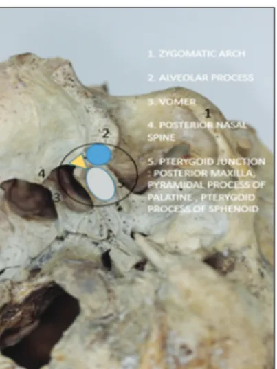
The tuberosity of maxilla is composed of Type III and Type IV cancellous bone. The pyramidal process of palatine and pterygoid process of the sphenoid are mainly composed of dense cortical bone. The pterygoid fossa is bordered by median and lateral pterygoid plates.[12] In Figure 1, the pterygopalatine fossa (PPF) is considered as a key area in the deep space, which needs to evaluated carefully during head and neck imaging. The PPF is confined by the junction of three bones (maxilla, palatine, and sphenoid). Fat, the pterygopalatine ganglion, the maxillary division (V2) of the trigeminal nerve and its branches, the Vidian (pterygoid) nerve, the distal branches of the maxillary artery, and a few emissary veins are the contents of the PPF.[13,14] The thickest buttress of bone is medial to the alveolar ridge. The ideal placement for the implant is through the pterygoid process into the pterygoid fossa.[12]
The thickest area of supporting bone is located in the middle part of the pterygoid process between plates. This 3–4 mm medial to the alveolar ridge, the implant should angle slightly medially to bisect dense point of bone in the pterygoid region. The hamular process on the medial pterygoid plate is palpable in the oropharynx. Implants are placed laterally to this landmark.[12] The pterygoid implants, when used in full arch rehabilitation, eliminate distal cantilevers, an extension of posterior occlusion, and the finest distribution of functional loads.[15-18]
Classification of the posterior maxillary implant-based on anatomic location by Reiser[11]
• Tuberosity – pyramidal process • Tuberosity – pterygoid process
• Tuberosity – pyramidal process – pterygoid process • Pyramidal process – pterygoid process
• Maxillary tuberosity.
PARP (pterygoid anatomic radiographic prediction)
Luis et al. proposed the classification of diagnostic prediction PARP for implantology in the pterygomaxillary region. Through the PARP, the choice of implant is individualized for each patient, acting as a guide to make implantology accessible in the pterygomaxillary region to the greatest number of specialists. From the degree of sinus invasion obtained after a three-dimensional (3D) computerized tomography (CT), the PARP establishes the prediction of the difficulty implied by implantology in this anatomical region, as well as the appropriate choice of the type of implant and length with which
to approach it.
The PARP classification allows working only in the pterygomaxillary region with retromolar implants[19] [Table 1]. • PARP 1. It is the simplest scenario when there is no sinus
invasion and we have a bone in all its route. In these cases, the length of the implant depends on the bone density.
• PARP 2. The patient presents with a sinus invasion but still has >10 mm of the remaining bone. In case of having good bone density, it would be more appropriate to place a conventionally conceptualized retromolar implant.
• PARP 3. This is a case of medium-high difficulty, with sinus invasion leaving a bone surface between 5 mm and 9 mm of remaining bone. In these cases, due to the scarce remnant of alveolar bone and the air of the sinus invasion, the pterygoid anchor will always be used in the apophasis of the same name, with a suitable density.
• PARP 4. In the majority of cases of a large sinus invasion, leaving only a remaining bone smaller than 5 mm, the possibility of using long pterygoid implants or opting for other surgical approaches will be evaluated.
Discussion
Treatment options for posterior maxillary rehabilitation 1. Maxillary sinus floor elevation.
2. Zygomatic implants. 3. Short implants. 4. Tilted implants.
Maxillary sinus floor elevation
The reduced vertical bone height in the posterior maxillary region is often a major obstacle to the placement of dental implants. Elevation of the maxillary sinus floor with or without grafting is the only solution for this problem. Various surgical techniques such as endoscopically controlled technique,[20] hydraulic pressure technique,[21] and antral membrane balloon elevation technique[22] have been presented to access the sinus cavity and elevate the sinus membrane.[23,24]
Zygomatic implants
The technique sensitive zygoma implants are indicated for severely resorbed maxilla this engages zygomatic bone for anchorage.[8] These implants are screw-shaped in commercially pure titanium of variable lengths of 30– 52.5 mm.[25] The concept can be expanded when required by inserting two zygomatic implants in a more anterior position (quad zygoma).
Short implants
The short implant is considered as an alternative in a situation which is characterized by limited vertical bone height. It used to avoid bone augmentation procedures in maxillary and mandibular posterior regions. Standard implants have a length of approximately >8 mm, while short implants are usually referred designed with intrabony lengths of ≤8 mm.[8]
Tilted implants
Since the 19th century, tilted concept in the posterior region of the maxilla was demonstrated as one of the alternatives to bone grafting. Using tilted implants, distribution of axial, shear, and transverse forces would not be harmful due to greater anterior-posterior coverage of the design, which has been proven by 3D finite element analysis of stress levels.[26,27] Tilting of the implants reduces the cantilever length by increasing the inter-implant distance and decreasing compressive stress. Multiple studies have suggested the use of tilted implants for maxillary rehabilitation using immediate loading.[26,27]
Protocol for pterygoid implant placement
A. Diagnostic level
Preclinical record:
a. Clinical assessment summary/relevant medical history: b. Pretreatment Photographs: Extraoral: Frontal, lateral,
oblique
Intraoral: Frontal, right, left, upper, and lower occlusal
View
c. Radiographs: OPG, CBCT, RVG.
B. Surgical level
Presurgical stereolithography model
Pterygoid implants are suited to all age groups and systemic conditions unless there is a frank surgical contraindication. Patients with diabetes Type 2 (HbA1c <7 %) are suitable for
pterygoid implant. To take clinical advantage of pterygoid implants surgical guides and stereolith models are necessary, fabrication of which is done by conversion of patients CT scan images (Dicom) to STL format. Virtual planning of implant placement and clinical angle measurement should be done. Surgical metal template is fabricated with markings of point of entry and drilling angulation as per the planned sites. The use of a surgical guide avoids perforations into adjacent anatomical sites (palatal or buccal). Models help to identify the patient-specific anatomy, point of entries, exit and mesiodistal, and buccpalatal angulations.
Surgical phase
The pterygoid implant is placed using a tilted concept, i.e., TTPHIL technique, surgical metal guide fabricated using the stereolith model. The implant is placed for the second to third molar edentulous space toward the junction formed by the posteroinferior projection of the sphenoid, palatine process and the maxillary surface with distal angulation of 25–45 degree depending on the maxillary floor and height of tuberosity. The implant site is prepared using a pilot drill and a final tapered drill (Single drill concept) in a superior, posterior, distal, and palatal direction. The entry point and angulations of drills are guided by the metal template. The implant used is 18–25 mm long and 3.75 mm or 4.2 mm in diameter. To check for the stability of the implant torque value of >40N cm are to be obtained if immediate loading is desired. Multiunit abutments with varying lengths (3–5 mm) and angulations (30°, 40°, and 50°) are placed. Parallelism was obtained on the same day of surgery. Post-operative panoramic radiographs confirmed the position of the implants. Tulasne[7] proposed the pterygoid implant technique using a 22 mm long implant, which was anchorage to the pterygoid plate through maxilla and palate with distal angulation between 35° and 55°. The osteotome technique minimizes surgical risk, preserves bone, with better tactile control, whereas
Table 1: Classification of PARP
PARP 1 PARP 2 PARP 3 PARP 4
Without sinus invasion With or without minimal sinus invasion Moderate sinus invasion Critical sinus invasion
Bone >13 mm 10–13 mm remaining bone 5–9.99 mm remaining
bone <5 mm remaining bone
Retromolar/pterygoid Retromolar/pterygoid Pterygoid Pterygoid
the drills facilitate the formation of the implant bed, especially in the dense cortical bone area.[28-31]
C. Prosthetic level
For partial or full arch rehabilitation with posterior pterygoid implants and anterior implants, the prosthetic protocol was as follows. A two-step open tray direct impression technique is used with putty and light body material after splinting of multiunit impression copings. Multiunit implant analogs are attached to the impression copings; gingival mask is poured around implant analog and die stone is poured to form the final cast. Jig trail, Jaw relation, and bite registration are done, recorded and sent to the lab for CAD CAM designing screw-retained fixed prosthesis. Before final cementation metal trial and bisque trial are done.
Conclusion
Extremely atrophic maxillae are the most challenging task for restorative dentists. Pterygoid implant provides a reasonable alternative to 3D maxillary reconstruction, sinus lifts, and bone augmentation technique. Many authors have reported success rates of pterygoid implants ranging from 90% to 100% after follow-up period ranging from 1 to 12 years with minimal complications. Avoidance of a prosthetic distal cantilever with good stability fit for immediate loading is possible with this technique.
References
1. Albrektsson T, Zarb G, Worthington P, Eriksson AR. The long-term efficacy of currently used dental implants: A review and proposed criteria of success. Int J Oral Maxillofac Implants 1986;1:11-25.
2. Cucchi A, Vignudelli E, Franco S, Corinaldesi G. Minimally invasive approach based on pterygoid and short implants for rehabilitation of an extremely atrophic maxilla: Case report. Implant Dent 2017;26:639-44.
3. Procacci P, Lora V, Rossetto A, Gelpi F, Marconcini S, Armani L, et al. Success of bone grafts in strophic posterior edentulous mandible: A Literature review. Minerva Stomatol 2014;62:59-62. 4. De Santis D, Trevisiol L, D’Agostino A, Cucchi A, De Gemmis A, Nocini PF, et al. Guided bone regeneration with autogenous block grafts applied to le fort I osteotomy for treatment of severely resorbed maxillae: A 4 to 6-year prospective study. Clin Oral Implants Res 2012;23:60-9.
5. Balaji VR, Lambodharan R, Manikandan D, Deenadayalan S. Pterygoid implant for atrophic posterior maxilla. J Pharm Bioallied Sci 2017;9:S261-S263.
6. Tulasne JF. Implant treatment of missing posterior dentition. In: Albrektsson T, Zarb GA, editors. The Branemark Osseointegrated Implant. Chicago: Quintessence; 1989. p. 103. 7. Tulasne JF. Osseointegrated fixtures in the pterygoid region.
In: Worthington P, Branemark PI, editors: Advanced Osseointegration Surgery: Applications in the Maxillofacial Region. Chicago: Quintessence; 1992. p. 182.
8. Ali SA, Karthigeyan S, Deivanai M, Kumar A. Implant rehabilitation for atrophic maxilla: A review. J Indian
Prosthodont Soc 2014;14:196-207.
9. Laney WR, editor. Glossary of Oral and Maxillofacial Implants. Chicago: Quintessence; 2007. p. 182-8.
10. Park YJ, Cho SA. Retrospective chart analysis on survival rate of fixtures installed at the tuberosity bone for cases with missing unilateral upper molars: A study of 7 cases. J Oral Maxillofac Surg 2010;68:1338-44.
11. Reiser GM. Implant use in the tuberosity, pterygoid, and palatine region: Anatomic and surgical considerations. In: Nevins M, Mellonig JT, editors. Implant Therapy Clinical Approaches and Evidence of Success. Vol. 2. Chicago: Quintessence; 1998. p. 197.
12. Graves SL. The pterygoid plate implant: A solution for restoring the posterior maxilla. Int J Periodontics Restorative Dent 1994;14:512-23.
13. Tashi S, Purohit BS, Becker M, Mundada P. The pterygopalatine fossa: Imaging anatomy, communications, and pathology revisited. Insights Imaging 2016;7:589-99.
14. Tanoue S, Kiyosue H, Mori H, Hori Y, Okahara M, Sagara Y,
et al. Maxillary artery: Functional and imaging anatomy for
safe and effective transcatheter treatment. Radiographics 2013;33:e209-24.
15. Anandakrishna GN, Rao G. Pterygomaxillary implants: A graftless solution to deficient maxillary bone. J Indian Prosthodont Soc 2012;12:182-6.
16. Bidra AS, Huynh-Ba G. Implants in the pterygoid region: A systematic review of the literature. Int J Oral Maxillofac Surg 2011;40:773-81.
17. Curi MM, Cardoso CL, Ribeiro Kde C. Retrospective study of pterygoid implants in the atrophic posterior maxilla: Implant and prosthesis survival rates up to 3 years. Int J Oral Maxillofac Implants 2015;30:378-83.
18. Candel E, Peñarrocha D, Peñarrocha M. Rehabilitation of the atrophic posterior maxilla with pterygoid implants: A review. J Oral Implantol 2012;38 Spec No:461-6.
19. Luis SS, de Barutell CA, Elena T. PARP: Diagnostic Prediction for the Choice of Clinical Strategies in the Pterygomaxillary Region; 2016. p. 2-11.
20. Engelke W, Schwarzwäller W, Behnsen A, Jacobs HG. Subantroscopic laterobasal sinus floor augmentation (SALSA): An up-to-5-year clinical study. Int J Oral Maxillofac Implants 2003;18:135-43.
21. Sotirakis EG, Gonshor A. Elevation of the maxillary sinus floor with hydraulic pressure. J Oral Implantol 2005;31:197-204. 22. Soltan M, Smiler DG. Antral membrane balloon elevation.
J Oral Implantol 2005;31:85-90.
23. Irinakis T, Wiebe C. Clinical evaluation of the Nobel active implant system: A case series of 107 consecutively placed implants and a review of the implant features. J Oral Implantol 2009;35:283-8.
24. Kumar AB, Anand U. Maxillary sinus augmentation. J Int Clin Dent Res Organ 2015;7:81-93.
25. Shah R, John LE, Mitra D, Rodrigues S, Prithyani S. Graftless implants in the posterior maxilla. Int Educ Res J 2017;3:19-22. 26. Bhering CL, Mesquita MF, Kemmoku DT, Noritomi PY,
immediate loading restoration. Int J Oral Implantol Clin Res 2014;5:12-23.
28. Nag PV, Sarika P, Pavankumar A. TTPHIL-ALL TILTTM concept
an innovative technique in immediate functional loading implant placement in maxilla. Sch J Dent Sci 2017;4:397-9. 29. Nag PV, Sarika P, Khan R, Bhagwatkar T. Immediate implantation
and loading in just two days with TTPHIL technique using
CAD/CAM prosthesis. Int J Appl Dent Sci 2018;4:209-13. 30. Nag V, Sarika P, Addanki P, Bhagwatkar T. Bite reconstruction
in aesthetic zone using TTPHIL technique. Natl J Integr Res Med 2018;9:51-2.
31. Nag PV, Sarika P, Khan R, Bhagwatkar T. Tall and tilted pin hole immediately loaded implants (TTPHIL) technique for maxillary arch rehabilitation. Int J Res Rev 2018;5:104-10.