R E V I E W A R T I C L E
Determination of the internal anatomy of a permanent
dentition: A review
Abhinav Diwan1, C. R. Sreedevi1, Tejavathi Nagaraj2, Vijay Raghava3, Pooja Sinha2, C. B. Moushmi1
1Department of Conservative Dentistry and Endodontics, Sri Rajiv Gandhi College of Dental Sciences and Hospital, Bengaluru, Karnataka, India, 2Department
of Oral Medicine and Radiology, Sri Rajiv Gandhi College of Dental Sciences and Hospital, Bengaluru, Karnataka, India, 3Department of Periodontics, Sri Rajiv
Gandhi College of Dental Sciences and Hospital, Bengaluru, Karnataka, India
Abstract
The human dental pulp takes on numerous confi gurations and shapes. The primary
requirements for the successful treatment outcome are a thorough knowledge of the tooth morphology, careful interpretation of radiographs, proper access preparation and a detailed exploration of the interior of the tooth. Various aids like magnifi cation and illumination must be utilized to achieve this goal. The following article is an attempt to describe and illustrate the various methods, which are used to determine the internal anatomy of the teeth. It is essential to understand the complexity of the root canal system and it is equally essential to understand the principles and problems associated with shaping and cleaning for determining working length and for determining the apical limits of canal preparations for performing microsurgical procedures.
Keywords: Endodontics, internal anatomy, pulp chamber, root canal
Correspondence:
Dr. C. R. Sreedevi, Department of Conservative Dentistry and Endodontics, Sri Rajiv Gandhi College of Dental Sciences and Hospital, Bengaluru, Karnataka, India.
Phone: +91-9739900068, E-mail: [email protected]
Received 12 January 2015; Accepted 16 February 2015
doi: 10.15713/ins.ijcdmr.36
How to cite the article:
Abhinav Diwan, C. R. Sreedevi, Tejavathi Nagaraj, Vijay Raghava, Pooja Sinha, C. B. Moushmi, “Determination of internal anatomy of a permanent dentition: A review,” Int J Contemp Dent Med Rev, vol.2015, Article ID: 110115, 2015. doi: 10.15713/ins.ijcdmr.36
Introduction
“Endodontics is that branch of dentistry that is concerned with the morphology, physiology and pathology of the human dental pulp and peri-radicular tissues. Its study and practice encompass the basic clinical sciences including biology of the normal pulp; the etiology, diagnosis, prevention and treatment of diseases and injuries of the pulp; and associated periradicular conditions.”[1]
American association of endodontics
Successful root canal therapy requires through knowledge of the tooth anatomy and root canal morphology, which may be quiet variable within the norm.[2]
The root canal morphology of teeth is often extremely complex and highly variable. Hess and Zurcher’s studies demonstrating the complexities of the root canal system have been established that a root with a tapering canal and single foramen is an exception rather than the rule.[1]
Even the incidence of the various anomalous root anatomy forms within the adult human dentition can be quiet variable. For each tooth in the permanent dentition, there is a wide range of
variation reported in the literature with respect to the frequency of occurrence of the number and the shape of the canals in each root, the number of roots, and the incidence of molar root fusion. Variations also occur due to ethnic background and age and the gender of the population studied.[3]
The external morphologic features of the crowns of teeth vary according to the shape and size of the head. The length of the crown diff ers with the size and sex of the person and is generally shorter in females than in males. As the external morphology of the teeth varies from person to person, so does the internal
morphology of the crown and the root.[4]
It is very important for practitioners to have thorough knowledge of the internal anatomy of the teeth. They must be able to visualize the internal anatomic relationships of teeth before undertaking endodontic therapy it is often said that “access is success,” for optimal endodontic results cannot be achieved if access cavity is properly planned and prepared.[4]
History
1842, Carabelli in hisfi rst comprehensive and systemic description of R.C, published drawings of sectioned teeth detailing the root
canal system. In 1965, Rankine-Wilson and Henry reported
a high incidence of 2 canals in mandibular incisors. In 1870 Muhlreiter who is the fi rst one to investigate root canal anatomy, sectioned teeth in all planes and described the internal anatomy with details. In 1890, Black contributed with the study of the root canal anatomy in the 1st edition of his book. In 1892, Alfred Gysi
presented impressive pictures of histological sections of the tooth showing the complexity of the internal anatomy. In 1969, Weine
et al. presented a paper indicating that the incidence of 4 canals was more than (51.5%) than three canals (48.5%) in maxillary
fi rst molar. In 1907, Fischer obtained internal anatomy using
celluloid but it was fragile, and the ramifi cations broke easily. In 1991, Bram and Fleisher reported a case of four distinct canals in
the mandibular second premolar.[1]
Methods of Determining Pulpal Anatomy
1. Clinical methods: a. Text book knowledge b. Exploration
c. Visualization endogram d. Fiberoptic endoscope
e. Magnetic resonance imaging (MRI) f. Dental operating microscope 2. In vitro methods:
a. Clearing study b. Radiography
c. Scanning electron microscope (SEM) d. Sectioning
e. Microcomputed tomography
f. Cone beam computed tomography (CBCT)
Text book knowledge
Many researches have conducted extensive studies using various methods to study the internal anatomy of teeth, and on this basis,
their fi ndings have been published. This knowledge of the work
done is very useful and important as the basic anatomy of the teeth, and the frequent variation(s) should be understood by the clinician before starting endodontic procedure.[5]
Exploration
During pulp space preparation, it is very important to at least have the basic knowledge of pulp space anatomy. Exploration is means of analyzing the anatomy in the hands of an experienced clinician. Once the pulpal fl oor is reached dark lines connecting the orifi ces and the grooves can be seen it is called the “dental map” [Figure 1]. Careful exploration of these grooves helps in identifying the orifi ces easily.[5]
Visualization endogram
Visualizing endogram is a technique that utilizes a new intracanal irrigant that has been formulated to provide a breakthrough
in clinical endodontic treatment called Ruddle’s solution. It consists of 17% EDTA, 5% sodium hypochlorite and hypaque which is an aqueous solution of iodized salts, viz. ditrizoate and sodium iodide [Figure 2].[5]
Mechanism of action
Hypaque is used to visualize root canal system anatomy because it is water soluble and radiopaque contrast solution. Sodium hypochlorite acts as a solvent. Hypaque acts as a radiopaque medium, and it is as radiopaque as gutta percha. The improved penetration is due to EDTA. After the access cavity is prepared, the Ruddle’s solution is injected passively into the root canal system. The sodium hypochlorite dissolves the pulp and eliminates the bacteria and other irritants within the root canal
system. The iodine portion of the Ruddle’s solution fl ows into
the vacated spaces which are cleared by the solvent action of the solution.[6]
Fiberoptic endoscope
It is a new method of magnifi ed intra canal visualization using
fi bro-optic endoscopes (orascope) or rod lens endoscopes.
Diff erence between an orascope and an endoscope is that
Figure 1: Th e determination of internal anatomy by exploration
orascope is made up of fi ber optics and an endoscope is made of glass rods. Both work in conjunction with a camera light source and a monitor. The option of a printer or a digital recorder can
be added to the system for documentation of the procedure.[1]
Fiberoptics are transparent fi bers made up of glass or plastics and therefore are small, light in weight, and very fl exible. The
0.7 mm fl exible fi beroptic endoscope helps to view canal
morphology and biomechanical preparations and helps to identify fractures, accessory canals, and remnant pulpal tissue. Orascopic endodontics is the use of orascopy for visualization in conventional or surgical endodontic treatment. A 0.8 mm lens
diameter, 0°, 15 mm in length, 10 K fi ber optic orascope and a
4 mm lens diameter, 30°, 4 cm rod-lens in length endoscope are used for visualization in conventional endodontics. The 2.7 mm lens diameter, 70°, 3 cm long rod-lens endoscope is used for visualization in surgical endodontics. Each probe consists of a
fl exible fi ber optic bundle with fi bers that emit light generated by the metal halide light source and others that transmit the
image. The digital signal processing unit fi lters the image to
produce an enhanced, consistent image. Storage and retrieval of images can be accomplished with connection to the clinical chair side workstation. Infection control is provided by placing disposable, optical-grade, plastic sheaths over the distal end of the probe.[3]
MRI
It’s a non-destructive method, which does not use ionizing radiation. Computer processing of the data from the MRI permits creation of two and three-dimensional (3D) reconstructions. The spatial reconstruction of an individual canal confi guration would be of great enlightenment for endodontic treatment. Since teeth are MR-invisible, the basic principle of this technique is that the teeth and jaw can be observed indirectly through contrast with a surrounding MR-visible medium. For this purpose, the oral cavity is fi lled with a nontoxic substance, such as water or
MR contrast media, which gives a high MR signal. Tutton et al.,
in 2002 were able to determine the roots of multi rooted teeth; smaller branches of the neurovascular bundle could be clearly identifi ed entering apical foramina.[1]
Dental operating microscope
Nylen, at the University of Stockholm, was the fi rst to develop a monocular microscope in 1922. In 1978, Apotheker developed
the fi rst device, the dentiscope, commercially available
for dental surgery and other procedures. DOM [Figure 3]
gives ×20 magnifi cation, has initiated a “new age” of surgical
endodontics. It enables us to take photos of high quality and magnifi cation for use in presentations and articles and also for
documentation.[1]
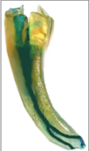
Clearing study
Many researchers have used clearing techniques to study the tooth’s internal anatomy. First the tooth is decalcifi ed using either
5% nitric acid or 10% hydrochloric acid and then dehydrated with varying concentrations of alcohol and immersed in various clearing agents like xylene, benzene, methyl salicylate, etc. The tooth becomes transparent, and then a dye is injected and the internal pulp space can be analyzed [Figure 4].[7]
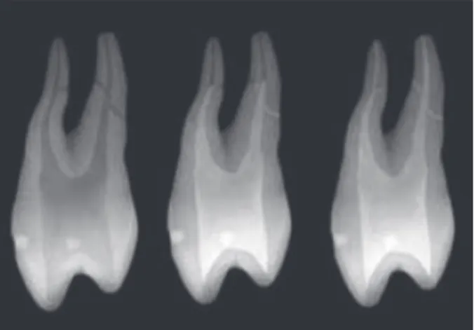
Endodontic radiography
Radiographs are the “eyes” of the dentists when performing many procedures. In 1895, Wilhelm Konard Roentgen discovered the cathode rays, which have contributed greatly to improve the dental health. Radiography is one of the most common methods of analyzing the pulp space by a clinician, but one must remember that this is only a two-dimensional image of a 3D object hence the clinician should analyze the pulp space, three-dimensionally,
and this comes through experience.[1]
Two basic type of X-ray machines are used in the dental offi ce. One has a range of kilo voltage with 2 mAmp settings with which the long16 inch cone is frequently used. The other type
Figure 3: A dental operating microscope
off ers only one kilo voltage and mAmp setting and only the short 8 inch cone.
Overlying canals may be separated using the “Clark rule” or the “SLOB” rule - it states that the object that moves in the same direction as the cone is located toward the lingual and the object that moves in the opposite direction of the cone lies in the buccal side.[3]
When extra canal is suspected, X-ray should be taken in mesial or distal 15° angulations.
When a radiograph shows that the canal suddenly stops in the radicular region the assumption is that it has bifurcated
or trifurcated into much fi ner diameters. To confi rm this, a
second radiograph is taken from a mesial angulation of 10-30°. In the radiograph, lateral radiolucency might suggest the presence of lateral canals. Knob like image may indicate apex that curves toward or away from the beam of the X-ray machine. Multiple vertical lines indicate the possibility of a thin root. The biomechanical preparation of the curved canals is an important
consideration in endodontic treatment.[1]
Digital radiography
Digital radiography utilizes computer technology in the capture, display, enhancement, and storage of radiographic images. The two major technologies presently used in intra oral digital X-ray systems are as follows: [1]
1. Solid state detectors
a. Charge coupled device (CCD)
b. Complementary metal oxide semi-conductor. 2. Storage phosphor detectors
a. Photostimulable phosphor. Advantages:
1. Lower radiation dose.
2. Environmental waste production. 3. Elimination of dark room cost. 4. Work fl ow benefi ts.
5. Electronic image transfer. Disadvantages:
1. High initial investment cost.
2. Issues of infection control as the detectors cannot be autoclaved.
3. Solid- state detectors are somewhat thicker and more rigid.
4. As they are wired it creates patient psycological discomfort.
5. It takes time to master the software.
Radiovisiography (RVG)
First developed by Mouyen et al. in 1989. Digital RVG is
equipped with special image analysis software, and in digital technology, it is possible to enlarge the images and their saving in the patient’s fi le. It is usually used in root length determination but it can also be used to study the anatomy of the root canal space in certain angulations. The image of the apical structures can be seen or length determined. These radiographs provide
additional visual information more easily with advantages of less exposure to radiation and elimination of chemical processing.[1]
SEM
A SEM uses focused beam of electrons for scanning a sample to produce an image. The SEM is a microscope that uses electron instead of light to form an image. The SEM has many advantages over traditional microscopes. It has large depth of fi eld and that it can achieve resolution better than 1 nm. Specimens can be observed in high vacuum, in low vacuum, in wet conditions, and at a wide range of cryogenic or elevated temperatures. SEM is one of the most useful instrument in research today.[8]
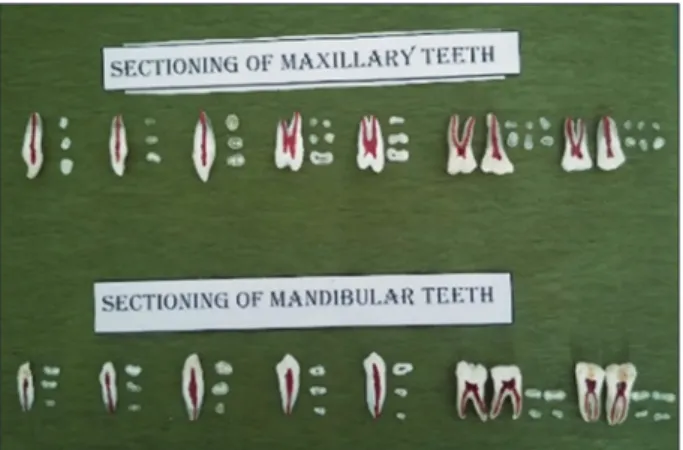
Sectioning
Longitudinal and cross-section is important to understand the pulp space anatomy as cross sectioning specimens will allow
us to understand the pulp space at diff erent levels. The study
sections will give the clinician a better understanding of the pulp space [Figure 5].
Micro computed tomography
Micro CT is based on multi-slice X-ray images that are grouped into a 3D image. There is no need to section the samples. The small voxels size of micro-CT result in higher resolution than CBCT. Micro CT evaluates the volume and area using scanning pre and post endodontic treatment. To analyze the internal anatomy, tooth is positioned into a pattern and scanned with a desktop X-ray micro focus CT scanner (Skyscan). The
equipment scan using 50 KV X-ray tube voltages, 800 μA anode
current, and a detector based on CCD camera of 1.3 MP. The whole system is connected to a computer used in the control, data acquisition, reconstruction and analysis of the attributes of the images.[9]
CBCT
This technique allows 3D imaging of root canals, utilizing computer image processing. The CBCT scanner obtains up to
nearly 600 distinct images by rotating around patients head. The patient is positioned off set of the table so the region of interest is scanned. It is a big boon in studying the root canal anatomy. This is a very accurate method of studying the root canal anatomy. Also, the model can be rotated in any plane in space and analyzed internally and externally, can be sectioned transversally and longitudinally. The areas and perimeter of the cross section can
be determined easily. The canal volume can also be evaluated.[10]
Conclusion
A thorough knowledge of the root canal anatomy, careful interpretation of radiographs, and adequate access to exploration of the tooth’s interiors are the prerequisites for a successful endodontic treatment.
With recent advances, studying the pulp morphology has
become much more effi cient. The clinician must be aware of the
latest developments so that he/she can render quality treatment to the patient.
References
1. Ingle J, Bakland L, Baumgartner C. Endodontics. 6th ed. USA: Peoples Medical Publishing House; 2013.
2. Cohen S, Hargreaves KM. Pathways of the Pulp. 9th ed. India: Mosby Publishers; 2010.
3. Weine FS. Endodontic Th erapy. 6th ed. India: Mosby Publications, Elsevier; 2009.
4. Chandra SB, Krishna GV. Grossman’s Endodontic Practice. 12th ed. India: Wolters Kluwer; 2010.
5. Garg N, Garg A. Textbook of Endodontics. 3rd ed. India: Jaypee Brothers; 2014.
6. Ruddle CJ. Advanced Endodontics. Place Unknown: Publisher Unknown; 2012. Available from: http://www.endoruddle.com. 7. Mahesh M, Rajiv SD, Shrinivas SV, Rudrayya SP, Chaitra H.
Demonstration of root canal morphology of human permanent teeth using transparent tooth model system. Int J Contemp Dent IJCD 2010;1:18-22.
8. Unknown Author. Scanning Electron Microscope. Place Unknown: Wikimedia Foundation. Available from: http://www. en.org/wiki/Scanning_electron_microscope. [Last updated on 2015 Jan 12].
9. Marciano MA, Duarte MA, Ordinola-Zapata R, Del Carpio-Perochena A, Cavenago BC, Villas-Bôas MH, et al. Applications of micro-computed tomography in endodontic research. Current Microscopy Contributions to Advances in Science and Technology (A. Méndez-Vilas, Ed.). 2012 FORMATEX.