INTRODUCTION
The prevalence of dental caries is steadily increasing, and periodontal diseases are amongst the most common
Antimicrobial efficacy of aqueous and ethanolic extracts of
Triphala on primary plaque colonizers: An in vitro study
Ruchika Gupta, BR Chandrashekar*, Pankaj Goel, Vrinda Saxena,
Sudheer Hongal, Manish Jain, Rahul Ganavadiya
Department of Public Health Dentistry, JSS Dental College and Hospital, JSS University, Mysore, Karnataka, India
ABSTRACT
Objective: The aim was to assess the antimicrobial efficacy of the aqueous and ethanolic extracts of Triphala at various concentrations against primary plaque colonizers. MaterialsandMethods: Preparation of the herbal extracts for this in vitro study was done using cold infusion method. Ethanol and Millipore water were used as solvents for extraction. A stock solution was prepared by adding 1000 mg of dried extract in 1 ml of dimethyl sulfoxide (DMSO). The stock solution was further diluted to obtain 6.25%, 12.5%, 25% and 50% concentrations of the extract. The antimicrobial efficacy testing of Triphala extracts against these bacteria was done by agar well diffusion method. 0.2% chlorhexidine was used as a positive control, while DMSO acted as a negative control. One-way analysis of variance and independent sample t-test were used for comparing mean diameter of inhibition zones. Results: All the concentrations of ethanolic and aqueous extracts of Triphala inhibited the growth of Streptococcus mutans,Streptococcus sanguisandStreptococcus salivarius. In general, the efficacy increased with increasing concentration with maximum inhibition at 50% concentration. There was no statistically significant difference in the mean diameter of inhibition zone between the ethanolic and aqueous extracts of Triphala against S.mutans. Conclusion: Both aqueous and ethanolic extracts of Triphala have the potential to be used antiplaque agents.
Key words: Antimicrobial efficacy, dental caries, minimum inhibitory concentration, periodontal diseases,
Streptococcus mutans, Triphala
*Address for correspondence:
Dr. BR Chandrashekar, Department of Public Health Dentistry, JSS Dental College and Hospital, JSS University, Mysore, Karnataka, India. E-mail: [email protected]
afflictions of mankind.1-4 Dental plaque is a microbial
ecosystem mainly consisting of densely packed microbial products and microbial byproducts.5 It is an important etiological factor for these two dental diseases.6
The current concept of disrupting the biofilm through professional and or home care practices are based on this association between dental plaque and periodontal diseases. The efficient plaque control demands a high degree of motivation and skill, which is beyond the scope of most individuals especially in rural settlements which are less educated, less aware about the importance of good oral hygiene.
Access this article online Journal Sponsor
Website:
www.jyoungpharm.org
DOI:
Antimicrobial mouth rinses have been suggested as adjuncts for mechanical plaque control methods. The most commonly used antiplaque agent is chlorhexidine gluconate. The use of chlorhexidine has some potential drawbacks like altered taste sensation, staining of teeth and development of resistant microorganisms that incapacitate their long term use. This necessitates the development of innovative strategies. One such strategy would be to verify the enormous wealth of medicinal plants abundantly available in nature.
“Triphala” has been described as a classic Ayurveda remedy.7 It comprised of fruits of three medicinal trees,
Amalaki - Emblica officinalis, Vibhitaki - Terminalia belerica, Haritaki - Terminalia chebula. It has antibacterial, antiseptic, and anti-inflammatory properties along with many other properties.5
The studies assessing the beneficial effects of Triphala in preventing oral diseases is scanty. Hence, the present study was undertaken to assess the antimicrobial efficacy of the aqueous and ethanolic extracts of Triphala at various concentrations against primary plaque colonizers.
MATERIALS AND METHODS
This in vitro study was conducted at Center for Scientific Research and Development, People’s University, Bhopal over a period of 2 months (December 2013 and January 2014).
Preparation of Triphala extracts
Ethanolic extract
The preparation of the herbal extract was done using cold infusion method. Two grams of commercially available Triphala Churna powder was dissolved in 10 ml of ethanol in a glass bottle. The mixture was then subjected to intermittent stirring for 48 h. The solution was filtered with Whatman filter paper. The filtrate was allowed to dry at room temperature. A stock solution was prepared by adding 1000 mg of dried extract in one ml of dimethyl sulfoxide (DMSO). The stock solution was further diluted to obtain 6.25%, 12.5%, 25% and 50% concentrations of the extract.
Aqueous extract
The procedure described above was employed to prepare 6.25%, 12.5%, 25% and 50% aqueous extract of Triphala using distilled water as a solvent for the extraction process.
Antimicrobial efficacy testing of ethanolic and aqueous extracts
Three primary plaque colonizers were used for antimicrobial efficacy testing in the present study. The pure laboratory
cultures of these bacteria were procured from American type culture collection (ATCC), USA. The antimicrobial efficacy testing of the ethanolic and aqueous extracts of Triphala against these bacteria was done by agar well diffusion method using Brian Heart Infusion agar. The zone of inhibition was measured using a metal scale at the end of 48 h of incubation. 0.2% chlorhexidine was used as a positive control, while DMSO acted as a negative control.
Minimal inhibitory concentration (MIC)
The MIC of ethanolic and aqueous extracts of Triphala
on Streptococcus mutans, Streptococcus sanguis and Streptococcus salivarius was determined using the agar well diffusion method described earlier. The required number of serial dilutions was prepared by serially diluting a standard stock solution. The lowest concentration of the extract that inhibited the growth
of S.mutans,S.sanguis and S.salivarius was considered the MIC of the extract against the particular bacteria.
Data analysis
The statistical analysis was performed using Statistical Package for Social Sciences version 20 (IBM, Chicago, USA). The mean inhibition zone between different concentrations of Triphala extract (ethanolic and aqueous) against each bacteria was compared using one-way analysis of variance and Tukey’s post-hoc test. The mean inhibition zone between ethanolic and aqueous extracts of Triphala was compared using independent sample t-test. The statistical significance was fixed at 0.05.
RESULTS
The details of the plant ingredients in Triphala churna and their yield are presented in Table 1. The yield was more with aqueous extract than the ethanolic extract. The details of the bacteria used in the present study are denoted in Table 2.
Antimicrobial efficacy against S. mutans
Table 1: Details of the plant extracts used in the present study
Extract Botanical name Common name Family Solvent
used Dry weight of extract (G) Yield %
Triphala churna Terminalia chebula Retz Haritaki Combretaceae Ethanol 0.295 14.75
Terminalia bellirica Bahera (Bibhitaka) Combretaceae
Emblica officinalis Amla or Indian
gooseberry Euphorbiaceae Millipore water 0.684 34.2
Table 2: Details of the bacteria used for antimicrobial efficacy testing in the present study
Bacteria ATCC
number Selective media used for revival Type of haemolysis on blood agar plate
S. mutans 25175 Brain heart infusion agar with 5% sheep blood
Gamma haemolysis
S. sanguis 10556 Brain heart infusion agar with 5% sheep blood
Alpha haemolysis
S. salivarius 13419 Brain heart infusion agar with 5% sheep blood
Gamma haemolysis
ATCC: American type culture collection, S. mutans: Streptococcus mutans, S. sanguis: Streptococcus sanguis, S. salivarius: Streptococcus salivarius
Table 3: Antimicrobial efficacy of ethanolic and aqueous extracts of triphala on S. mutans
Concentration Mean diameter of inhibition zone in millimeters (SD)*
Ethanolic
extract Aqueous extract Statistical inference
6.25% 24.17 (3.84) 20.50 (2.43) t value: 1.976
P value: 0.076 12.5% 24.75 (2.04) 25.08 (1.07) t value: −0.354
P value: 0.731 25% 25.58 (3.61) 26.33 (1.17) t value:‑0.484
P value: 0.639 50% 29.17 (0.68) 29.50 (0.63) t value: −0.877
P value: 0.401 0.2% chlorhexidine 16.25 (0.27) 16.25 (0.27)
Statistical
inference F20.72 value:
P value: 0.001
F value: 91.26
P value: 0.001 *SD: Standard deviation
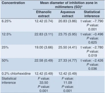
Table 4: Antimicrobial efficacy of ethanolic and aqueous extracts of triphala on Streptococcus sanguis
Concentration Mean diameter of inhibition zone in millimeters (SD)*
Ethanolic
extract Aqueous extract Statistical inference
6.25% 12.42 (0.74) 20.83 (3.86) t value: −7.790
P value: 0.001 12.5% 22.83 (3.11) 23.75 (5.95) t value: −0.496
P value: 0.625 25% 19.00 (3.66) 25.50 (4.41) t value: −2.780
P value: 0.019 50% 22.58 (0.49) 27.33 (4.77) t value: −2.426
P value: 0.036 0.2% chlorhexidine 12.42 (0.49) 12.42 (0.49)
Statistical
inference F33.50 value:
P value: 0.001
F value: 11.09
P value: 0.001 SD: Standard deviation
Antimicrobial efficacy against S. sanguis
The maximum inhibition was observed at 12.5% concentration (22.8 ± 3.1 mm) while using ethanolic extract and at 50% concentration (27.3 ± 4.8 mm) using aqueous extract. All the concentrations showed a significantly higher zone of inhibition compared to chlorhexidine, while using the ethanolic and aqueous extracts (P = 0.001, Table 4). However, there was no significant difference in the mean diameter of inhibition between 6.25% ethanolic extract
and chlorhexidine. The aqueous extract of Triphala yielded significantly higher mean diameter of inhibition zones compared with ethanolic extracts against S.sanguis at 6.25%
(P = 0.001), 25% (P = 0.019) and 50% concentrations (P = 0. 036, Table 4). However, the difference in the mean diameter of inhibition zone between the aqueous and ethanolic extracts of Triphala at 12.5% was not statistically significant (P = 0.625) though aqueous extract produced a marginally higher efficacy.
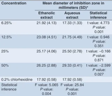
Antimicrobial efficacy against S. salivarius
to antibiotics. Current advancement in drug discovery technology and search for novel chemical diversity has intensified the efforts of exploring herbal medicines. Triphala is one such herbal medicine that exhibits number of health benefits.8
The antimicrobial efficacy of Triphala at various concentrations against three dental plaque microorganisms considered to be the initial colonizers in the process of plaque formation was assessed.9
We found the ethanolic and aqueous extracts of Triphala at all concentrations to be effective against S.mutans,S.sanguis and S.salivarius.
T.chebula Retz contains polyphenols, terpenes, anthocyanins, flavonoids, alkaloids and glycosides.9 Emblica officinalis contains vitamin C and vitamin C complex.10 T. bellirica
fruit contains chemical constituents such as tannins; viz gallic acid, ellagic acid, and Phylemblin.11 Some components including a cardenolide, glycoside, oil containing palmitooleolinolein, 16-hentriacontanone, hexahydroxydiphenic acid ester, Friedelin, B-sitosterol, chebulagic acid are isolated from the fruit extract.12-15
The inhibitory effect of Triphala on S. mutans could be
attributed to these phytochemical constituents.
Jagadeesh et al., (2009)16 demonstrated the antioxidant
and antimicrobial activity of Triphala against S. mutans. Jagtap and Karkera (1999)17 reported that extracts of T. chebula strongly inhibited the growth and adherence of S.mutans. Thomas etal., (2011)8 found the mean inhibition
zone for the aqueous extract of Triphala at 50%, 25% and 12.5% against S. mutans to be 30 mm, 28 mm, and 24 mm respectively using microbial type culture collection (MTCC) strains. The mean inhibition zone for the aqueous extract of Triphala at 50%, 25% and 12.5% against clinical isolates of S.mutans was found to be 34 mm, 30 mm, and 28 mm, respectively. There was no significant difference in the efficacy with increasing concentrations. Although, the mean inhibition zones found in our study at various concentrations were comparable to the mean zones in this study, we found a significantly higher zone of inhibition at 50% concentration compared to other concentrations, contradictory to the findings of this study. A study by Prajapathi and Raol (2014)18 found the aqueous extract of
Triphala to inhibit S.mutans. The mean inhibition zone was 17 mm against MTCC strains and 19 mm against clinical isolates. Here, 100 µl of 2% extract was used while we used 50 µl of 10% extract. They found variable results in terms of mean inhibition zone with maximum inhibition observed with acetone extract. The minor variations in the mean zone between our findings and these studies Table 5: Antimicrobial efficacy of ethanolic and aqueous extracts
of triphala on Streptococcus salivarius
Concentration Mean diameter of inhibition zone in millimeters (SD)*
Ethanolic
extract Aqueous extract Statistical inference
6.25% 21.92 (4.13) 17.33 (1.33) t value: 4.773
P value: 0.001 12.5% 23.08 (4.51) 21.75 (4.49) t value: 0.946
P value: 0.351 25% 25.17 (4.06) 25.50 (2.78) t value: −0.166
P value: 0.871 50% 26.25 (2.88) 29.33 (0.41) t value: −2.599
P value: 0.027 0.2% chlorhexidine 17.92 (0.58) 17.92 (0.58)
Statistical
inference F value: 5.065P value: 0.004
F value: 25.84
P value: 0.001 SD: Standard deviation
Table 6: Minimum inhibitory concentration of ethanolic and aqueous extracts of triphala on S. mutans, S. sanguis and S. salivarius
Bacteria Milligram/ml (%)
Ethanolic extract Aqueous extract
S. mutans 1 mg/ml (0.1) 1 mg/ml (0.1)
S. sanguis 0.5 mg/ml (0.05) 0.5 mg/ml (0.05)
S. salivarius 0.5 mg/ml (0.05) 2 mg/ml (0.2)
S. mutans: Streptococcus mutans, S. sanguis: Streptococcus sanguis, S. salivarius: Streptococcus salivarius
higher mean inhibition zone compared to ethanolic extract
at 50% (P = 0.027). There was no significant difference in the mean diameter of inhibition zones between aqueous and ethanolic extracts at 12.5% (P = 0.351) and 25% concentrations (P = 0.871, Table 5). The inhibition zone produced by the ethanolic and aqueous extracts of Triphala
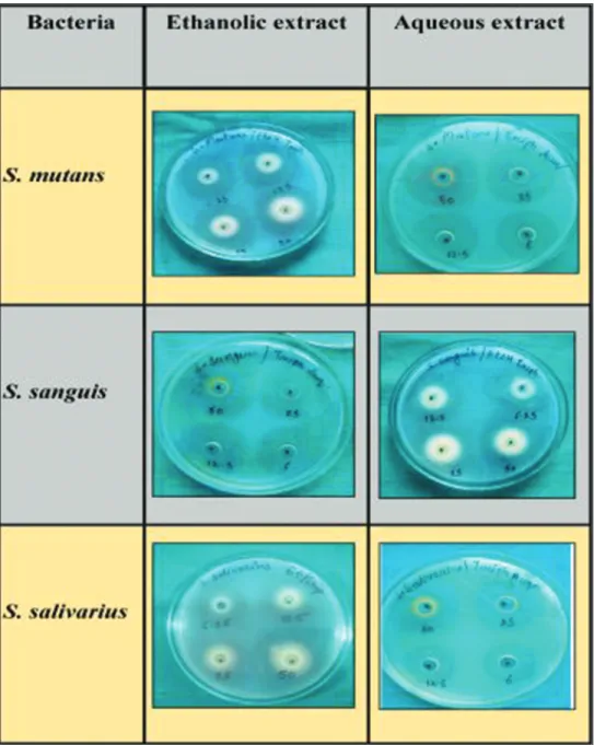
on S.mutans,S.sanguisandS.salivarius is presented in Figure 1.
MIC
The ethanolic extract of Triphala inhibited S. mutans,
S. sanguis and S. salivarius at 1 mg/ml, 0.5 mg/ml and 0.5 mg/ml, respectively. The MIC of aqueous extract of Triphala on S.mutans,S.sanguisandS.salivarius was 1 mg/ml, 0.5 mg/ml and 2 mg/ml, respectively (Figure 2, Table 6).
DISCUSSION
The aggregates of bacterial cell embedded in a polysaccharide and protein matrix adhering to the teeth is termed dental plaque.8 Several anti-plaque agents are available in the
could be attributed to methodological differences involved in the extraction process, antimicrobial efficacy testing, differences in the bacterial strains, concentration and volume of the extract used. The inhibition zone in our study is computed after subtracting the diameter of the well (7 mm) from the actual diameter of inhibition zone. Thomas etal., (2011)8 found the MIC of ethanolic extract
of Triphala on MTCC strains and clinical isolates of S.mutans to be 6.25% and 3.12%, respectively. These MIC values against S.mutans are high compared to MIC values in our study. The difference in the bacterial strains between our study and this study may explain these differences. The literature comparing the antimicrobial efficacy of aqueous and ethanolic extracts of Triphala on oral bacteria is
non-existent. Hence, our results could not be compared with any previous published literature. The present invitro study was conducted using ethanol and aqueous extracts of Triphala on ATCC strains of S.mutans,S.sanguis and S.salivarius. The antimicrobial efficacy testing using other solvent systems and clinical isolates of these bacteria could offer different results. Moreover, further studies on secondary and tertiary plaque colonizers could add to beneficial effects of Triphala before considering this for clinical use.
CONCLUSION
Based on the results of the present study, the following conclusions are drawn:
Figure 1: Mean diameter of inhibition zone by ethanolic and aqueous extracts of Triphala on Streptococcus mutans, Streptococcus sanguis and
• Both aqueous and ethanolic extracts of Triphala inhibited
the growth of S.mutans,S. sanguis and S.salivarius. Hence, they could be considered as alternates to chlorhexidine. • The mean diameter of inhibition zone in general increased
with increasing concentration with maximum efficacy observed at 50% concentration and least at 6.25%. • There was no statistically significant difference in the
mean diameter of inhibition zone between the ethanolic
and aqueous extracts of Triphala against S.mutans. • The aqueous extract of Triphala showed a higher mean
diameter of inhibition zone against S.sanguis compared
with ethanolic extracts.
• The ethanolic extract of Triphala produced a significantly higher mean diameter of inhibition zone against S. salivarius compared to aqueous extracts at 6.25% concentration, while aqueous extract produced a significantly higher mean inhibition zone compared to ethanolic extract at 50% concentration.
REFERENCES
1. Botelho MA, Santos RA, Martins JG, Carvalho CO, Paz MC, Azenha C, etal. Efficacy of a mouth rinse based on leaves of the neem tree (Azadirachta Indica) in the treatment of patients with chronic gingivitis: A double-blind, randomized, controlled trial. J Med Plants Res 2008;2:341-6. Figure 2: Minimum inhibitory concentration of the ethanloic and aqueous extracts of Triphala on Streptococcus mutans, Streptococcus sanguis
2. Patro BK, Ravi Kumar B, Goswami A, Mathur VP, Nongkynrih B. Prevalence of dental caries among adults and elderly in an urban resettlement colony of New Delhi. Indian J Dent Res 2008;19:95-8.
3. Dhar V, Bhatnagar M. Dental caries and treatment needs of children (6-10 years) in rural Udaipur, Rajasthan. Indian J Dent Res 2009;20:256-60. 4. Dhar V, Jain A, Van Dyke TE, Kohli A. Prevalence of gingival diseases,
malocclusion and fluorosis in school-going children of rural areas in Udaipur district. J Indian Soc Pedod Prev Dent 2007;25:103-5.
5. Desai A, Anil M, Debnath S. Clinical trial to evaluate the effects of Triphala as a mouthwash in comparison with chlorhexidine in chronic generalized periodontitis patient. Indian J Dent Adv 2010;2:243-7.
6. Singh S, Chaknis P, DeVizio W, Petrone M, Panagakos FS, Proskin HM. A clinical investigation of the efficacy of three commercially available dentifrices for controlling established gingivitis and supragingival plaque. J Clin Dent 2010;21:105-10.
7. Tandon S, Gupta K, Rao S, Malagi KJ. Effect of Triphala mouthwash on the caries status. Int J Ayurveda Res 2010;1:93-9.
8. Thomas B, Shetty S, Vasudeva A, Shetty V. Comparative evaluation of Antimicrobial Activity of Triphala and commercially available toothpastes: An in-vitro study. Int J Public Health Dent 2011;2:8-12.
9. Nield-Gehrig JS. Dental Plaque Biofilms. Available from: http//www. dentalcarestamford.com. [Last cited on 2014 Apr 07].
10. Emblica officinalis. Natural Remedies – Research Center. Available from: http//www.thaiherbinfo.com/herb/Phyllanthus%20 emblica./embblica%20officinalis. [Last cited on 2014 Mar 25].
11. Jadon A, Bhadauria M, Shukla S. Protective effect of Terminalia belerica Roxb. and gallic acid against carbon tetrachloride induced damage in albino rats. J Ethnopharmacol 2007;109:214-8.
12. Lekha KM, Awasthi KI, Bola N. Chemical examination of Terminaliabellirica Roxb. Indian Chem Soc 1968;45:913-7.
13. Singh BK, Kumar A. Chemical examination of the fruits of Terminalia
bellirica Roxb. Indian Acad Sci 1946;23A:379-83.
14. Bhutani KK. Occurence of hexahydroxydiphenic acid ester in Terminalia
bellirica fruits. Indian J Nat Prod 1991;7:16-7.
15. Bajpai N, Tiwari JS. A note on phytochemical investigation of Terminalia genus. J Indian Chem Soc 1994;71:643.
16. Jagdeesh LA, Kumar A, Kaviyarasan V. Effect of triphala on dental biofilm. Indian J Sci Technol 2009;2:30-3.
17. Jagtap AG, Karkera SG. Potential of the aqueous extract of Terminalia chebula as an anticaries agent. J Ethnopharmacol 1999;68:299-306.