LOAD CHARACTERISTICS AND RESPONSES IN MALE ATHLETES WITH PATELLAR TENDINOPATHY
Laura Stanley Pietrosimone
A dissertation submitted to the faculty at the University of North Carolina at Chapel Hill in partial fulfillment of the requirements for the degree of Doctor of Philosophy in Interdisciplinary
Human Movement Science.
Chapel Hill 2018
Ó2018
iii ABSTRACT
Laura Stanley Pietrosimone: Loading Characteristics and Responses in Male Athletes with Patellar Tendinopathy
(Under the direction of Darin A. Padua)
Context: Clinical management of tendinopathy is difficult, as tendon pathoetiology occurs on a continuum with inconsistent associations between structural pathology and pain. Tendon is highly responsive to mechanical load; however, load mismanagement can trigger homeostatic imbalances that lead to the development of tendinopathy. There is a need to characterize loading profiles and responses of individuals along the tendinopathic continuum to inform improved clinical management strategies. Objective: To evaluate differences in biomechanical and loading volume profiles and to determine the effects of an acute bout of patellar tendon isometric loading exercise on lower extremity landing biomechanics in male athletes with patellar tendinopathy.
iv
v
DEDICATION
This dissertation is dedicated to two of my greatest role models: Richard “Poppy” Joseph Crowder, Sr.
Richard “Uncle Rick” Joseph Crowder, Jr.
For the way they overcame obstacles. For their enthusiasm for life, both things big and small. For the gratitude with which they approached every day and every experience. For the way they
put family before everything else. For their smiles, their laughs, and their six-foot-seven-inch bear hugs. For always strongly supporting their granddaughter and niece to pursue her goals. Poppy and Uncle Rick, we miss you dearly, but your legacy lives on in the eyes of the people fortunate enough to know you. In my personal and professional life, I pledge to strive to leave a
vi
ACKNOWLEDGEMENTS
To my advisor, Dr. Darin Padua. Thank you for the honor of working with you for the last four years. I can confidently say that coming back to school was one of the best decisions of my professional life, and I am indebted to you for providing me this unbelievable opportunity. Thank you for your time, energy, patience, loyalty, and humor. From you I have learned how to be a clinical scientist, but I have also learned how to do so as a strong leader and professional. Thank you for modeling and supporting the belief that family comes first. Thank you for being willing to take a gal who bleeds the darker shade of blue and making her a part of the UNC family. I am privileged to have been your student and promise to continue to model your lessons as I move forward in my journey.
To Dr. Troy Blackburn, thank you for your support during my time at UNC, particularly in your mentoring with statistics, laboratory techniques, and all things technical. Thank you for being willing to teach a statistics seminar to four of us on your own time. Your attention to detail in your work is something that I respect and will carry with me as a scientist.
To Dr. Erik Wikstrom, thank you for the time and expertise that you have brought to this project. Thank you for the many random conversations in the lab about knees and ankles and what matters clinically. Your passion for your work is truly inspiring.
vii
Thank you for sharing the UTC and for willingly providing me so many unique opportunities. Clinicians like you inspire me to work hard to pursue answers to questions that will make a difference for our patients.
To Dr. Sean Docking, thank you for all that you’ve done to mentor me from across the pond. Your expertise in ultrasound and willingness to collaborate has been truly impactful in my learning process. Thank you for your openness to share thoughts and programs, and your overall enthusiasm for what you do.
To Dr. Jill Cook, you are a role model of a strong woman in science, and it has been a true honor to have you involved in my doctoral training. I will always remember hearing you speak at the APTA meeting in Anaheim, after which I became increasingly excited to study tendon health. Thank you for sharing your time and expertise on this project, and your inspiring career as a clinical scientist.
To my husband, Brian, thank you for being by my side along this journey. You inspire me every day by your passion for your work, but most importantly by your love for family. You make me laugh. You make me better. I could not have travelled this road without you. It has been a true adventure, but I wouldn’t want it any other way. I am so thankful that we are teammates for life and cannot wait for the many memories to come.
amazing of a spouse, parent, colleague, and friend as both of you. I love you and am forever thankful for you.
For all of my extended family, life is never boring with the crew we have! Thank you for your energy, support, and high fives along this journey. To be surrounded with such a network of strong family makes life so full.
To my labmates, thank you for all of your support over the past four years. Our shared excitement for learning and our natural camaraderie made coming to work each day motivating and fun. Thank you for always being there to pick me up through the rough moments,
professionally and personally, and always being eager to celebrate the joyful moments. The garden level of Fetzer created a bond that will never be broken.
To the HMSC program and faculty, thank you for the opportunity to study at such a fantastic University and renowned program. Each individual has taught me something meaningful along the way that I will carry with me into the next chapter. Thank you for continuing to strive to train well-rounded scientists.
To my clinical mentor, Dr. Chuck Thigpen, thank you for first teaching me what clinical research was all about, and inspiring me to take the leap to pursue my PhD. You have been a coach to me through PT school, residency, clinical work, and my doctoral training. Thank you for always reminding me that we do research for our patients, and to pursue questions that will move the needle forward. You have truly taught me the power that a strong mentor can have on someone’s life, and I hope to pay that forward as I carry forward.
always ensure that my research will center around improving the quality of your lives in meaningful ways.
TABLE OF CONTENTS
LIST OF TABLES ...xii
LIST OF FIGURES ... xiv
LIST OF ABBREVIATIONS ... xvi
CHAPTER 1: INTRODUCTION ... 1
Specific Aims, Research Question Objectives, & Hypotheses ... 3
Independent Variable ... 17
Dependent Variables ... 17
CHAPTER 2: REVIEW OF LITERATURE ... 18
SECTION 1: Injury Epidemiology & Pathoetiology ... 21
Epidemiology of Patellar Tendinopathy ...21
Models of Tendon Pathoetiology ...23
The Continuum of Tendon Pathology ...25
Differential Diagnosis: A Critical Feature of Evaluation of Anterior Knee Pain ...29
Defining Tendon Pathology: Evidence from Previous Literature ...30
Symptom Characteristics Associated with Patellar Tendinopathy ...30
Structural Characteristics Associated with Patellar Tendinopathy ...33
SECTION 2: Intrinsic and Extrinsic Factors Associated with Patellar Tendinopathy ... 37
Intrinsic Factors ...38
Ankle, Knee, and Hip Kinematic & Kinetic Characteristics Associated with Symptomatic Patellar Tendinopathy ...41
Ankle, Knee, and Hip Kinematic & Kinetic Characteristics Associated with Patellar Tendon Structural Pathology ...47
Patellar Tendon Biomechanical Loading Characteristics Associated with Patellar Tendinopathy ...48
Extrinsic Factors ...53
Physical Activity and Training Load Monitoring in Sport ...53
Physical Activity and Training Load Monitoring of Individuals with Lower Extremity Musculoskeletal Injury Conditions ...55
SECTION 3: Exercise-Based Intervention Paradigms for Patellar Tendinopathy ... 60
Mechanotherapy: Implications for Treatment of Tendinopathies ...60
Historical Perspective: Eccentric Exercise for the Treatment of Tendinopathies ...62
Treating Symptomatic Patellar Tendinopathy: Evidence for Isometric Loading Exercise ...63
CHAPTER 3: EXPERIMENTAL DESIGN & METHODS ... 67
SUBJECTS ... 67
DATA COLLECTION ... 69
Procedures ...69
Overall Study Design ...69
xi
Instrumentation ...87
Three-Dimensional Motion Capture Biomechanical Data Collection Instrumentation ...87
Physical Activity Monitoring Data Collection Instrumentation ...88
DATA PROCESSING & REDUCTION... 88
Laboratory Biomechanics: Three-Dimensional Motion Capture Data ...88
Marker Identification & Processing ...88
Joint Center Calculations ...89
Kinematic Calculations...89
Kinetic Calculations ...89
Data Reduction...90
Dependent Variable Calculation ...91
Real-World Physical Activity: Cumulative External Load Monitoring ...92
Dependent Variable Calculation ...93
STATISTICAL ANALYSIS ... 93
POWER ANALYSIS ... 95
CHAPTER 4: RESULTS ... 97
Specific Aim 1 ... 97
Specific Aim 2 ... 116
Specific Aim 3 ... 125
CHAPTER 5: MANUSCRIPT 1 ... 134
CHAPTER 6: MANUSCRIPT 2 ... 154
CHAPTER 7: MANUSCRIPT 3 ... 176
APPENDICES ... 203
Appendix 1. Pubertal Development Scale ... 203
Appendix 2. Tegner Activity Level Scale. ... 204
Appendix 3. International Physical Activity Questionnaire (IPAQ) ... 205
Appendix 4. Knee Injury History Form. ... 207
Appendix 5. Percentage of predicted mature height calculation ... 208
Appendix 6. Victorian Institute of Sport Assessment-Patellar Tendon questionnaire. ... 209
Appendix 8. Algorithm for patellar tendon abnormality diagnosis. ... 212
Appendix 9. ActiGraph Wear Position ... 213
Appendix 10. Supplementary material for manuscript 3. ... 214
xii
LIST OF TABLES
Table 1.1. Summary of Specific Aim 1 ... 7
Table 1.2. Summary of Specific Aim 2 ... 11
Table 1.3. Summary of Specific Aim 3 ... 15
Table 2.1. Characteristics of stages of tendon pathology described
in the continuum model ... 27
Table 2.2. Criteria to characterize symptomatic patellar tendinopathy in
current study ... 32
Table 4.1. Descriptive characteristics of the study population (mean ± sd) ... 119
Table 4.2. Descriptive characteristics for study population for load frequency and duration metrics (mean ± sd, 95% CI) ... 119
Table 4.3. Descriptive characteristics (mean ± sd, 95% CI) for load volume variables (based on # of stepsMVPA and involved limb biomechanics during
the double-limb landing task) ... 120
Table 4.4. Group comparisons for load volume variables (based on # of stepsMVPA
and involved limb biomechanics during the double-limb landing task) ... 121
Table 4.5. Group comparisons for load magnitude variables for the involved
(SYM & ASYM) and dominant (CON) limbs during the double limb landing task. ... 121
Table 4.6. Effect size calculations for group comparisons for load magnitude
variables for the involved (SYM & ASYM) and dominant (CON) limbs during the
double limb landing task. ... 122
Table 4.7. Comparison of groups for biomechanical energetic variables for
involved limb during the single-limb landing task... 122
Table 4.8. Effect size calculations for group comparisons for the involved
(SYM & ASYM) and dominant (CON) limbs during the single-limb landing task. ... 123
Table 4.9. Single-limb landing task limb symmetry indices (means ± sd and 95% confidence intervals) for the involved (SYM & ASYM) and dominant
Table 4.10. Double-limb landing task limb symmetry indices (means ± sd and 95% confidence intervals) for the involved (SYM & ASYM) and dominant
(CON) limbs ... 124
Table 4.11. Descriptive characteristics of the study population. ... 127
Table 4.12. Single leg decline squat (SLDS) pain scores (NRS: 0-10) during
each testing session. ... 127
Table 4.13. Descriptive characteristics (mean difference, standard deviation, 95% CI) for each biomechanical variable change score for the symptomatic and asymptomatic groups for the isometric and sham-TENS
intervention conditions. ... 128
Table 4.14. Cohen’s d effect sizes for mean differences (pre-post) within
xiv
LIST OF FIGURES
Figure 1.1. Study design overview... 16
Figure 2.1. The continuum model of tendon pathology. ... 29
Figure 2.2. The Single Leg Decline Squat (SLDS)... 33
Figure 2.3. Algorithm for criteria to characterize patellar tendon structural pathology. ... 36
Figure 2.4. Dye’s Envelope of Function. ... 60
Figure 3.1. Overall study design diagram. ... 69
Figure 3.2. HUMAC Norm Dynamometer set-up for intervention protocol. ... 73
Figure 3.3. Testing procedures for Sessions 2 & 3. ... 80
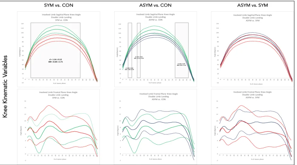
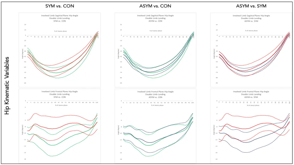
Figure 4.1: Mean and 95% confidence interval waveforms for involved limb knee kinematic variables during the double-limb jump landing task. ... 101
Figure 4.2: Mean and 95% confidence interval waveforms for involved limb hip kinematic variables during the double-limb jump landing task. ... 102
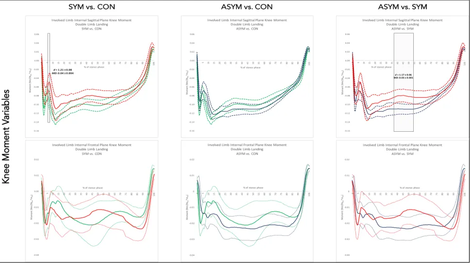
Figure 4.3: Mean and 95% confidence interval waveforms for involved limb knee internal moment variables during the double-limb jump landing task. ... 103
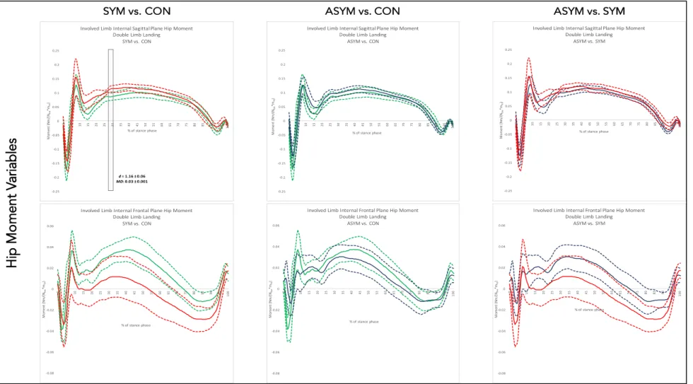
Figure 4.4: Mean and 95% confidence interval waveforms for involved limb hip internal moment variables during the double-limb jump landing task. ... 104
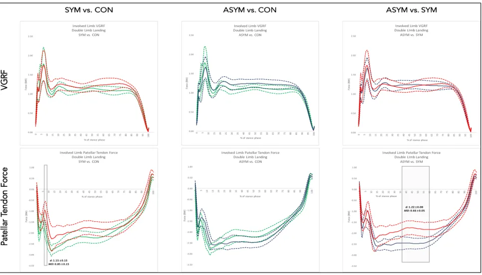
Figure 4.5: Mean and 95% confidence interval waveforms for involved limb vertical ground reaction force and patellar tendon force during the double-limb jump landing task. ... 105
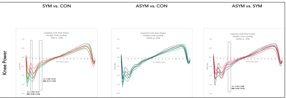
Figure 4.6: Mean and 95% confidence interval waveforms for involved limb knee power during the double-limb jump landing task. ... 106
Figure 4.7: Mean and 95% confidence interval waveforms for vertical ground reaction force (vGRF) limb symmetry indices during the double-limb jump landing. ... 107
Figure 4.8: Mean and 95% confidence interval waveforms for internal knee extension moment limb symmetry indices during the double-limb jump landing. ... 108
Figure 4.10: Mean and 95% confidence interval waveforms for involved limb
knee kinematic variables during the single-limb jump landing task. ... 110
Figure 4.11: Mean and 95% confidence interval waveforms for involved limb
hip kinematic variables during the single-limb jump landing task. ... 111
Figure 4.12: Mean and 95% confidence interval waveforms for involved limb
knee internal moment variables during the single-limb jump landing task. ... 112
Figure 4.13: Mean and 95% confidence interval waveforms for involved limb
hip internal moment variables during the single-limb jump landing task. ... 113
Figure 4.14: Mean and 95% confidence interval waveforms for involved limb vertical ground reaction force and patellar tendon force during the
single-limb jump landing task. ... 114
Figure 4.15: Mean and 95% confidence interval waveforms for involved limb
knee power during the single-limb jump landing task. ... 115
Figure 4.16: Study CONSORT Diagram. ... 130
Figure 4.17: Individual participant SLDS pain (NRS: 0-10) change scores following the isometric (blue open circles) and sham-TENS
(open red circles) interventions with median group
change (black horizontal line). ... 131
Figure 4.18: Mean and 95% confidence intervals for change scores for isometric
and sham-TENS conditions for the SYM and ASYM groups. ... 131
Figure 4.19: Individual participant pre- and post-isometric intervention SLDS pain scores (NRS 0-10) with mean (dark blue line) and95% confidence
bounds (shaded area). ... 132
Figure 4.20: Individual participant pre- and post-sham-TENS intervention
SLDS pain scores (VAS 0-10) with mean (dark red line) and 95% confidence
xvi
LIST OF ABBREVIATIONS
ASYM: Asymptomatic
cFPT: Cumulative Patellar Tendon Force CON: Control
FPT: Peak Patellar Tendon Force FPTI: Patellar Tendon Force Impulse HSR: Heavy Slow Resistance KEM: Knee Extension Moment
KEMI: Knee Extension Moment Impulse KP: Knee Power
KW: Negative Knee Work NRS: Numeric Rating Scale PT: Patellar Tendinopathy
PTA: Patellar Tendon Abnormality SYM: Symptomatic
US: Ultrasonography
CHAPTER 1: INTRODUCTION
Patellar tendinopathy is prevalent in individuals who are physically active, particularly athletes who participate in sports with repetitive jumping manueveurs.1–5 While some athletes are able to maintain sport participation, the long-term consequences of chronic tendinopathy include reduced physical activity and quality of life,6,7 with up to 53% of individuals with symptomatic patellar tendinopathy quitting their sport due to chronic tendon pain.8 Clinical management of tendinopathy is difficult, as tendon pathoetiology occurs on a continuum with inconsistent associations between structural pathology and pain.9–11 Tendon is highly responsive to
mechanical load; however, load mismanagement can trigger homeostatic imbalances that lead to the development of structural pathology and/or symptoms.10 While laboratory-based assessments have established some evidence of altered biomechanics in adults with a history of patellar tendinopathy, there is a lack of literature directly comparing biomechanical movement profiles of symptomatic and asymptomatic individuals with patellar tendon structural pathology.
Furthermore, the laboratory environment cannot account for the influence of cumulative external load incurred during real-world physical activity on variables associated with the development of patellar tendinopathy.
and consequent reduction in physical activity. Additionally, while eccentric-based strength-training protocols have traditionally been utilized in the treatment of chronic
tendinopathies,13–15 emerging evidence supports the use of isometric exercise for individuals with symptomatic patellar tendinopathy. Isometric exercise has recently been shown to improve pain and self-reported function in adults with patellar tendinopathy,16–18 resulting in excellent patient compliance and tolerance when implemented in-season.16 However, the acute effects of
isometric patellar tendon loading on landing biomechanics is unknown. Determining the effects of this novel exercise intervention on movement characteristics in clinical populations may allow for improved subgrouping of patients into impairment-based rehabilitation programs and
subsequently improve clinical effectiveness.
The overall objective of this study is to determine the effects of an acute bout of patellar tendon isometric loading exercise on lower extremity landing biomechanics, and to evaluate differences in biomechanical profiles of individuals at varying stages of the tendon pathology continuum using both laboratory and real-world movement assessments. Our approach will utilize a randomized cross-over study design to assess acute intervention effects on lower extremity kinetic and kinematic biomechanical variables during landing, and a cross-sectional quantification of one-week cumulative external load using wearable technology.
Aim 1. To ascertain the impact of symptomatic PTA and asymptomatic PTA on lower extremity landing kinematics and kinetics.
Aim 3. To investigate whether an acute isometric patellar tendon loading exercise protocol changes lower extremity landing kinematics and kinetics in individuals with symptomatic and asymptomatic PTA.
The proposed project is innovative because it will be the first to establish the effects of patellar-tendon specific loading exercise on biomechanical movement profiles of individuals along the continuum of tendon pathology, and the first to monitor cumulative external load in a tendinopathic population. Long-term, an efficacious real-world monitoring system will enhance clinical practice by allowing for timely identification of trends in loading that may influence the development of structural pathology and altered biomechanics in multiple patient populations.
Specific Aims, Research Question Objectives, & Hypotheses
Specific Aim 1. To ascertain the impact of symptomatic PTA and asymptomatic PTA on lower extremity landing kinematics and kinetics.
Research Questions
1.1 Do individuals with symptomatic PTA and asymptomatic PTA demonstrate different sagittal and frontal plane knee and hip joint angles during the loading phase of each landing task compared to individuals who are asymptomatic and without PTA (healthy control group)?
1.2 Do individuals with symptomatic PTA and asymptomatic PTA demonstrate different internal sagittal and frontal plane knee and hip joint moments during the loading phase of each landing task compared to individuals who are asymptomatic and without PTA (healthy control group)?
loading rates during the loading phase of each landing task compared to individuals who are asymptomatic and without PTA (healthy control group)?
1.4 Do individuals with symptomatic PTA and asymptomatic PTA demonstrate different peak patellar tendon force magnitudes and patellar tendon force loading rates during the loading phase of each landing task compared to individuals who are asymptomatic and without PTA (healthy control group)?
1.5Do individuals with symptomatic PTA and asymptomatic PTA demonstrate
differences in inter-limb symmetry for kinetic variables during the loading phase of each landing task compared to individuals who are asymptomatic and without PTA (healthy control group)?
Hypotheses
Hypothesis 1: Individuals with symptomatic PTA and asymptomatic PTA will demonstrate different lower extremity landing kinematics and kinetics compared to healthy controls.
1.1 Individuals with symptomatic PTA will demonstrate lesser sagittal plane knee and hip flexion displacement on the involved limb, while individuals with asymptomatic PTA will demonstrate greater sagittal plane knee and hip flexion displacement on the involved limb compared to the matched limb of healthy controls.
1.2 Individuals with symptomatic PTA will demonstrate lesser net sagittal plane knee and hip internal extension moment on the involved limb, while individuals with
extension moment on the involved limb compared to the matched limb of healthy controls.
1.3 Individuals with symptomatic PTA will demonstrate lesser peak vertical ground reaction force on the involved limb, while individuals with asymptomatic PTA will demonstrate greater peak vertical ground reaction force on the involved limb compared to the matched limb of healthy controls.
1.4 Individuals with symptomatic PTA will demonstrate lesser peak patellar tendon force on the involved limb, while individuals with asymptomatic PTA will demonstrate greater peak patellar tendon force on the involved limb compared to the matched limb of healthy controls.
1.5 Individuals with symptomatic PTA will demonstrate greater magnitude inter-limb asymmetry in kinetic variables during the loading phase of the landing tasks compared to individuals with asymptomatic PTA and healthy controls.
Rationale
patellar tendon structure and tendinopathy symptomology, and simultaneously include a healthy control group (no structural pathology or symptomology). This study is the first to compare biomechanical profiles of three distinct groups: individuals who are asymptomatic with a PTA, symptomatic with a PTA, and asymptomatic without PTA (health control). Through this design, we will be able to assess the independent effects of both structural pathology and pain on
biomechanical profiles during sport-specific tasks. The clinical relevance of this design is that strives to determine if biomechanical profiles are different between individuals at differing stages along the continuum of tendon pathology, which could inform the development of enhanced impairment-based, individualized treatment programs.
Target Journal
The manuscript reporting the results of this specific aim will be prepared for submission to Medicine & Science in Sports & Exercise (MSSE) (Impact Factor: 4.041). Previous studies reporting biomechanical profiles of tendinopathic populations have been published in
MSSE,23,25,26 so the results of our study, which uniquely controls for both structural and
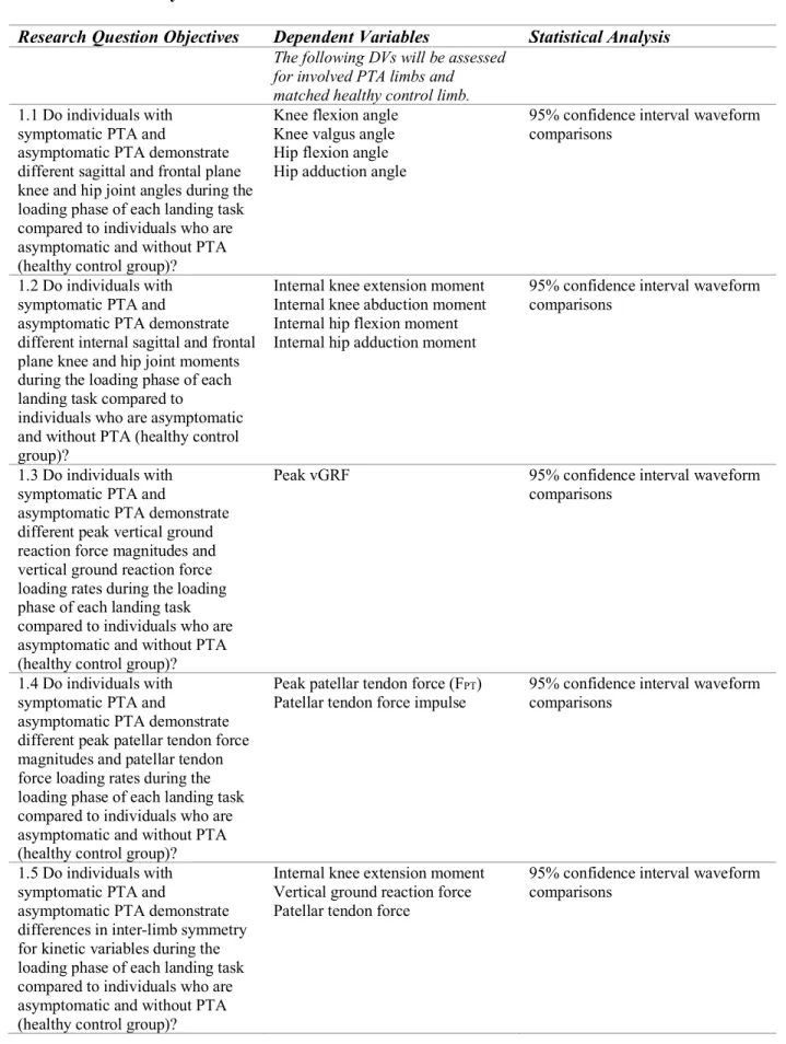
Table 1.1. Summary of Aim 1
Research Question Objectives Dependent Variables Statistical Analysis
The following DVs will be assessed for involved PTA limbs and matched healthy control limb. 1.1 Do individuals with
symptomatic PTA and
asymptomatic PTA demonstrate different sagittal and frontal plane knee and hip joint angles during the loading phase of each landing task compared to individuals who are asymptomatic and without PTA (healthy control group)?
Knee flexion angle Knee valgus angle Hip flexion angle Hip adduction angle
95% confidence interval waveform comparisons
1.2 Do individuals with symptomatic PTA and
asymptomatic PTA demonstrate different internal sagittal and frontal plane knee and hip joint moments during the loading phase of each landing task compared to
individuals who are asymptomatic and without PTA (healthy control group)?
Internal knee extension moment Internal knee abduction moment Internal hip flexion moment Internal hip adduction moment
95% confidence interval waveform comparisons
1.3 Do individuals with symptomatic PTA and
asymptomatic PTA demonstrate different peak vertical ground reaction force magnitudes and vertical ground reaction force loading rates during the loading phase of each landing task compared to individuals who are asymptomatic and without PTA (healthy control group)?
Peak vGRF 95% confidence interval waveform comparisons
1.4 Do individuals with symptomatic PTA and
asymptomatic PTA demonstrate different peak patellar tendon force magnitudes and patellar tendon force loading rates during the loading phase of each landing task compared to individuals who are asymptomatic and without PTA (healthy control group)?
Peak patellar tendon force (FPT)
Patellar tendon force impulse 95% confidence interval waveform comparisons
1.5 Do individuals with symptomatic PTA and
asymptomatic PTA demonstrate differences in inter-limb symmetry for kinetic variables during the loading phase of each landing task compared to individuals who are asymptomatic and without PTA (healthy control group)?
Internal knee extension moment Vertical ground reaction force Patellar tendon force
Specific Aim 2. To ascertain the impact of symptomatic PTA and asymptomatic PTA on cumulative external load during a one-week monitoring period.
Research Questions
2.1 Do individuals with symptomatic PTA and asymptomatic PTA demonstrate different average steps-per-day during a one-week monitoring period compared to individuals who are asymptomatic and without PTA (healthy control group)?
2.2 Do individuals with symptomatic PTA and asymptomatic PTA demonstrate different cumulative patellar tendon loads compared to individuals who are asymptomatic and without PTA (healthy control group)?
Hypotheses
Hypothesis 2: Individuals with symptomatic PTA and with asymptomatic PTA will demonstrate different average daily and cumulative loading volume during a one-week monitoring period than healthy controls, such that:
2.1 Individuals with symptomatic PTA will demonstrate less loading volume than both individuals with asymptomatic PTA and healthy controls.
2.2 Individuals with asymptomatic PTA will demonstrate greater cumulative patellar tendon loads than both individuals with symptomatic PTA and healthy controls. Rationale
controlled loading in certain athletic populations.29–31 However, loading frequency, intensity, and duration may influence tendon response, particularly in the presence of maladaptation (structural pathology and/or symptomology).10 Therefore, quantification of key objective physical activity characteristics, as outlined by the FITT principle32,33 may provide critical information on tendon adaptation to load that is currently not systematically evaluated in standard clinical practice around patellar tendinopathy.
A limitation of traditional biomechanical assessments in the area of musculoskeletal injury and sports medicine is the exclusive use of laboratory measures (i.e. three-dimensional motion capture) to study associations between movement characteristics and injury. While considered the gold-standard movement assessment tool, three-dimensional motion capture only provides a brief, controlled snapshot of an individual’s biomechanical profile, which does not account for cumulative loading repetition or the influence of overall physical activity exposure. Therefore, real-world physical activity monitoring is a critical missing piece in the study of overuse injury development. Previous literature has demonstrated associations between high training and competition workloads and injury.34,35 Specifically, high training load volume (training hours/week, match/week) increases the risk of patellar tendinopathy in adolescent male and female volleyball athletes (OR: 1.72-3.38).36 Additionally, in a cohort of collegiate female volleyball athletes, cumulative season training load was negatively association with post-season VISA-P scores (r=-0.512, p=0.043), indicating that athletes with higher loads during the
competitive season reported more post-season patellar tendon pain. (Stanley et al, in progress) The utility of quantifying physical activity outcomes in this population is to obtain a more objective understanding of the associations between cumulative external load and clinical
quantified physical activity metrics in various pathologic populations. Individuals with chronic ankle instability were found to participate in 24 less minutes of physical activity per day than healthy counterparts37, and individuals following ACLR took on average 2000 less steps-per-day than healthy controls.38 In a population of adults with osteoarthritis (OA), combined laboratory- and real-world based knee joint loading metrics were better able to distinguish between
individuals with and without OA than traditional laboratory-based assessments alone.39
Therefore, based on the foundational knowledge of tendon as a mechano-responsive tissue, and growing evidence of altered real-world loading in pathological populations, measuring and monitoring real-world loading metrics is a critical component in the study of overuse injuries.
Target Journal
Table 1.2. Summary of Aim 2
Research Question Objectives Dependent Variables Statistical Analysis
2.1 Do individuals with symptomatic PTA and
asymptomatic PTA demonstrate different average steps-per-day during a one-week monitoring period compared to individuals who are asymptomatic and without PTA (healthy control group)?
Average steps-per-day Average MVPA-per-day
One-way ANOVA
2.2 Do individuals with symptomatic PTA and
asymptomatic PTA demonstrate different cumulative patellar tendon loads compared to individuals who are asymptomatic and without PTA (healthy control group)?
Specific Aim 3. To investigate whether an acute isometric patellar tendon loading exercise protocol changes lower extremity landing kinematics and kinetics in individuals with symptomatic and asymptomatic PTA.
Research Questions
3.1 Does an acute isometric patellar tendon loading exercise protocol change sagittal and frontal plane knee and hip joint angles during the loading phase of the
double-limb landing task in individuals with symptomatic PTA compared to those with asymptomatic PTA?
3.2 Does an acute isometric patellar tendon loading exercise protocol change net internal sagittal and frontal plane knee and hip joint moments during the loading phase of the double-limb landing task in individuals with symptomatic PTA compared to the asymptomatic PTA?
3.3 Does an acute isometric patellar tendon loading exercise protocol change peak vertical ground reaction force during the loading phase of the double-limb
landing task in individuals with symptomatic PTA compared to the asymptomatic PTA?
Hypotheses
Hypothesis 3. The acute isometric patellar tendon loading exercise protocol will elicit different changes in lower extremity landing kinematics and kinetics in individuals with symptomatic PTA compared to the asymptomatic PTA.
3.1 The acute isometric patellar tendon loading exercise protocol will increase sagittal plane knee and hip joint angles in the symptomatic PTA group, relative to the asymptomatic PTA.
3.2 The acute isometric patellar tendon loading exercise protocol will alter sagittal plane knee and hip joint moments, specifically greater internal knee extension moment and lesser internal hip flexion moment, in the symptomatic PTA group, relative to the asymptomatic PTA.
3.3 The acute isometric patellar tendon loading exercise protocol will increase peak vertical ground reaction force in the symptomatic PTA group relative to the asymptomatic PTA.
3.4 The acute isometric patellar tendon loading exercise protocol will increase peak patellar tendon force in the symptomatic PTA group relative to the asymptomatic PTA.
Rationale
treatment strategies is critical. Tendon is a visco-elastic tissue that readily responds to loading via mechanotransduction processes;12 therefore, using exercise-based therapies to promote positive adaptation when tendon is structurally compromised both before and after symptom onset is supported. Isometric patellar tendon loading exercise has recently been shown to both acutely and chronically decrease tendon pain, improve quadriceps strength, and improve self-reported knee function during sport in individuals with symptomatic patellar tendinopathy.16–18
However, the effects of this targeted tissue-specific loading protocol on lower extremity biomechanics has not yet been investigated. Athletes with symptomatic patellar tendinopathy demonstrate load avoidance movement strategies during sport-specific tasks, including
reductions in sagittal plane knee displacement and mechanical energy absorption, lesser vertical ground reaction force, and lesser internal knee extension moment.19,21,22 This study will be the first to test the acute effects of an isometric patellar tendon loading exercise protocol17 on
landing biomechanics in individuals across the tendon pathology continuum (both asymptomatic and symptomatic individuals with structural abnormalities). Using isometric loading
interventions to acutely change movement biomechanics may provide an important next step in rehabilitation paradigms for tendinopathy as a method to promote improve load-tolerance during and stimulate positive mechano-transductive responses in individuals with tendon pathology. Target Journal
The manuscript reporting the results of this specific aim will be prepared for submission to the Journal of Orthopedics and Sports Physical Therapy (JOSPT) (Impact Factor: 2.55). Tissue-specific loading interventions are common in clinical practice for the treatment of tendinopathies. Therefore, the results of this specific aim have the potential to aid in the
appropriate for JOSPT, as this journal seeks to publish clinically-relevant studies specific to common orthopedic conditions. Additionally, this journal is housed within my professional organization, the American Physical Therapy Association (APTA).
Table 1.3. Summary of Aim 3
Research Question Objectives Dependent Variables Statistical Analysis
3.1 Does an acute isometric patellar tendon loading exercise protocol change sagittal and frontal plane knee and hip joint angles during the loading phase of the double-limb landing taskin individuals with symptomatic PTA compared to those with asymptomatic PTA?
The following kinematic variables will be calculated for initial contact, peak, and displacement across the loading phase of the landing task for the involved PTA limb: Knee flexion angle Knee valgus angle Hip flexion angle Hip adduction angle
Between groups: 2x2 mixed-model repeated-measures analysis of variance ANOVA on change scores for involved limbs from pre- to post-intervention
3.2 Does an acute isometric patellar tendon loading exercise protocol change net internal sagittal and frontal plane knee and hip joint moments
during the loading phase of the double-limb landing task in individuals with symptomatic PTA compared to those with
asymptomatic PTA?
The following kinetic variables will be calculated across the descending phase of the landing task for the involved PTA limb:
Peak Internal knee extension moment
Peak Internal knee abduction moment
Peak Internal hip flexion moment Peak Internal hip adduction moment
Between groups: 2x2 mixed-model repeated-measures analysis of variance ANOVA on change scores for involved limbs from pre- to post-intervention
3.3 Does an acute isometric patellar tendon loading exercise protocol change peak vertical ground reaction force during the loading phase of the double-limb landing task in individuals with
symptomatic PTA compared to those with asymptomatic PTA?
Peak vGRF a. 2x2 mixed-model repeated measures ANOVA on change scores for involved limbs peak vGRF from pre- to post-intervention
3.4 Does an acute isometric patellar tendon loading exercise protocol change peak patellar tendon force during the loading phase of the double-limb landing task in individuals with symptomatic PTA compared to those with
asymptomatic PTA?
Peak patellar tendon force (FPT)
Patellar tendon force impulse
a. 2x2 mixed- model repeated measures ANOVA on change scores for involved limbs peak FPT
and FPT impulse from pre- to
Figure 1.1. Overview of the proposed study methodology to assess the acute effects an isometric patellar tendon loading exercise protocol on lower extremity landing biomechanics, and to quantify cumulative external load metrics over a one-week training period.
1-week wash-out period
Session 1 US Imaging Diagnostic Assessment + PTA - PTA Symptom Assessment SYM ASYM Group Assignment
PTA + SYM PTA + ASYM
CONTROL Ses sio n 2 Ses sio n 3 MVIC Assessment
1-week load monitoring period
3D Biomechanics
Assessment
Pain
Assessment Intervention Protocol
ISOMETRIC or CONTROL
3D Biomechanics
Assessment Pain
Assessment AssessmentMVIC Informed Consent 3D Biomechanics Assessment Pain
Assessment Intervention Protocol
ISOMETRIC or
3D Biomechanics
Assessment Pain
Assessment AssessmentMVIC
CONTROL Randomized
Cross-Over Study Design to Isometricor
Control
Independent Variable
1. Patellar Tendon Structural and Symptom Profile
a. Symptomatic / PTA vs. Asymptomatic / PTA vs. Asymptomatic / No PTA
Dependent Variables
1. Biomechanical Variable Change Scores following the Isometric Patellar Tendon Loading Exercise Intervention collected over the loading phase of the double-limb landing task; Baseline Biomechanical Variables 95% Confidence Interval Waveform Comparisons over the entire stance phase of the double-limb landing task:
i. Sagittal plane knee joint angle ii. Frontal plane knee joint angle iii. Sagittal plane hip joint angle iv. Frontal plane hip joint angle
v. Net internal sagittal plane knee joint moment vi. Net internal frontal plane knee joint moment vii. Net internal sagittal plane hip joint moment viii. Net internal frontal plane hip joint moment
ix. Vertical ground reaction force x. Patellar tendon force
xi. Patellar tendon force impulse
2. Cumulative External Load Variables from 1-Week Load Monitoring Period a. Average daily steps-per-day
CHAPTER 2: REVIEW OF LITERATURE
The Landscape of Musculoskeletal Injury in Youth Sports
The last half-century has witnessed a steady rise in sports participation among youth athletes.44 In the United States, high school sports participation increased by approximately 80% between 1971 and 2005,44 which has been attributed to growing opportunities for females in sport and the growing emphasis on health promotion in youth.45,46 In a large cross-sectional, nationally representative sample, approximately 62% of high school students (70% of males; 53% of females) reported participating in at least one sport.47 Nearly 8 million boys and girls participated in organized high school sports during the 2014-2015 school year.48 The importance of physical activity in youth populations has been recognized by national and international governing bodies as a priority from health behavior and economic perspectives. The World Health Organization recommends at least sixty-minutes of moderate to vigorous physical activity (MVPA) per day for youths aged 5-17, and notes that greater than sixty minutes-per-day may provide additional health benefits.49 Physical activity and sports participation in the youth population has been associated with numerous positive health and social behaviors47 and
continued physical activity into adulthood,46 lowering the risk of a variety of disease conditions, including cardiovascular disease, diabetes, and various cancers.50
adolescent sports-, recreation-, and exercise-related (SRE) injuries, due to both acute and chronic mechanisms.51 During the 2014-2015 school year, high school athletes in the United States suffered an estimated 1.2 million injuries.52
Of particular concern is the high prevalence of musculoskeletal (MSK) injury in youth athletes. MSK injury diagnoses constitute the majority of self-reported SRE injuries in both high school and collegiate populations.53–55 In a large-scale national survey, 64% of reported sport-related injury episodes occurred among individuals aged 5-24, and were reported as
approximately 41% higher than national estimates of injuries that require emergency department visits.53 Additionally, the majority of reported SRE injuries were to the upper (31.2%) and lower (38.9%) extremities.53 The consequences of MSK injuries sustained at a young age are
numerous, including economic, social, and long-term outcomes. In the United States, MSK SRE injuries comprise up to 64% of all emergency department visits by individuals 19 years old and younger.56 While there is limited data describing long-term health impacts of injuries sustained in young athletes, this is an area gaining increasing interest due to the rise in youth sport
participation. In fact, recent evidence demonstrates that athletes with a history of MSK injury or a current MSK injury score lower than uninjured counterparts on validated quality of life
measures, including health problems and social functioning, and report less perceived physical capability.57–60
While MSK injuries sustained through acute, traumatic mechanisms, such as anterior cruciate ligament (ACL) injury, are debilitating, costly, and result in time loss for sport participation,61–63 chronic, overuse MSK injuries are often equally as challenging and
tendinopathy, a common overuse injury in athletes, is a challenging condition to treat due to its varied clinical presentation, specifically the inconsistently-present clinical indicators of pain and structural pathology. Tendinopathies result from mismanagement of external load, typically in the direction of tissue overloading.9,64 Traditional management of lower limb tendinopathies emphasized notable reductions in external load until full resolution of symptoms was achieved.65 Eccentric exercise protocols, particularly for chronically symptomatic tendinopathies, constitute the common standard of care for tendinopathy,14,66 and are likely one of the most widely-spread implemented treatment paradigms in rehabilitative musculoskeletal clinical practice. However, evidence supporting a continuum of tendon pathology, described by progressive stages of structural pathology, suggests that one-size-fits-all eccentric exercise protocols may not be appropriate to prescribe for all stages of tendinopathy.10 Emerging evidence demonstrates
This review will focus on three primary areas around the topic of patellar tendinopathy to support the current study: 1) epidemiology and pathoetiology, 2) intrinsic and extrinsic factors, and 3) exercise-based intervention paradigms. Overall, the aims of this study seek to contribute to the current understanding of the continuum of tendon pathology,10 and inform our
understanding of modifiable factors that can be directly applied to the clinical management of tendinopathy.
SECTION 1: Injury Epidemiology & Pathoetiology
Epidemiology of Patellar Tendinopathy
Patellar tendinopathy is a chronic, overuse injury condition resulting from excessive tissue load.67,68 Epidemiological studies have demonstrated a 2.5 – 14.4% prevalence of patellar tendinopathy in a diverse group of sports requiring high loading rates and power demands.4,5 In particular, individuals participating in sports involving repetitive jumping and landing have been shown have the highest rates of patellar tendinopathy, due to the repetitive load placed on the tendon tissue. The prevalence in elite and recreational adult basketball athletes has been reported to be as high as 32% and 12%, respectively; a similar prevalence has been noted in elite (45%) and recreational (14%) volleyball athletes.4,5 Athletes participating in other sports, such as track and field (running and high/long jump athletes), tennis, and skiing, have also been shown to readily develop tendinopathy.67
studied as adult populations, an approximate 7% prevalence of patellar tendinopathy in adolescent (ages 14-18) athletes has been noted.1 Moreover, in young elite volleyball athletes, boys have been shown to have four-times higher risk of developing PT than girls, independent of training and match exposure.36 While the sex discrepancy appears to be consistent in different age groups and sports, due to increased sport participation among young females,46 continued evaluation of both sexes should be pursued. For example, a high prevalence (26.6%) of anterior knee pain has been documented in female adolescent athletes assessed over three-years during pre-participation screenings, including higher prevalence in high-school (34.4%) versus middle-school (23.5%) aged athletes,70 suggesting that young females should be monitored closely during sport participation. Additionally, as athlete’s progress from junior to senior sporting levels, the incidence of patellar tendinopathy increases,71 likely due to the cumulative chronic load from aggregated years of sport participation. Moreover, Hall et al (2015) demonstrated an increased risk of patellar tendinopathy in youth athletes who specialize in a single sport (OR: 1.27 – 4.0).72
pathoetiology of tendinopathy, however, it is thought that the effects of chronic tendinopathy on performance, self-perceived function, and quality of life are likely underestimated.36
The prevalence of patellar tendinopathy in youth athletes, particularly in those who participate in sports involving high frequency and intensity of repetitive jumping and landing, and are exposed to high cumulative training volumes, necessitates continued attention from the sports medicine clinical and research communities. This may be of particular importance during a period when youth sports participation is increasing exponentially in the United States, not only to reduce onset and progression of pathology, but to also ensure that young individuals are able to maintain healthy levels of physical activity as they mature. Knowledge of risk factors for patellar tendinopathy provides avenues to improve treatment strategies to decrease the burden of this condition in young athletes, which in turn will promote lifelong physical activity.
Models of Tendon Pathoetiology
The management of tendinopathy in clinical settings can be challenging, due largely to the varied clinical presentation, specifically inconsistent relationships between tendon structural pathology, function, and pain.10 Multiple models are represented in existing literature that outline the pathoetiology of tendinopathy. These models all suggest that the development of
tendinopathy occurs along a continuum, but that distinct factors initiate the pathoetiologic cascade. Specifically, collagen matrix disruption, inflammation, and tendon cell response have been identified in previous literature as key factors driving tendinopathic processes.73
pathology.73,74 This model suggests that degenerative pathology is irreversible, and is considered end-stage pathology. A limitation to this model is that it does not consistently describe phases of tendon adaptation that may precede degeneration, which is a critical facet when identifying injury prevention strategies in high-risk population.
Further, inflammatory models are aligned around the notion that inflammatory substances drive tendinopathic processes. While widely accepted amongst clinical and scientific groups, there are several limitations to this model. One of the key limitations is the lack of cellular inflammation that is present in pathological tendons. For decades, it was thought that the primary source of pain in tendinopathy was due to inflammation within the tendon (i.e. ‘tendinitis’). However, numerous studies have clearly demonstrated that there is a lack of intra-tendinous inflammation present in tendinopathy. Two key investigations conducted by Khan et al (1996) and Sanchis-Alfonso (2001) provide strong evidence to support this shift.75,76 In both of these studies, biopsies of patellar tendons from individuals with recalcitrant symptoms were taken at the time of surgery. Histochemical analysis indicated consistent changes to the structural matrix, including poorly aligned irregular collagen fibers, shift from Type I to Type III collagen,
increased swollen tenocytes, fibrils, and fluid, and heightened expression of matrix-degrading cytokines (i.e. TNF-alpha). Interestingly, there was a complete lack of inflammatory cells and biomarkers. Instead, both studies noted the presence of neuronal-sprouting and increased vascularity within the pathologic tendon. Other studies have reported similar findings,
In summary, early models of tendon pathology were defined by two distinct
classifications of tendon status: tendinitis (acute) and degenerative tendinosis (chronic).65 The distinction between these two classifications was based largely on duration of symptoms and key aspects of the clinical exam. Specifically, tendinitis was classified based on the following key constructs:
• Acute onset with short duration of symptoms
• Inflammatory processes within the tendon proper (-itis), resulting in pain • Lack of obvious structural changes to the extracellular matrix (ECM)
• Notable tenderness to palpation and observable/palpable focal swelling on physical exam Degenerative tendon pathology (tendinosis) was classified based on the following key
constructs:
• Chronic, recalcitrant symptomology (>6+ months)
• Notable changes to the extra-cellular matrix, including collagen disorganization or a complete loss of matrix integrity
• Irreversible structural changes that are unable to respond to load-based treatments • +/- tenderness to palpation and observable/palpable focal swelling on physical exam The Continuum of Tendon Pathology
different stages of the pathologic continuum may require different treatment approaches.10 In fact, the prescription of inappropriate interventions to a tendon may lead to a worse outcome.77
Therefore, a shift in the model of tendon pathology to that of a continuum model has been largely supported over the last decade.9,10 This model defines tendon pathology across three continuous stages, and contends that the movement from one stage to another is largely
determined by imposed or external loading stimulus. There is considerable evidence
demonstrating that external load is one of the primary etiological factors that influences tendon structural properties.9,78,79 Biological tissue homeostasis has been described by Dr. Scott Dye (1996) as the envelope of function, or “the range of load that can be applied across an individual tissue in a given period without supraphysiologic overload or structural failure.”64 Tendon health is intimately related to mechanical homeostasis. Tendon adaptation occurs through
mechanotransduction, the physiological process by which the body translates mechanical load into a cellular response that leads to structural change.12 (see Section 3) Evidence describing structural changes that occur when a tendon is mechanically stimulated, or increasing the capacity of the tendon,80,81 supports the continuum model that tendon pathology does develop along a continuum, without discrete “onset” and “resolution” points. Since external loading is a modifiable construct, understanding how tendon responds to both acute and cumulative loading, particularly in athletes at an elevated risk of developing overuse injuries, may improve tailored intervention delivery focused on load-based management.
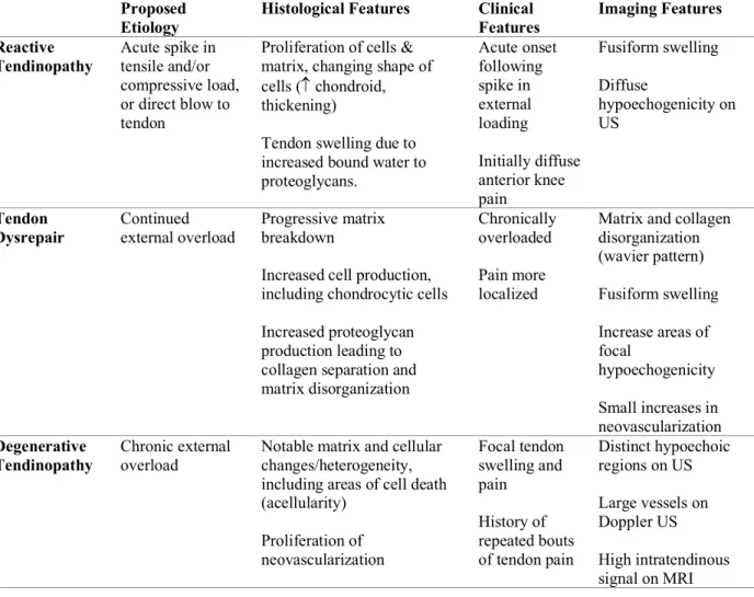
the difficulty of clinical management. The three stages described by Cook et al. in the continuum model are: reactive, dysrepair, and degenerative tendinopathic stages (Table 2.1).
Table 2.1. Characteristics of stages of tendon pathology described in the continuum model from Cook et al (2009).9
Proposed
Etiology Histological Features Clinical Features Imaging Features Reactive
Tendinopathy
Acute spike in tensile and/or compressive load, or direct blow to tendon
Proliferation of cells & matrix, changing shape of cells ( chondroid, thickening)
Tendon swelling due to increased bound water to proteoglycans. Acute onset following spike in external loading Initially diffuse anterior knee pain Fusiform swelling Diffuse hypoechogenicity on US Tendon
Dysrepair Continued external overload Progressive matrix breakdown Increased cell production, including chondrocytic cells Increased proteoglycan production leading to collagen separation and matrix disorganization
Chronically overloaded Pain more localized
Matrix and collagen disorganization (wavier pattern) Fusiform swelling Increase areas of focal
hypoechogenicity Small increases in neovascularization Degenerative
Tendinopathy
Chronic external overload
Notable matrix and cellular changes/heterogeneity, including areas of cell death (acellularity) Proliferation of neovascularization Focal tendon swelling and pain History of repeated bouts of tendon pain
Distinct hypoechoic regions on US Large vessels on Doppler US High intratendinous signal on MRI
optimal levels, a catabolic pathway is promoted, stress-shielding occurs, and, over time, it is more likely to enter a degenerative stage. However, once in the degenerativestage, continued overload to the tendon has become catabolic, as the magnitude/frequency/duration of load has exceeded tendon’s tolerance and allowed inadequate time to recover, remodel, and repair. There is an approximate four-fold increased risk of symptom development in individuals with a patellar tendon abnormality.43 Evidence suggests that there is little capacity for tendon structure to
reverse back to a normal structural state once in a degenerative state.10,83,84
However, the emergence of an additional tendinopathic stage on the continuum,
‘reactive-on-degenerative tendinopathy’ has been supported by evidence demonstrating portions of degenerative tendon that are surrounded by structurally intact tendon.10 The hypothesis around the mechanism of ‘reactive-on-degenerative tendinopathy’ is that structurally intact tendon assumes the bulk of loading (as degenerative tendon is mechanically dormant) via stress-shielding, and therefore may pass through intermittent stages of reactivity. Recent work by Docking et al (2015) demonstrates that degenerative tendon compensates for its inert structure by increasing cross-sectional area to maintain adequate aligned fibrillar structure around the
periphery of the degenerative region.80 As a result, the tendon maintains its capacity for load-transmission and can be targeted through load-based therapies. Importantly, sound progressive loading paradigms that address both pain and load-capacity in reactive-on-degenerative
tendinopathies should be prescribed in order to maximize tendon resilience and prevent progressive structural pathology and functional disability (Figure 2.1).
of pain.86,87 Therefore, clearly defining the symptom and structural characteristics that may influence the progression of tendinopathy is of critical importance in the management of tendinopathy.
Figure 2.1. The continuum of tendon pathology (image from: Cook et al, Br J Sports Med, 2016)10
Differential Diagnosis: A Critical Feature of Evaluation of Anterior Knee Pain
One of the key components to identifying appropriate treatment pathways for individuals with patellar tendinopathy is a sound differential diagnosis. In the context of the proposed study, clearly defining diagnostic criteria for patellar tendinopathy is a critical feature of pursuing the study aims to compare landing biomechanics between distinct pathologic and healthy
populations.
Common Differential Diagnoses for Non-Traumatic Anterior Knee Pain
Patellar tendinopathy is one of several conditions that presents clinically as non-traumatic anterior knee pain. The breadth of differential diagnoses is expected, due to the multiple
chondromalacia, pes anserine or supra/infrapatellar bursitis, iliotibial band syndrome, fat pad impingement, and osteoarthritis.88,89 Furthermore, in pediatric and adolescent populations, additional differential diagnoses must be considered, including osteochondroses
(Osgood-Schlatters disease and Sinding-Larsen-Johansen disease, osteochondritis dissecans, inflammatory disorders, referred pain (i.e. slipped capital femoral epiphysis or Perthe’s disease),
osteosarcomas, or patellofemoral instability.88,89 In both clinical and research environments, a thorough clinical exam to rule-out conditions that are non-mechanical in nature and require referral should always occur first. If patellar tendinopathy is suspected, a systematic assessment of signs and symptoms should be confirmed.
Defining Tendon Pathology: Evidence from Previous Literature
Symptom Characteristics Associated with Patellar Tendinopathy
Patellar tendinopathy is characterized by several hallmark features that have been well-described in previous literature11,67,90: 1) localized pain at the proximal patellar tendon, just inferior to the inferior patellar pole11,91, and 2) load-dependent pain that is provoked with high demands on the knee extensor mechanism.5,92,93 Specifically, load-related tendon pain typically occurs immediately upon the initiation of loading, and resolves once the load is removed or at rest. Tendon pain also increases with increasing load magnitude and loading rates of activities involving high energy storage and release across the extensor mechanism, such as during deep squatting or repetitive hopping.11,94 Occasionally, during the course of an exercise bout, tendon pain may subside, as the tendon “warms-up”; however, pain typically reemerges following cessation of activity and may last for several hours to days.94
patellofemoral pain syndrome (PFPS) or infrapatellar fat pad (IFP) impingement. Unlike patellar tendinopathy, PFPS is characterized by diffuse anterior knee pain under or around the patella, provocation with lower-loading activities and prolonged knee flexion positioning, and reduction of symptoms with joint alignment correction, such as via taping or manual
repositioning.11,70,89,95,96 Additionally, while there is evidence of tissue communication between the IFP and the patellar tendon97 (i.e. cytokine production,98 neovascularization,76,99,100 and structural connections75,101102), pain derived from IFP impingement is typically located adjacent to the tendon and is more commonly provoked with knee hyperextension or direct palpation.
Finally, in the context of evaluating young athletes with suspected patellar tendinopathy, developmental conditions involving tendon-growth plate interfaces, such as Osgood-Schlatters disease (tibial tuberosity) and Sinding-Larsen-Johansson disease (inferior patellar pole), should be considered. These two developmental, traction-apophysitis conditions are most common in pre-pubertal cohorts during periods rapid growth.89,103 In the present study, the pubertal status of invited participants will be confirmed using validated measures104–106, with the goal of
minimizing the potential that either of the aforementioned developmental conditions may be the source of patellar tendon pain in the symptomatic group.
The criteria selected to characterize patellar tendon pain and delineate a symptomatic group assignment for participants in the current study are supported by previous literature (Table 5). Tendon pain is most commonly utilized clinically to diagnose patellar tendinopathy,
tendinopathy. However, the sensitivity (68%) and specificity (9%) of palpation is poor, and thus palpation is not considered as a robust diagnostic tool.108 In addition to patient history, including detailed questioning of activity-related stimulants of symptoms, pain maps109 and provocation tests110 are commonly used to differentiate patellar tendon pain from other conditions.
Table 2.2. Criteria to characterize symptomatic patellar tendinopathy in current study
Criteria for Symptomatic Patellar Tendinopathy
Key Previous Literature Notes
1. Localized
Load-Dependent Pain
Blazina et al. (1973) Roels et al. (1978)
Stages 1-4 (pain location/provocation) Cook et al. (2000) Junior basketball athletes Rudavsky & Cook (2014) Topical review
2. Single-limb decline squat (SLDS) pain that remains localized to tendon
Purdam et al. (2003) Adolescent (14-18 years) male and female basketball athletes
Malliaras et al. (2006) Adult volleyball athletes Cook et al. (2005) Adult volleyball athletes
emphasize that when using the SLDS provocation test, the test should be conducted with participants squatting between 50-60 degrees of knee joint flexion,110 where maximum force development through the patellar tendon has been reported.111 Additionally, larger magnitudes of knee flexion excursion (70-80 degrees) engage the patellofemoral joint and result in peak
patellofemoral compressive forces112; therefore, avoiding a knee flexion angle greater than 60 degrees during the SLDS test should be confirmed. The SLDS has been reported to have a low standard error of 5% on repeated assessment.110
Figure 2.2. The Single Leg Decline Squat (SLDS). Participants are instructed to squat to approximately 60 degrees of knee joint flexion on a 25-degree decline board and rate the magnitude and location of pain. (image from: Malliaras et al, J Orth Spor Phys Ther, 2015)11
In summary, clear definitions for inclusion criteria to define symptomatic patellar
tendinopathy are critical to determine the independent influence of pain and structural pathology on primary biomechanical outcomes of interest, and, furthermore, the effectiveness of the isometric patellar tendon loading exercise protocol.
Structural Characteristics Associated with Patellar Tendinopathy
imaging should always be accompanied by a sound history, clinical exam, and evaluative testing. The goal of diagnostic musculoskeletal imaging is to visualize bodily tissues in order to
objectively characterize features that may be indicative of pathology. Overview of Tendon Composition
Healthy tendon is primarily comprised of Type I collagen which is highly organized and well-aligned in parallel longitudinally within the tendon, providing tendon with high tensile strength.115 Small amounts of Type III collagen and Type X collagen are also present in tendon, primarily at tendon-bone interfaces.116 Tendon fibroblasts, or tenocytes, are spindle-shaped cells located along collagen fibers, and are the primary cell modulating the tendon structural
environment via mechanotransduction.12,117 Proteoglycans (primarily decorin and biglycan) support the tendon extracellular matrix, aid in regulation of collagen fiber formation, and control fibril diameter.118 Proteoglycans and water constitute a large majority of the tendon extracellular matrix. In healthy tendon, the biochemical contrast between collagen and water in tendon results in little to no signal on MRI, and a homogenous, parallel orientation of well-organized fibrillar structure on US.119
Compositional Changes Associated with the Development of Tendinopathy
study, the presence or absence of neovascularization will not be utilized as an inclusion criteria for a patellar tendon abnormality. Detection of neovascularization via Doppler signal on US can demonstrate poor inter-day reliability,121 and as it is easily influenced by exercise.122
Additionally, the selection of US for use in the current study is attributed to its accessibility, feasibility, and ease of interpretation. While MRI is reported to have excellent reproducibility, soft tissue contrast, and captures multi-planar images,113 it is costly, inaccessible, and not
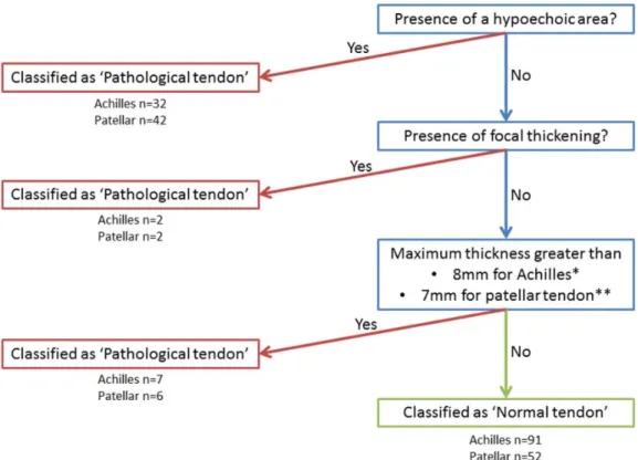
required for the purposes of the study aims. Furthermore, advances in ultrasonographic imaging, including probe technology and image processing software that allows thorough visualization of tendon fibrillar structure, suggest that US imaging is an efficacious tool in tendon imaging.114 Ultrasound Assessment of Tendon Structure
Historically, conventional ultrasound imaging techniques have been utilized to evaluate tendon status, through measurement and grading of cross-sectional area (CSA) and
echogenicity.119,123,124 Hypoechoic regions are suggestive of poor tendon quality, and have been linked to symptomatology in numerous populations.125,126 Cook et al.1 demonstrated that in junior basketball players, 79% of the patellar tendons categorized on clinical evaluation as having “current tendinopathy” also had a hypoechoic region on US imaging. Additional ultrasonographic assessments, including Doppler sonography, have been utilized to investigate neovascularization of tendon, and have been shown to associated with the presence of
symptomatic Achilles tendinopathy.3,121,127
two-year patient-reported function. Additionally, in a group of competitive club runners, while conventional US detected increased tendon thickness, there were no associations with self-reported symptoms of Achilles tendinopathy.129 Limitations to conventional US include error related to probe placement and handling during scan acquisition, and slight changes or error in transducer position (tilt and rotation) can generate anisotropy artifact that may mimic images visualized with actual pathology.114 However, a recent systematic review found that diagnostic US assessments for tendon size demonstrate acceptable inter- and intra-rater reliability.131 Therefore, based on the evidence, the use of US to classify tendon pathology (Figure 2.3) in the current study is well-supported.