PEARLS
The microbiome and the hallmarks of cancer
Laura E. Fulbright1, Melissa Ellermann1, Janelle C. Arthur1,2,3*
1 Department of Microbiology and Immunology, University of North Carolina at Chapel Hill, Chapel Hill, North Carolina, United States of America, 2 Center for Gastrointestinal Biology and Disease, University of North Carolina at Chapel Hill, Chapel Hill, North Carolina, United States of America, 3 Lineberger Comprehensive Cancer Center, University of North Carolina at Chapel Hill, Chapel Hill, North Carolina, United States of America
Introduction
The “Hallmarks of Cancer,” proposed by Hanahan and Weinburg in 2001 and updated in 2011, logically define how a normal cell progresses to a tumorigenic state within a complex neoplastic environment [1]. These hallmark capabilities have given us remarkable insight into the multistep changes that occur within the tissue microenvironment during cancer develop-ment. However, it has become well established that host-associated microbial communities, termed microbiota, also play integral roles in modulating various aspects of host physiology. This includes host processes such as cellular metabolism and immune function that become highly dysregulated during carcinogenesis. Perturbations to the microbiota also disrupt these homeostatic processes, promoting the development of numerous diseases including inflamma-tory bowel diseases (IBD) and colorectal cancer (CRC).Helicobacter pyloriserved as the initial link between bacteria and cancer, when it was discovered that infection predisposed humans to gastric cancer [2]. More recently, fast and inexpensive next-generation sequencing methods combined with research initiatives to support multi-investigator research teams (for example, the National Institutes of Health (NIH)-funded Human Microbiome Project) have revolution-ized our understanding of the microbiota and human disease. In parallel, animal models have demonstrated a causal relationship between particular microbes and cancer development through fecal transplants from cancer-bearing mice or inoculation of cancer-associated microbes into formerly germ-free mice. Together, these studies have shown that our resident microbes likely influence the initiation and progression of tumorigenesis by modulating most, if not all, established host factors that comprise the hallmarks of cancer. Further knowledge defining how the microbiota modulates host physiology and disease pathogenesis, particularly in the context of cancer, will provide a framework for the holobiont concept of cancer develop-ment and enable the identification of novel microbial targets for preventative and therapeutic strategies. This review will explore how specific members of the microbiota, summarized in
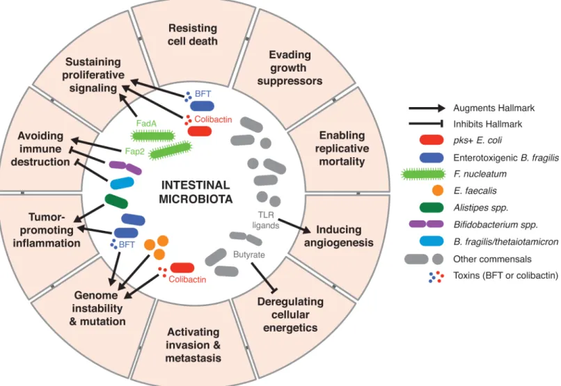
Fig 1andTable 1, influence the hallmarks of cancer.
How does the microbiota influence cellular proliferation and host cellular
energetics?
Normal tissues tightly regulate growth-promoting and death-inducing signals to maintain homeostatic cell densities, tissue architecture, and function. Dysregulation of these signaling pathways can lead to sustained cellular proliferation. The intercellular adhesion molecule, E-cadherin, is a common target engaged by intestinal bacteria that promotes epithelial prolifera-tion by activating the Wnt/ß-catenin pathway. For example, enterotoxigenicBacteroides fragi-lis(ETBF), resident among the microbiota of some individuals, secretesB.fragilistoxin (BFT)
a1111111111 a1111111111 a1111111111 a1111111111 a1111111111
OPEN ACCESS
Citation: Fulbright LE, Ellermann M, Arthur JC
(2017) The microbiome and the hallmarks of cancer. PLoS Pathog 13(9): e1006480.https://doi. org/10.1371/journal.ppat.1006480
Editor: John M. Leong, Tufts Univ School of
Medicine, UNITED STATES
Published: September 21, 2017
Copyright:©2017 Fulbright et al. This is an open access article distributed under the terms of the
Creative Commons Attribution License, which permits unrestricted use, distribution, and reproduction in any medium, provided the original author and source are credited.
Funding: This manuscript was supported by
funding from NIH NIDDK 1K01DK103952-01. The funders had no role in study design, data collection and analysis, decision to publish, or preparation of the manuscript.
Competing interests: The authors have declared
that promotes cleavage of E-cadherin [3]. This enables the nuclear translocation of ß-catenin, subsequent transcription of proto-oncogene c-Myc, and colonic epithelial hyperplasia [3]. Through a similar mechanism,Fusobacterium nucleatumenhances epithelial proliferation through engagement of its adhesin FadA with E-cadherin [4]. Neutralizing FadA abrogated the tumor-promoting activities ofF.nucleatumin a murine xenograft cancer model [4], dem-onstrating the potential of targeting bacterial interactions with E-cadherin as a novel strategy in mitigating cancer progression. Taken together, these studies demonstrate that the micro-biota can be a source of activating signals for aberrant epithelial proliferation as an initiating step in cancer development.
Cellular senescence—when cells cease to divide—is often considered a barrier for prolifera-tion. However, senescent cells secrete growth factors that enable tumor growth, and intestinal bacteria may induce this pathway to malignancy. Colibactin-producing (pks+) Escherichia coli induce a senescence-associated secretory phenotype (SASP) in which senescent cells secrete growth factors that stimulate epithelial proliferation and enhance tumor growth [5]. Thus, microbial-induced cellular senescence and bystander proliferation provide additional mecha-nisms by which malignancy can arise from host–microbial interactions.
Fig 1. Microbial-derived signals modulate numerous hallmarks of cancer through diverse mechanisms.
Perturbations to the local metabolic environment can also favor or inhibit sustained cancer cell proliferation and tumorigenesis. For example, the microbial metabolome has long been established as a modulator of host cellular metabolism. Short-chain fatty acids such as butyrate are generated through microbial fermentation of dietary fibers and are a preferred primary energy source for colonocytes. In contrast, cancer cells preferentially utilize glucose as a carbon source through glycolysis—a phenomenon known as the Warburg effect. Butyrate not only exerts an anticancer effect by starving cancer cells, but impaired butyrate metabolism increases intracellular concentrations of butyrate, which acts as a histone deacetylase inhibitor and pro-motes apoptosis and inhibition of cellular proliferation through epigenetic modifications [6]. Given the complexity of the microbial metabolome, it will be important to broaden our inves-tigation beyond individual metabolites and consider the impact of the metabolome as a whole on cellular energetics and other hallmarks of cancer.
How does the microbiota shape the local tumor microenvironment?
The microbiota influences cancer development by modulating the local tumor microenviron-ment through its effects on tissue remodeling and mucosal immunity. Angiogenesis, one aspect of tissue remodeling that occurs during tumorigenesis, enables adequate blood flow,
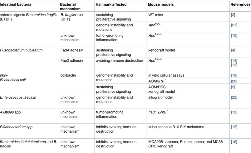
Table 1. Members of the intestinal microbiota associated with cancer development and resistance.
Intestinal bacteria Bacterial mechanism
Hallmark affected Mouse models References
enterotoxigenic Bacteroides fragilis (ETBF)
B. fragilis toxin
(BFT)
sustaining
proliferative signaling
WT mice [3]
genome instability and mutations
ApcMin/+ [21]
unknown mechanism
tumor-promoting inflammation
ApcMin/+ [10]
Fusobacterium nucleatum FadA adhesin sustaining
proliferative signaling
xenograft model [4]
Fap2 adhesin avoiding immune destruction ApcMin/+ [14]
[13]
pks+
Escherichia coli
colibactin genome instability and mutations
in vitro cellular assays [19]
AOM/Il10-/- [20]
sustaining
proliferative signaling
AOM/DSS xenograft model
[5]
Enterococcus faecalis unknown
mechanism
genome instability and mutations
allograft model [22]
Alistipes spp. unknown
mechanism
tumor-promoting inflammation
Il10-/-Lcn2-/- [12]
Bifidobacterium spp. unknown
mechanism
inhibits avoiding immune destruction
subcutaneous B16.SIY melanoma [15]
Bacteroides thetaiotamicron and B. fragilis
unknown mechanism
inhibits avoiding immune destruction
MCA205 sarcoma, Ret melanoma, and MC38 CRC xenograft
[16]
Abbreviations: AOM, azoxymethane; Apc, adenomatosis polyposis coli; CRC, colorectal cancer; DSS, dextran sodium sulfate; Il10, interleukin 10; Lcn2, lipocalin2; Min, multiple intestinal neoplasia
which is integral for tumor persistence and proliferation. Although direct links between endogenous bacteria and tumor-associated angiogenesis have not been reported, the micro-biota is required for normal development of the vasculature within the intestines [7]. More-over, in the context of infection, microbial products such as lipopolysaccharide engage with Toll-like receptors to promote angiogenesis, an effect that is augmented by damage-associated molecular patterns that may also be present within the tumor microenvironment [8]. Further studies will determine whether specific microbes influence angiogenesis and tumor-associated remodeling of the vasculature.
The close proximity of the microbiota and mucosal immune system also provides the potential for endogenous bacteria to impact the tumor microenvironment by stimulating a variety of protumorigenic immune responses. T-helper-17 (Th17) immunity is generally pro-tumorigenic, associated with worse prognosis in CRC, and driven by microbes and microbial products [9]. Colonization of tumor-susceptible adenomatosis polyposis coli–multiple intesti-nal neoplasia (ApcMin/+) mice with ETBF enhances Th17-driven inflammation and colonic tumor development [10] [11]. Blocking the interleukin-(IL)-17 signaling axis reduces down-stream signal transducer and activator of transcription factor 3 (STAT3) signaling in tumor and nontumor cells, thus preventing inflammation and tumorigenesis [10] [11]. Similarly, the carcinogenic potential of the intestinal commensalAlistipesis associated with enhanced IL-6 production, STAT3 activation, epithelial hyperplasia, and epithelial barrier dysfunction [12]. Thus, specific members of the microbiota stimulate Th17-driven inflammation and aid in establishing a tumor-permissive inflammatory environment.
While Th17 immune responses promote tumor development, others involving cytotoxic immune cells are essential for identifying and destroying precancerous and malignant cells.F. nucleatumdampens this arm of cancer immunity through 2 distinct mechanisms to enable tumor progression and persistence.F.nucleatumutilizes its Fap2 adhesion to silence the tumor-killing capabilities of cytotoxic immune cells through direct interaction with the immune inhibitory receptor T-cell immunoreceptor with immunoglobulin and immunore-ceptor tyrosine-based inhibitory motif domains (TIGIT) [13].F.nucleatumabundance is also correlated in clinical and animal studies with an enrichment of myeloid-derived suppressor cells and tumor-associated macrophages, both of which inhibit antitumor T-cell responses [14].
Does the microbiota promote genome instability and mutations?
The breakdown of genome maintenance within the host, whether through DNA damage accu-mulation or failure to properly segregate chromosomes, allows premalignant and malignant cells to both retain and accelerate the rate of mutations. Several gut microbes are a potential source of DNA mutagens. In vitro studies first demonstrated thatpks+ E.coliinduce DNA double-strand breaks, aneuploidy, cell-cycle arrest, and improper cellular division [19]. Multi-ple animal models of CRC have demonstrated thatpks+ E.colipromote DNA damage in vivo, yet inflammation remains unaffected and unlikely to be a driving force behind this damage [20] [5]. In contrast, other resident microbes can induce DNA damage by promoting inflam-mation and a pro-oxidant microenvironment. ETBF induces colonic epithelial expression of spermine oxidase (SMO), an enzyme that generates the DNA-damaging agent peroxide. Inhi-bition of SMO prevents ETBF-induced DNA damage, which corresponds with a decrease in ETBF-induced inflammation and tumorigenesis [21].Enterococcus faecalisinfected macro-phages promote DNA double-strand breaks, aneuploidy, and chromosomal instability in murine colonic epithelial cells, which, once transformed, initiate tumor formation in a murine allograft model [22]. The ability of microbes to both directly and indirectly cause DNA damage and genomic instability make the microbiome both a potential risk factor and therapeutic target.
Conclusions
The densest populations of endogenous microbes are found within the intestines and are in close proximity to the epithelium and underlying mucosal immune system. As a result, the ear-liest observations linking the microbiota with the hallmarks of cancer have primarily focused on gastric cancers and CRC. Nonetheless, more recent studies have also implicated the micro-biota in cancers at distal sites as a potential predictor of successful response to cancer therapy and as a means to augment the efficacy of existing anticancer therapeutics. Furthermore, the well-established link between several viruses and human cancers (i.e., Human papillomavirus and cervical, genital, anal, and oral cancers; Epstein-Barr virus and lymphomas; hepatitis C virus and hepatocellular carcinoma; Kaposi’s sarcoma–associated herpesvirus and Kaposi’s sarcoma) provides a strong rationale to investigate the role of nonbacterial members of the microbiota (virus, fungi, and archaea) in modulating the hallmark capabilities and cancer development. Finally, the cancer microenvironment itself can enhance the procarcinogenic activities of the microbiota [23], which further demonstrates the importance of the crosstalk between host and microbe in modulating cancer progression. In summary, because of the extensive capacity of the microbiota to influence many hallmarks of cancer, treatment for a variety of cancers may soon involve personalized medicine targeting the microbiota.
References
1. Hanahan D, Weinberg RA. Hallmarks of cancer: the next generation. Cell. Elsevier; 2011; 144: 646– 674.https://doi.org/10.1016/j.cell.2011.02.013PMID:21376230
2. Marshall BJ. The 1995 Albert Lasker Medical Research Award. Helicobacter pylori. The etiologic agent for peptic ulcer. JAMA: The Journal of the American Medical Association. 1995; 274: 1064–1066. https://doi.org/10.1001/jama.274.13.1064PMID:7563460
3. Rhee KJ, Wu S, Wu X, Huso DL, Karim B, Franco AA, et al. Induction of Persistent Colitis by a Human Commensal, Enterotoxigenic Bacteroides fragilis, in Wild-Type C57BL/6 Mice. Infect Immun. 2009; 77: 1708–1718.https://doi.org/10.1128/IAI.00814-08PMID:19188353
5. Cougnoux A, Dalmasso G, Martinez R, Buc E, Delmas J, Gibold L, et al. Bacterial genotoxin colibactin promotes colon tumour growth by inducing a senescence-associated secretory phenotype. Gut. BMJ Publishing Group; 2014; 63: 1932–1942.https://doi.org/10.1136/gutjnl-2013-305257PMID:24658599 6. Donohoe DR, Holley D, Collins LB, Montgomery SA, Whitmore AC, Hillhouse A, et al. A gnotobiotic
mouse model demonstrates that dietary fiber protects against colorectal tumorigenesis in a microbiota-and butyrate-dependent manner. Cancer Discovery. American Association for Cancer Research; 2014; 4: 1387–1397.https://doi.org/10.1158/2159-8290.CD-14-0501PMID:25266735
7. Stappenbeck TS, Hooper LV, Gordon JI. Developmental regulation of intestinal angiogenesis by indige-nous microbes via Paneth cells. Proc Natl Acad Sci USA. National Acad Sciences; 2002; 99: 15451– 15455.https://doi.org/10.1073/pnas.202604299PMID:12432102
8. Osherov N, Ben-Ami R. Modulation of Host Angiogenesis as a Microbial Survival Strategy and Thera-peutic Target. Sheppard DC, editor. PLoS Pathog. 2016; 12: e1005479–8.https://doi.org/10.1371/ journal.ppat.1005479PMID:27078259
9. Grivennikov SI, Wang K, Mucida D, Stewart CA, Schnabl B, Jauch D, et al. Adenoma-linked barrier defects and microbial products drive IL-23/IL-17-mediated tumour growth. Nature. 2012; 10: 789–15. https://doi.org/10.1038/nature11465
10. Wu S, Rhee K-J, Albesiano E, Rabizadeh S, Wu X, Yen H-R, et al. A human colonic commensal pro-motes colon tumorigenesis via activation of T helper type 17 T cell responses. Nat Med. Nature Publish-ing Group; 2009;: 1–8.https://doi.org/10.1038/nm0109-1
11. Housseau F, Wu S, Wick EC, Fan H, Wu X, Llosa NJ, et al. Redundant Innate and Adaptive Sources of IL17 Production Drive Colon Tumorigenesis. Cancer Research. American Association for Cancer Research; 2016; 76: 2115–2124.https://doi.org/10.1158/0008-5472.CAN-15-0749PMID:26880802 12. Moschen AR, Gerner RR, Wang J, Klepsch V, Adolph TE, Reider SJ, et al. Lipocalin 2 protects from
inflammation and tumorigenesis associated with gut microbiota alterations. Cell Host Microbe. 2016; 19: 455–469.https://doi.org/10.1016/j.chom.2016.03.007PMID:27078067
13. Gur C, Ibrahim Y, Isaacson B, Yamin R, Abed J, Gamliel M, et al. Binding of the Fap2 Protein of Fuso-bacterium nucleatum to Human Inhibitory Receptor TIGIT Protects Tumors from Immune Cell Attack. Immunity. 2015; 42: 344–355.https://doi.org/10.1016/j.immuni.2015.01.010PMID:25680274 14. Kostic AD, Chun E, Robertson L, Glickman JN, Gallini CA, Michaud M, et al. Fusobacterium nucleatum
Potentiates Intestinal Tumorigenesis and Modulates the Tumor-Immune Microenvironment. Cell Host Microbe. 2013; 14: 207–215.https://doi.org/10.1016/j.chom.2013.07.007PMID:23954159
15. Sivan A, Corrales L, Hubert N, Williams JB, Aquino-Michaels K, Earley ZM, et al. Commensal Bifidobac-terium promotes antitumor immunity and facilitates anti-PD-L1 efficacy. Science. American Association for the Advancement of Science; 2015; 350: 1084–1089.https://doi.org/10.1126/science.aac4255 PMID:26541606
16. Vetizou M, Pitt JM, Daillere R, Lepage P, Waldschmitt N, Flament C, et al. Anticancer immunotherapy by CTLA-4 blockade relies on the gut microbiota. Science. 2015; 350: 1079–1084.https://doi.org/10. 1126/science.aad1329PMID:26541610
17. Mazmanian SK, Liu CH, Tzianabos AO, Kasper DL. An Immunomodulatory Molecule of Symbiotic Bac-teria Directs Maturation of the Host Immune System. Cell. 2005; 122: 107–118.https://doi.org/10.1016/ j.cell.2005.05.007PMID:16009137
18. Mazmanian SK, Round JL, Kasper DL. A microbial symbiosis factor prevents intestinal inflammatory disease. Nature. 2008; 453: 620–625.https://doi.org/10.1038/nature07008PMID:18509436 19. Nougayrède J-P, Homburg S, Taieb F, Boury M, Brzuszkiewicz E, Gottschalk G, et al. Escherichia coli
induces DNA double-strand breaks in eukaryotic cells. Science. 2006; 313: 848–851.https://doi.org/10. 1126/science.1127059PMID:16902142
20. Arthur JC, Perez-Chanona E, Mu¨hlbauer M, Tomkovich S, Uronis JM, Fan T-J, et al. Intestinal inflam-mation targets cancer-inducing activity of the microbiota. Science. American Association for the Advancement of Science; 2012; 338: 120–123.https://doi.org/10.1126/science.1224820PMID: 22903521
21. Goodwin AC, DeStefano Shields CE, Wu S, Huso DL, Wu X, Murray-Stewart TR, et al. Polyamine catabolism contributes to enterotoxigenic Bacteroides fragilis-induced colon tumorigenesis. Proceed-ings of the National Academy of Sciences. National Acad Sciences; 2011; 108: 15354–15359.https:// doi.org/10.1073/pnas.1010203108PMID:21876161
22. Wang X, Yang Y, Huycke MM. Commensal bacteria drive endogenous transformation and tumour stem cell marker expression through a bystander effect. Gut. BMJ Publishing Group; 2015; 64: 459–468. https://doi.org/10.1136/gutjnl-2014-307213PMID:24906974