Small Animal Models for Human Immunodeficiency Virus (HIV),
Hepati-tis B, and Tuberculosis: Proceedings of an NIAID Workshop
Ramesh Akkina
1, Daniel L. Barber
2, Moses T. Bility
3, Karl-Dimiter Bissig
4, Benjamin J. Burwitz
5,
Katrin Eichelberg
2, Janice J. Endsley
6, J. Victor Garcia
7, Richard Hafner
2, Petros C. Karakousis
8,
Brent E. Korba
9, Rajen Koshy
2, Chris Lambros
2, Stephan Menne
9, Eric L. Nuermberger
8,
Alexander Ploss
10, Brendan K. Podell
1, Larisa Y. Poluektova
11, Brigitte E. Sanders-Beer
2,*,
Selvakumar Subbian
12and Angela Wahl
71
Department of Microbiology, Immunology and Pathology, Colorado State University, Fort Collins, Colorado, USA; 2
National Institute of Allergy and Infectious Diseases (NIAID), National Institutes of Health, Bethesda, Maryland, USA; 3Department of Infectious Diseases and Microbiology, University of Pittsburgh, Pittsburgh, Pennsylvania, USA; 4Department of Pediatrics, Duke University, Durham, North Carolina, USA; 5Vaccine and Gene Therapy Institute, Ore-gon Health and Science University, Beaverton, OreOre-gon, USA; 6Department of Microbiology and Immunology, Univer-sity of Texas Medical Branch, Galveston, Texas, USA; 7Division of Infectious Diseases, Center for AIDS Research, Uni-versity of North Carolina at Chapel Hill, North Carolina, USA; 8Division of Infectious Diseases, Center for Tuberculo-sis Research,Johns Hopkins University School of Medicine, Baltimore, Maryland, USA; 9Department of Microbiology & Immunology, Georgetown University, Washington, DC, USA; 10Department of Molecular Biology, Princeton Univer-sity, Princeton, New Jersey, USA; 11Department of Pharmacology and Experimental Neuroscience and Translational Mouse Model Core Facility, University of Nebraska Medical Center, Omaha, Nebraska, USA; 12The Public Health Re-search Institute Center of New Jersey Medical School, Rutgers University, Newark, New Jersey, USA
A R T I C L E H I S T O R Y
Received: October 20, 2019 Revised: November 27, 2019 Accepted: December 11, 2019
DOI:
10.2174/1570162X18666191223114019
Abstract: The main advantage of animal models of infectious diseases over in vitro studies is the gain in the understanding of the complex dynamics between the immune system and the pathogen. While small animal models have practical advantages over large animal models, it is crucial to be aware of their limitations. Although the small animal model at least needs to be susceptible to the pathogen under study to obtain meaningful data, key elements of pathogenesis should also be re-flected when compared to humans. Welldesigned small animal models for HIV, hepatitis viruses and tuberculosis require, additionally, a thorough understanding of the similarities and differences in the immune responses between humans and small animals and should incorporate that knowl-edge into the goals of the study. To discuss these considerations, the NIAID hosted a workshop on ‘Small Animal Models for HIV, Hepatitis B, and Tuberculosis’ on May 30, 2019. Highlights of the workshop are outlined below.
Keywords: HIV, AIDS, co-infections, HBV, tuberculosis, animal models.
1. INTRODUCTION
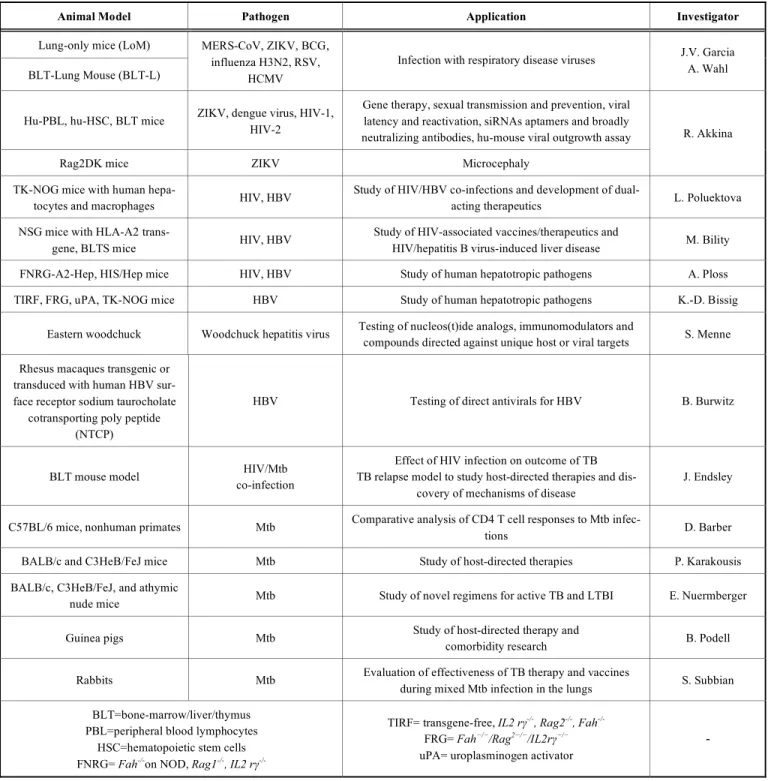
The meeting was convened to review the spectrum of small animal models for HIV and its frequently associated co-infections, hepatitis viruses and Mycobacterium tubercu-losis (Mtb), which are in the portfolio of the Division of AIDS, NIAID. The purpose of the meeting was to gain in-sight into the types of research that can be carried out in small animals (Table 1) to generate meaningful data and to understand when it would be appropriate to switch to larger animals, such as nonhuman primates (NHP).
*Address correspondence to this author at the Division of AIDS, National Institute of Allergy and Infectious Diseases, National Institutes of Health, 5601 Fishers Lane, Bethesda, MD 20892-9830, USA; Tel: (240) 627-3209; E-mail: [email protected]
2. SESSION 1: HIV AND EMERGING INFECTIONS
J. V. Garcia introduced new mouse models that can
Table 1. Small Animal Models Presented for HIV, HBV, and Mycobacterium tuberculosis infection.
Animal Model Pathogen Application Investigator
Lung-only mice (LoM)
BLT-Lung Mouse (BLT-L)
MERS-CoV, ZIKV, BCG, influenza H3N2, RSV,
HCMV
Infection with respiratory disease viruses J.V. Garcia A. Wahl
Hu-PBL, hu-HSC, BLT mice ZIKV, dengue virus, HIV-1, HIV-2
Gene therapy, sexual transmission and prevention, viral latency and reactivation, siRNAs aptamers and broadly neutralizing antibodies, hu-mouse viral outgrowth assay
Rag2DK mice ZIKV Microcephaly
R. Akkina
TK-NOG mice with human
hepa-tocytes and macrophages HIV, HBV
Study of HIV/HBV co-infections and development of
dual-acting therapeutics L. Poluektova
NSG mice with HLA-A2
trans-gene, BLTS mice HIV, HBV
Study of HIV-associated vaccines/therapeutics and
HIV/hepatitis B virus-induced liver disease M. Bility
FNRG-A2-Hep, HIS/Hep mice HIV, HBV Study of human hepatotropic pathogens A. Ploss
TIRF, FRG, uPA, TK-NOG mice HBV Study of human hepatotropic pathogens K.-D. Bissig
Eastern woodchuck Woodchuck hepatitis virus Testing of nucleos(t)ide analogs, immunomodulators and
compounds directed against unique host or viral targets S. Menne
Rhesus macaques transgenic or transduced with human HBV sur-face receptor sodium taurocholate
cotransporting poly peptide (NTCP)
HBV Testing of direct antivirals for HBV B. Burwitz
BLT mouse model HIV/Mtb
co-infection
Effect of HIV infection on outcome of TB TB relapse model to study host-directed therapies and
dis-covery of mechanisms of disease
J. Endsley
C57BL/6 mice, nonhuman primates Mtb Comparative analysis of CD4 T cell responses to Mtb
infec-tions D. Barber
BALB/c and C3HeB/FeJ mice Mtb Study of host-directed therapies P. Karakousis
BALB/c, C3HeB/FeJ, and athymic
nude mice Mtb Study of novel regimens for active TB and LTBI E. Nuermberger
Guinea pigs Mtb Study of host-directed therapy and
comorbidity research B. Podell
Rabbits Mtb Evaluation of effectiveness of TB therapy and vaccines
during mixed Mtb infection in the lungs S. Subbian
BLT=bone-marrow/liver/thymus PBL=peripheral blood lymphocytes
HSC=hematopoietic stem cells FNRG= Fah-/-on NOD, Rag1-/-,IL2 rγ
-/-TIRF= transgene-free, IL2 rγ-/-, Rag2-/-, Fah-/-
FRG= Fah−/−/Rag2−/−/IL2rγ−/−
uPA= uroplasminogen activator
-
the Lung mouse. Similar to the BLT model [3-5], BLT-Lung mice became reconstituted with human CD45 cells in the peripheral blood and tissues. For comparison, LoM and BLT-Lung mice were injected with luciferase-encoding HCMV directly into the lung organoid, and the infection was monitored through bioluminescence imaging. BLT-Lung mice, due to the presence of cellular immune responses, were able to control CMV infection whereas LoM were not. CD8+ T cells expressed IFNγ and CD107α, and CD4+ T cells ex-pressed IFNγ and TNFα following CMV peptide stimulation demonstrating that the T cells were functional. In addition,
HCMV neutralizing activity was detected in the plasma of the HCMV-exposed BLT-Lung mice. The BLT-Lung model could also potentially be used for HIV co-infections with TB or CMV.
Fig. (1). BLT/hu-Lung Mice. The BLT-Lung mouse model was created by combining the features of the Lung-only mouse (LoM) with the features of the BLT mouse. Thus, the BLT-Lung mouse model consists of both a systemic human immune system and non-hematopoietic human target cells.
[6, 7]. Another example was the use of hu-mouse models for the testing of oral pre-exposure prophylaxis strategies (PrEP). Results demonstrated that synergistic as well as an-tagonistic pharmacokinetic (PK) aspects can be delineated when different combinations of drugs, such as maraviroc, raltegravir, and tenofovir, are administered [8]. While HIV-1 strains are extensively studied, pathogenesis and preclinical studies on HIV-2, which also induces AIDS, have been lim-ited due to the lack of a suitable animal model. Dr. Akkina’s work with HIV-2 infected humanized hematopoietic stem cell (hu-HSC) mice demonstrated that they experienced per-sistent viremia and CD4 T cell loss, i.e., hallmarks of AIDS. Results also revealed that combinatorial ART suppresses HIV-2 infection and latent virus re-emerges after treatment interruption [9]. Besides HIV, viral pathogenesis and im-mune responses to Zika virus (ZIKV) can also be evaluated in BLT mice [10]. Future work is directed towards develop-ing a model for dengue secondary infection, which is an im-portant risk factor for dengue hemorrhagic fever and shock syndrome.
3. SESSION 2: HEPATITIS VIRUSES (HBV, HCV) AND HIV-HEPATITIS VIRUS CO-INFECTIONS
L. Poluektova focused on HIV-HBV coinfection models
for therapy. HIV and HBV display similarities in their repli-cation cycles as they both have a reverse transcription (RT) step. Therapeutics approved for HBV mainly include nu-cleos(t)ide analogues inhibitors of RT, two of which (teno-fovir and lamivudine) also have anti-HIV activity. She re-ported that Long-Acting Slow Effective Release antiretrovi-ral therapeutics (LASER ART) control HBV replication in the liver of humanized mice [11, 12]. In rats, a single injec-tion of 75 mg/kg nanoformulated lamivudine persisted for 28 days in the peripheral blood with significant accumulation in spleen, liver, and lymph nodes. Similarly, a single injection of 75 mg/kg nanoformulated lamivudine suppressed HBV replication for 4 weeks in chronically HBV-infected TK-NOG mice transplanted with human hepatocytes. To
estab-lish an HIV/HBV co-infection model, TK-NOG mice were reconstituted with human hepatocytes as well as monocyte-derived macrophages [13]. It was found that the engrafted human cells suppressed HBV replication in hepatocyte transplanted mice; however, HIV infection counteracted the anti-HBV activity of the human cells. In summary, co-transplantation of hepatocytes and immune cells or combina-tions of antigen-presenting cells (APCs) and lymphocytes injected into mice can create a model for studying HIV/HBV co-infections, supporting the development of dual-acting therapeutics.
M. Bility presented a humanized mouse model that is suit-able for HBV infection by reconstituting immunodeficient A2/NSG mice [NOD.Cg-Prkdc(scid) Il2rγ (tm1Wjl)/ SzJ mice with human HLA-A2 transgene] with human hematopoietic stem cells and liver progenitor cells (A2/NSG-hu HSC/Hep mice) [14, 15]. He also discussed the bone marrow-liver-thymus-spleen (BLTS)-humanized mouse model, which rep-resents a significant improvement over existing models, with notably “improved” human myeloid cells, IgG secreting-B cells, and primary and secondary lymphoid tissues and hu-man immunity [16, 17]. Adding huhu-man skin and liver cell grafts to the BLTS mouse model facilitates the study of HIV vaccines and therapeutics and hepatitis B virus-induced liver disease, respectively. Further enhancements of human xenograft animal models include immunodeficient rat and rabbit models, which may provide tools for developing ro-bust small animal models for long-term studies of HIV co-morbidities and associated end-organ disease.
Modeling (HIV-exacerbated) viral hepatitis in humanized mice was discussed by A. Ploss [18]. As mentioned earlier, human liver chimeric mice are a versatile model for studying human hepatotropic pathogens [19, 20]. FNRG-A2 xenore-cipients support robust co-engraftment with HLA-matched human hepatocytes and hematopoietic cells, and both FNRG-A2-Hep and human immune system (HIS)/Hep mice are susceptible to HBV infection (Fig. 2). FNRG-A2 HIS-Hep dually engrafted mice mount an HBV-specific CD8+ T
+++++++++++++++++++++++++++++++++++++++++++++++++++++++++++++++++++++++++++ +++++++++++++
BLT Mice
Systemic Human Immune System
BLT-Lung Mice
Systemic Human Immune System Non-Hematopoietic Human Target Cells
Autologous tissues
LoM
Fig. (2). Hu-Liver and HIS Mice. Dually engrafted mice have been generated by combining characteristics of human liver HEP-mice with features of human immune system mice. The HIS/HEP mice, therefore, allow susceptibility to human hepatotropic pathogens (HBV, HCV, HDV, HEV, etc.) and human lymphotropic pathogens (e.g., EBV, HIV) and can be used for evaluating the pathogenesis and immune re-sponses of hepatotropic pathogens and for preclinical testing of vaccine candidates.
cell response, creating the possibility of immuno-phenotyping as hepatitis B core antigen (HBcAg)-specific CD8+ T cells acquire an activated phenotype. HBV viremia is consistently lower in these dually engrafted animals, sug-gesting some level of immune control by the co-engrafted HIS mice. Expectedly, at six weeks post-HBV infection, only minor liver damage was observed in HBV-infected FNRG-A2 HIS-Hep mice. However, further improvements are needed to mimic human immunity in these humanized mice.
K.-D. Bissig also presented studies of HBV replication in human liver chimeric mice [21, 22]. Three requirements need to be met for successful human hepatocyte transplanta-tion: selection advantage of transplanted cells or selection disadvantage of endogenous cells, regeneration of the liver, and immunological tolerance. Human liver chimeric mice can be divided into genetic models (uPA, FRG, TK-NOG, PiZ, AFZ, etc.) or chemical models that utilize drugs to de-stroy mouse hepatocytes before transplantation of human liver cells. Limitations of the uPA mouse [23] include bleed-ing and neonatal death, a narrow window for transplantation of human hepatocytes, and a short life span after humaniza-tion. In the FRG [21] or transgene-free TIRF [24] mouse model (transgene-free, Il2 rγ-/-, Rag2-/-, Fah-/-), which is based on a tyrosine metabolism defect, the drug NTBC (ni-tisinone) compensates for the FAH deficiency, and selection pressure for transplanted human hepatocytes can be applied by removing the compound [21]. These mice can achieve greater than 95% humanization of the liver [22] and can be used to study the pathogenesis of viral hepatitis and to evalu-ate the engraftment of stem cell-derived and diseased hepa-tocytes, hepatitis-related gene therapies, and the metabolism of hepatitis drugs [25]. Higher HBV titers correlate with higher human chimerism. Variability from mouse-to-mouse and the lack of a human immune system were mentioned as limitations of the human chimeric liver mice.
S. Menne introduced the Eastern woodchuck (Marmota
monax) as an immunocompetent model for developing new HBV therapies [26, 27]. The woodchuck hepatitis virus (WHV), as well as HBV, belong to the genus Orthohepad-navirus in the family Hepadnaviridae. The genome structure, morphology and lifecycle of WHV and HBV are similar. Woodchuck infants infected with the strain WHV7 originat-ing from Maryland became positive for WHV DNA and surface antigen (WHsAg), with peaks 8-12 weeks after in-oculation. The majority of animals progress to chronicity (60-75%) and develop hepatocellular carcinoma later in life. As in HBV infection, covalently closed circular DNA (cccDNA) is produced. Acute infection period during the first year is suitable for studying early pathogenic features. One to 2.5 years after infection is the ideal time frame for evaluating antiviral immunotherapies, and antitumor immu-notherapies are usually tested 2.5 years post-infection. FDA-approved drugs, such as IFNα-2b, lamivudine, adefovir, en-tecavir, telbivudine, tenofovir, and other nucleos(t)ide ana-logs have been evaluated during their development phase in the woodchuck model. At present, the woodchuck model is used for testing immunomodulators (toll-like and RIG-I-like receptor agonists, AAV-encoded and recombinant IFN-α), checkpoint inhibitors (αPD-1), and compounds directed against unique host or viral targets [28, 29].
B. Burwitz is developing a rhesus macaque model of
chronic HBV infection because chimpanzees are no longer available for this type of research. The HBV surface recep-tor, human sodium taurocholate cotransporting polypeptide (hNTCP), differs by five amino acids within the HBV bind-ing region between humans and macaques. Due to one of these amino acid differences (G158R), HBV cannot bind to macaque NTCP. Dr. Burwitz demonstrated that macaque hepatocytes expressing hNTCP can support full HBV repli-cation cycle following HBV binding and entry [30]. He con-cluded that his laboratory now has an HBV-susceptible pri-mate model for testing direct-acting antivirals.
A
Human
hepatocytes
B
Human
immune system
Human liver chimeric mice
HIS-mice
C
Human
immune system & liver
HIS/HEP-mice
Human hepatotropic pathogens(HBV, HCV, HDV, HEV, malaria causing parasites), DMPK,
toxicology
Human lymphotropic pathogens (e.g. EBV, HIV…)
4. SESSION 3: MYCOBACTERIUM TUBERCULOSIS
(MTB) INFECTION OR HIV-MTB CO-INFECTIONS
J. Endsley has employed the BLT humanized mouse
model for studying the complex molecular and immunologi-cal interactions of HIV/TB co-infection [31-33]. BLT mice develop poorly organized necrotic granulomas in response to Mtb infection. The subsequent induction of HIV co-receptors and proinflammatory response (IL-1β and TNFα) is associ-ated with HIV replication at the TB granuloma site. HIV/TB co-infected mice have increased mycobacterial growth in the lung, and histologically the granulomas are larger and more inflamed compared to mice infected with Mtb alone. While exploring the mechanisms of these pro-inflammatory out-comes, Dr. Endsley found that mycobacterial exposure acti-vates CLEC10a, a macrophage galactose-type lectin (MGL) selectively expressed on activated M2 macrophages and dendritic cells. However, CLEC10a is strongly downregu-lated in HIV/TB co-infected mice along with activation of proinflammatory response. Therefore, MGL may play an important immune role in TB through anti-inflammatory and/or antibacterial mechanisms because proinflammatory cytokines are induced in MGL knock-out mice infected with MTB [34] . Additionally, Dr. Endsley developed a small animal model of TB relapse as a result of HIV infection, in which BLT mice were infected with Mtb and, when they progressed to active disease, they were treated with rifampin and isoniazid for 2 months. Mice with paucibacillary Mtb infection following drug treatment were found to relapse upon infection with HIV-1 (JR-CSF).
D. Barber studied T cell migration/differentiation and
spatial localization of CD4 T cells at sites of bacterial repli-cation and identified new CD4 T cell molecules associated with protection against Mtb infection in mice and macaques. He pointed out that CD4 T cells are critical for the contain-ment of TB because they provide help to Mtb-infected macrophages. In mice, less-differentiated CXCR3+ Th1 cells migrate into the lung parenchyma and protect against Mtb infection, while CX3CR1+ terminal effector cells accumu-late in the blood vessels and do not contribute to control of the infection [35, 36]. However unlike in mice, in monkeys, Mtb-specific CD4 T cells are largely CXCR3+ and CXCR3+CCR6+ (Th1*) cells, and neither develop a CX3CR1+ terminal effector phenotype nor accumulate in the blood vessels [37]. Collectively, it seems that mice make an overly Th1-polarized T-cell response during Mtb infection. Another aspect of Dr. Barber’s research is the identification of new CD4 T-cell molecules that are associated with protec-tion against Mtb infecprotec-tion. In mice, CD153 (CD30L) was preferentially expressed by protective lung parenchymal CD4 T cells and was required for host survival of Mtb tion as CD153-deficient mice succumbed early to Mtb infec-tion [38]. Similar to mice, NHP and human Mtb-specific CD4 T cells expressed CD153 and its expression correlated with better outcomes. He concluded that there are major dif-ferences in the quality of T-cell polarization between mice and primates, leading to major differences in function and migration.
P. Karakousis showed interest in host-directed therapies (HDT) for TB since Mtb is known to subvert host responses and induce lung damage [39, 40]. Preclinical endpoints of
HDT include the assessment of bacterial clearance in the host and histological or radiographic assessment of lung in-flammation. Although vastly underutilized, invasive spi-rometry and unrestrained whole-body plethysmography can be used to measure lung function in small animals. The con-ventional mouse model of TB chemotherapy has been em-ployed to study the adjunctive activity of metformin [41], simvastatin [42, 43], and tofacitinib [44] (JAK inhibitor). Advantages include convenience, reduced cost, and avail-ability of immunological reagents; however, mouse TB le-sions (Fig. 3c) differ significantly from their human counter-parts, raising the question of whether this model is predictive for studying the activity of novel HDT agents against TB. Specifically, mouse TB lesions are similar in cellular compo-sition to human TB lesions, but tissue caseation and hypoxia and liquefaction/cavitation, pathological hallmarks of human TB, are absent. To overcome these deficiencies, the C3HeB/FeJ mouse model, which lacks the lpr1 gene, is in-creasingly being used as a preclinical drug screening model since these mice can develop necrotizing granulomas and even lung cavities upon Mtb infection [45, 46]. For example, ibuprofen adjunctive therapy resulted in significantly de-creased tissue bacillary loads and inde-creased survival in Mtb-infected C3HeB/FeJ mice. However, among all models pre-sented, the NHP model is believed to reproduce most accu-rately the clinical, histological, and microbiological charac-teristics of human active and latent Mtb infection (LTBI) (Fig. 3a).
E. Nuermberger also relies on mouse models for TB
regimen development with the goal of a) characterizing indi-vidual drug effects against Mtb, b) evaluating the activity of novel drug regimens (in models of active TB and LTBI), c) determining the contribution of individual drugs to multidrug regimens, and d) assessing the impact of caseous pathology (e.g., in C3HeB/FeJ mice) and immunodeficiency (e.g., in athymic nude mice). His laboratory developed a paucibacil-lary mouse model of LTBI to test the activity of clinical regimens, which accurately rank orders the regimens by rec-ommended treatment duration in clinical use [47, 48]. He also found that the duration of treatment needed to cure 50% of mice gave a good estimate of effective treatment durations in clinical use. The model predicts the excellent activity of short-course bedaquiline treatment for LTBI, including for close contacts of multidrug-resistant TB patients. Third, he showed that BALB/c mouse models of active TB demon-strated utility and predictive accuracy in the development of novel regimens for active TB. Moreover, C3HeB/FeJ mice offer additional value for assessing the impact of caseation (Fig. 3d) on drug and regimen efficacy and may be a useful model for studying cavitation [49, 50]. Progress has been made towards establishing a quantitative, PK/PD-based translational modeling and simulation platform for predicting clinical trial results using pre-clinical PK/PD and phase 1 PK data [51].
Fig. (3). Comparison of Mtb Lesions in Humans and Various Small Animal Models. The diversity of granulomas in various animal mod-els of TB is shown. Lesions in rhesus macaques (A) appear histopathologically remarkably analogous to human granulomas consisting of a central area of necrosis with a wall of macrophages surrounded by a cuff of leukocytes. Similar structures also develop in guinea pigs (B). In stark contrast, C57BL/6 mice (C) do not develop highly organized TB lesions, and C3HeB/FeJ mice (D) develop highly suppurative lesions with cavitations more closely resembling abscesses than caseation. Similar to NHP, rabbits (E) develop lesions with cavitation resembling human pathology.
lesions that form after development of the cell-mediated re-sponse and are dominated by IFNγ and regulatory cytokines, such as TGFβ [52, 53]. Due to necrotic lesions resembling human granulomas, this model has been primarily used for HDT, relapse models, and antimicrobial reduction [54]. In recent work, the guinea pig has also served as a model for comorbidity research to understand the mechanisms contrib-uting to a higher risk of active TB disease and the impact of risk factors on TB disease outcomes. Moreover, guinea pigs are also frequently used for metabolic research because, un-like other rodents, they have glucose and lipid metabolism similar to humans [55]. Accordingly, guinea pig has contrib-uted to the understanding of type 2 diabetes-TB comorbidity and represents an appropriate model for testing metformin or other repurposed metabolic drugs, which hold promise as
adjunctive TB therapies [41]. Despite the value of this spe-cies in the development of new therapeutic strategies, the guinea pig has limitations in that this species does not relia-bly develop LTBI or cavitation [56].
S. Subbian presented a pulmonary Mtb infection model
Fig. (4). Infectious Cycle of Mtb in Humans and Animal Models. This diagram shows the typical infectious cycle in mice, guinea pigs, rabbits, and nonhuman primates. All models develop primary TB and granulatomous responses ranging from granulomas resembling human pathology to pseudo-granulomas. Those from C3HeB/FeJ mice, rabbits, and NHP also evolve into cavitations that facilitate transmissions by aerosol. Paucibacillary infections and latency with the possibility of re-activation are also seen in rabbits and NHP.
developed LTBI spontaneously as defined by a lack of culti-vable bacteria in the lung homogenates. This latent infection can be reactivated by immune suppression with triamci-nolone (Kenalog®) or dexamethasone. Next, Dr. Subbian’s group set up a pulmonary infection model in rabbits with mixed Mtb strains (Mtb HN878 and CDC1551) to under-stand the effect of dual infection on the host response. Dr. Subbian’s team also examined the dose-dependent host re-sponse to MtbHN878 infection in rabbits with or without prior BCG-vaccination. His data demonstrated that the aero-sol inoculum dose of Mtb determined the course of initial infection towards either progressive disease or manifestation of infection and establishment of LTBI. The protective effi-cacy of BCG vaccines was also directly impacted by the Mtb challenge dose. In summary, the rabbit model of active TB and LTBI displayed distinct pathological features relevant to respective human conditions.
5. DISCUSSION
During the ‘HIV and Emerging Infections’ session, clari-fication was requested whether the human lung in the BLT-Lung model is ventilated and whether the movement of the cilia and mucus secretions are present. It was explained that lung organoids can be maintained for over one year and all normal lung functions are preserved. GvHD development is donor-dependent and is mainly seen when male mice are transplanted with female human tissues. The CD4/8 ratios in the lung tissue are more human-like than mouse-like.
During the ‘HBV’ session, questions pertained mostly to the generation of HBV humanized mouse models. Several transgenic and genetically modified immunodeficient mouse strains were presented that are invaluable for testing anti-HBV therapeutics. For the HIV/anti-HBV co-infection model, the
simultaneous transplantation of human hepatocytes and hu-man immune cells allows for modeling the interaction be-tween the two viruses and the interaction of the viruses with the immune system. Moreover, liver sections from human-ized mice carrying HBV and HIV are suitable for RNA tran-scriptome analysis, which can reveal the pathways of HBV replication control by immune cells. The drawback of a pos-sible allogeneic reaction was resolved with the FNRG-A2 mouse model where HLA-type prescreened donors were used for engraftment; HLA-A2-positive hepatocytes and hematopoietic stem cells can also be combined for dual in-fection.
For ‘Tuberculosis’ session, interest was expressed in fac-tors involved in Mtb re-activation in the HIV/TB co-infection model. Regarding the comparison of mouse and primate TB models, mice have IL-17 producing cells, but they are not bona fide Th17 cells. Also, the granulomas are histologically different between mice and primates. APC function in murine and primate systems is not well under-stood and was regarded as a potential gap area. Another issue raised was the timing of HDT and whether it should be ad-ministered before or after the TB-specific treatment. One observation that was made was that individuals with diabetes who were receiving metformin for at least three months be-fore TB treatment was initiated showed increased survival rates. In summary, the questions raised were mostly of tech-nical nature or regarded clarifications of scientific data pre-sented at the meeting.
Concerning the limitations of various animal models [59, 60], humanized mouse models and primates are the only choices for HIV/SIV and HBV due to their narrow host range. Humanized mouse models are less expensive than primate models, and HIV/HBV rather than SIV or surrogate hepatitis viruses can be used for infection. However, their
Infectious Cycle of
Mtb
in Humans and Animal Models
Exposure
(Mtb-Aerosol)
Infection
(All Models)
Paucibacillary/ Latency
Active Disease
(Mouse, Guinea pig, Rabbit, NHP)
Transmission
Cavitation
size, rate of metabolism, and longevity present some experi-mental challenges. More small animal model choices are available for TB, such as mouse, guinea pig, and rabbit mod-els, some of which recapitulate human pathogenesis, but they do not recapitulate human (HLA-restricted) immune re-sponses. However, TB infection in primates is still the model most comparable to humans.
CONCLUSION
Humanized mouse models are required for lympho- and hepatotropic infections, such as HIV and HBV and HIV/TB co-infections. Non-humanized animal models, such as C57BL6 and C3HeB/FeJ mice, guinea pigs, and rabbits can only accommodate Mtb infections, and not HIV or HBV/HCV.
AUTHOR CONTRIBUTIONS
R.A., D.L.B., M.T.B., K-D.B., B.J.B., J.J.E., J.V.G., P.C.K., S.M., E.L.N., A.P., B.K.P., L.Y.P., S.S., and A.W. presented original data at the workshop and edited the script. B.E.S. organized the workshop and wrote the manu-script. K.E., R.H., B.E.K., R.K., and C.L. chaired sessions, contributed to the discussions, and edited the manuscript.
CONSENT FOR PUBLICATION
Not applicable.
FUNDING
The workshop was funded with federal funds from the National Institute of Allergies and Infectious Diseases, Na-tional Institutes of Health, Department of Health and Human Services, under contract no. HHSN272201100001G.
CONFLICT OF INTEREST
R.A., D.L.B., M.T.B., K-D.B., B.J.B., J.J.E., J.V.G., P.C.K., S.M., E.L.N., A.P., B.K.P., L.Y.P., S.S., and A.W. receive NIH funds for generating the presented data. B.E.S, K.E., R.H., B.E.K., R.K., and C.L. do not have any conflicts of interest.
ACKNOWLEDGEMENTS
The authors would like to thank Cynthia Soriano for her support of the workshop and Dr. Sandra Bridges for helpful discussions. In addition, the authors would like to thank NI-AID Meet, in particular, Eric Delaney and Monica Washing-ton, for help in organizing the workshop.
REFERENCES
[1] Wahl A, De C, Abad Fernandez M, et al. Precision mouse models with expanded tropism for human pathogens. Nat Biotechnol 2019; 37(10): 1163-73.
http://dx.doi.org/10.1038/s41587-019-0225-9 PMID: 31451733 [2] Honeycutt JB, Garcia JV. Humanized mice: models for evaluating
NeuroHIV and cure strategies. J Neurovirol 2018; 24(2): 185-91. http://dx.doi.org/10.1007/s13365-017-0567-3 PMID: 28831774
[3] Kovarova M, Benhabbour SR, Massud I, et al. Ultra-long-acting removable drug delivery system for HIV treatment and prevention. Nat Commun 2018; 9(1): 4156.
http://dx.doi.org/10.1038/s41467-018-06490-w PMID: 30297889 [4] Denton PW, Garcia JV. Mucosal HIV-1 transmission and
preven-tion strategies in BLT humanized mice. Trends Microbiol 2012; 20(6): 268-74.
http://dx.doi.org/10.1016/j.tim.2012.03.007 PMID: 22503637 [5] Wahl A, Swanson MD, Nochi T, et al. Human breast milk and
antiretrovirals dramatically reduce oral HIV-1 transmission in BLT humanized mice. PLoS Pathog 2012; 8(6): e1002732.
http://dx.doi.org/10.1371/journal.ppat.1002732 PMID: 22737068 [6] Charlins P, Schmitt K, Remling-Mulder L, et al. A humanized
mouse-based HIV-1 viral outgrowth assay with higher sensitivity than in vitro qVOA in detecting latently infected cells from indi-viduals on ART with undetectable viral loads. Virology 2017; 507: 135-9.
http://dx.doi.org/10.1016/j.virol.2017.04.011 PMID: 28432928 [7] Schmitt K, Akkina R. Ultra-sensitive HIV-1 latency viral
out-growth assays using humanized mice. Front Immunol 2018; 9: 344. http://dx.doi.org/10.3389/fimmu.2018.00344 PMID: 29556230 [8] Thompson CG, Rosen EP, Prince HMA, et al. Heterogeneous
antiretroviral drug distribution and HIV/SHIV detection in the gut of three species. Sci Transl Med 2019; 11(499): eaap8758. http://dx.doi.org/10.1126/scitranslmed.aap8758 PMID: 31270274 [9] Hu S, Neff CP, Kumar DM, et al. A humanized mouse model for
HIV-2 infection and efficacy testing of a single-pill triple-drug combination anti-retroviral therapy. Virology 2017; 501: 115-8. http://dx.doi.org/10.1016/j.virol.2016.11.013 PMID: 27912079 [10] Schmitt K, Charlins P, Veselinovic M, et al. Zika viral infection
and neutralizing human antibody response in a BLT humanized mouse model. Virology 2018; 515: 235-42.
http://dx.doi.org/10.1016/j.virol.2017.12.026 PMID: 29310105 [11] Gendelman HE, McMillan J, Bade AN, Edagwa B, Kevadiya BD.
The promise of long-acting antiretroviral therapies: from need to manufacture. Trends Microbiol 2019; 27(7): 593-606.
http://dx.doi.org/10.1016/j.tim.2019.02.009 PMID: 30981593 [12] Edagwa B, McMillan J, Sillman B, Gendelman HE. Long-acting
slow effective release antiretroviral therapy. Expert Opin Drug De-liv 2017; 14(11): 1281-91.
http://dx.doi.org/10.1080/17425247.2017.1288212 PMID: 28128004
[13] Dagur RS, Wang W, Makarov E, Sun Y, Poluektova LY. Estab-lishment of the dual humanized TK-NOG mouse model for HIV-associated liver pathogenesis. J Vis Exp 2019; (151).
[14] Bility MT, Cheng L, Zhang Z, et al. Hepatitis B virus infection and immunopathogenesis in a humanized mouse model: Induction of human-specific liver fibrosis and M2-like macrophages. PLoS Pathog 2014; 10(3): e1004032.
http://dx.doi.org/10.1371/journal.ppat.1004032 PMID: 24651854 [15] Cheng L, Li F, Bility MT, Murphy CM, Su L. Modeling hepatitis B
virus infection, immunopathology and therapy in mice. Antiviral Res 2015; 121: 1-8.
http://dx.doi.org/10.1016/j.antiviral.2015.06.012 PMID: 26099683 [16] Samal J, Kelly S, Na-Shatal A, et al. Human immunodeficiency
virus infection induces lymphoid fibrosis in the BM-liver-thymus-spleen humanized mouse model. JCI Insight 2018; 3(18): 120430. http://dx.doi.org/10.1172/jci.insight.120430 PMID: 30232273 [17] Bility MT, Nio K, Li F, et al. Chronic hepatitis C infection-induced
liver fibrogenesis is associated with M2 macrophage activation. Sci Rep 2016; 6: 39520.
http://dx.doi.org/10.1038/srep39520 PMID: 28000758
[18] Winer BY, Ding Q, Gaska JM, Ploss A. In vivo models of hepatitis B and C virus infection. FEBS Lett 2016; 590(13): 1987-99. http://dx.doi.org/10.1002/1873-3468.12157 PMID: 27009462 [19] Douam F, Ploss A. The use of humanized mice for studies of viral
pathogenesis and immunity. Curr Opin Virol 2018; 29: 62-71. http://dx.doi.org/10.1016/j.coviro.2018.03.003 PMID: 29604551 [20] de Jong YP, Rice CM, Ploss A. New horizons for studying human
[21] Bissig KD, Le TT, Woods NB, Verma IM. Repopulation of adult and neonatal mice with human hepatocytes: A chimeric animal model. Proc Natl Acad Sci USA 2007; 104(51): 20507-11. http://dx.doi.org/10.1073/pnas.0710528105 PMID: 18077355 [22] Bissig KD, Wieland SF, Tran P, et al. Human liver chimeric mice
provide a model for hepatitis B and C virus infection and treatment. J Clin Invest 2010; 120(3): 924-30.
http://dx.doi.org/10.1172/JCI40094 PMID: 20179355
[23] Dandri M, Burda MR, Török E, et al. Repopulation of mouse liver with human hepatocytes and in vivo infection with hepatitis B vi-rus. Hepatology 2001; 33(4): 981-8.
http://dx.doi.org/10.1053/jhep.2001.23314 PMID: 11283864 [24] Barzi M, Pankowicz FP, Zorman B, et al. A novel humanized
mouse lacking murine P450 oxidoreductase for studying human drug metabolism. Nat Commun 2017; 8(1): 39.
http://dx.doi.org/10.1038/s41467-017-00049-x PMID: 28659616 [25] Bissig-Choisat B, Wang L, Legras X, et al. Development and
res-cue of human familial hypercholesterolaemia in a xenograft mouse model. Nat Commun 2015; 6: 7339.
http://dx.doi.org/10.1038/ncomms8339 PMID: 26081744 [26] Menne S, Cote PJ. The woodchuck as an animal model for
patho-genesis and therapy of chronic hepatitis B virus infection. World J Gastroenterol 2007; 13(1): 104-24.
http://dx.doi.org/10.3748/wjg.v13.i1.104 PMID: 17206759 [27] Alioto TS, Cruz F, Gomez-Garrido J, et al. The genome sequence
of the Eastern woodchuck (Marmota monax): A preclinical animal model for chronic hepatitis B. G3 (Bethesda) 2019; 9(12): 3943-52. [28] Suslov A, Wieland S, Menne S. Modulators of innate immunity as novel therapeutics for treatment of chronic hepatitis B. Curr Opin Virol 2018; 30: 9-17.
http://dx.doi.org/10.1016/j.coviro.2018.01.008 PMID: 29444493 [29] Korolowizc KE, Li B, Huang X, et al. Liver-targeted toll-like
re-ceptor 7 agonist combined with entecavir promotes a functional cure in the woodchuck model of hepatitis B virus. Hepatol Com-mun 2019; 3(10): 1296-310.
http://dx.doi.org/10.1002/hep4.1397 PMID: 31592075
[30] Burwitz BJ, Wettengel JM, Mück-Häusl MA, et al. Hepatocytic expression of human sodium-taurocholate cotransporting polypep-tide enables hepatitis B virus infection of macaques. Nat Commun 2017; 8(1): 2146.
http://dx.doi.org/10.1038/s41467-017-01953-y PMID: 29247188 [31] Calderon VE, Valbuena G, Goez Y, et al. A humanized mouse
model of tuberculosis. PLoS One 2013; 8(5): e63331.
http://dx.doi.org/10.1371/journal.pone.0063331 PMID: 23691024 [32] Nusbaum RJ, Calderon VE, Huante MB, et al. Pulmonary
tubercu-losis in humanized mice infected with HIV-1. Sci Rep 2016; 6: 21522.
http://dx.doi.org/10.1038/srep21522 PMID: 26908312
[33] Huante M, Nusbaum R, Endsley J. Co-infection with TB and HIV: converging epidemics, clinical challenges, and microbial synergy. 2019; pp. 123-53.
[34] Jondle CN, Sharma A, Simonson TJ, Larson B, Mishra BB, Sharma J. Macrophage galactose-type lectin-1 deficiency is associ-ated with increased neutrophilia and hyperinflammation in gram-negative pneumonia. 2016; 196(7): 3088-96.
http://dx.doi.org/10.4049/jimmunol.1501790
[35] Sakai S, Kauffman KD, Schenkel JM, McBerry CC, Mayer-Barber KD, Masopust D, et al. Cutting edge: control of Mycobacterium tuberculosis infection by a subset of lung parenchyma-homing CD4 T cells. Journal of Immunology (Baltimore, Md : 1950) 2014; 192(7): 2965-9.
[36] Sakai S, Mayer-Barber KD, Barber DL. Defining features of pro-tective CD4 T cell responses to Mycobacterium tuberculosis. Curr Opin Immunol 2014; 29: 137-42.
http://dx.doi.org/10.1016/j.coi.2014.06.003 PMID: 25000593 [37] Kauffman KD, Sallin MA, Sakai S, et al. Defective positioning in
granulomas but not lung-homing limits CD4 T-cell interactions with Mycobacterium tuberculosis-infected macrophages in rhesus macaques. Mucosal Immunol 2018; 11(2): 462-73.
http://dx.doi.org/10.1038/mi.2017.60 PMID: 28745326
[38] Sallin MA, Kauffman KD, Riou C, et al. Host resistance to pulmo-nary Mycobacterium tuberculosis infection requires CD153 expres-sion. Nat Microbiol 2018; 3(11): 1198-205.
http://dx.doi.org/10.1038/s41564-018-0231-6 PMID: 30202016 [39] Kolloli A, Subbian S. Host-directed therapeutic strategies for
tu-berculosis. Front Med (Lausanne) 2017; 4: 171.
http://dx.doi.org/10.3389/fmed.2017.00171 PMID: 29094039 [40] Frank DJ, Horne DJ, Dutta NK, et al. Remembering the host in
tuberculosis drug development. J Infect Dis 2019; 219(10): 1518-24.
http://dx.doi.org/10.1093/infdis/jiy712 PMID: 30590592
[41] Singhal A, Jie L, Kumar P, et al. Metformin as adjunct antitubercu-losis therapy. Sci Transl Med 2014; 6(263): 263ra159.
http://dx.doi.org/10.1126/scitranslmed.3009885 PMID: 25411472 [42] Dutta NK, Bruiners N, Pinn ML, et al. Statin adjunctive therapy
shortens the duration of TB treatment in mice. J Antimicrob Che-mother 2016; 71(6): 1570-7.
http://dx.doi.org/10.1093/jac/dkw014 PMID: 26903278
[43] Dutta NK, Bruiners N, Zimmerman MD, et al. Adjunctive host-directed therapy with statins improves tuberculosis-related out-comes in mice. J Infect Dis 2019; pii: jiz517.
http://dx.doi.org/10.1093/infdis/jiz517 PMID: 31605489
[44] Maiga M, Ahidjo BA, Maiga MC, et al. Efficacy of adjunctive tofacitinib therapy in mouse models of tuberculosis. EBioMedicine 2015; 2(8): 868-73.
http://dx.doi.org/10.1016/j.ebiom.2015.07.014 PMID: 26425693 [45] Vilaplana C, Marzo E, Tapia G, Diaz J, Garcia V, Cardona PJ.
Ibuprofen therapy resulted in significantly decreased tissue bacil-lary loads and increased survival in a new murine experimental model of active tuberculosis. J Infect Dis 2013; 208(2): 199-202. http://dx.doi.org/10.1093/infdis/jit152 PMID: 23564636
[46] Apt A, Kramnik I. Man and mouse TB: contradictions and solu-tions. Tuberculosis (Edinb) 2009; 89(3): 195-8.
http://dx.doi.org/10.1016/j.tube.2009.02.002 PMID: 19345146 [47] Nuermberger EL. Preclinical efficacy testing of new drug
candi-dates. Microbiol Spectr 2017; 5(3).
http://dx.doi.org/10.1128/microbiolspec.TBTB2-0034-2017 PMID: 28643624
[48] Zhang T, Li SY, Williams KN, Andries K, Nuermberger EL. Short-course chemotherapy with TMC207 and rifapentine in a murine model of latent tuberculosis infection. Am J Respir Crit Care Med 2011; 184(6): 732-7.
http://dx.doi.org/10.1164/rccm.201103-0397OC PMID: 21659613 [49] Dartois V. The path of anti-tuberculosis drugs: From blood to
le-sions to mycobacterial cells. Nat Rev Microbiol 2014; 12(3): 159-67.
http://dx.doi.org/10.1038/nrmicro3200 PMID: 24487820
[50] Lanoix JP, Ioerger T, Ormond A, et al. Selective inactivity of pyrazinamide against tuberculosis in C3HeB/FeJ mice is best ex-plained by neutral pH of caseum. Antimicrob Agents Chemother 2015; 60(2): 735-43.
http://dx.doi.org/10.1128/AAC.01370-15 PMID: 26574016 [51] Bartelink IH, Zhang N, Keizer RJ, et al. New paradigm for
transla-tional modeling to predict long-term tuberculosis treatment re-sponse. Clin Transl Sci 2017; 10(5): 366-79.
http://dx.doi.org/10.1111/cts.12472 PMID: 28561946
[52] Turner OC, Basaraba RJ, Orme IM. Immunopathogenesis of pul-monary granulomas in the guinea pig after infection with Mycobac-terium tuberculosis. Infect Immun 2003; 71(2): 864-71.
http://dx.doi.org/10.1128/IAI.71.2.864-871.2003 PMID: 12540568 [53] Ly LH, Russell MI, McMurray DN. Cytokine profiles in primary
and secondary pulmonary granulomas of Guinea pigs with tubercu-losis. Am J Respir Cell Mol Biol 2008; 38(4): 455-62.
http://dx.doi.org/10.1165/rcmb.2007-0326OC PMID: 18032570 [54] Kiran D, Podell BK, Chambers M, Basaraba RJ. Host-directed
therapy targeting the Mycobacterium tuberculosis granuloma: a re-view. Semin Immunopathol 2016; 38(2): 167-83.
http://dx.doi.org/10.1007/s00281-015-0537-x PMID: 26510950 [55] Fernandez ML. Guinea pigs as models for cholesterol and
lipopro-tein metabolism. J Nutr 2001; 131(1): 10-20. http://dx.doi.org/10.1093/jn/131.1.10 PMID: 11208932
[56] Ernst JD. The immunological life cycle of tuberculosis. Nat Rev Immunol 2012; 12(8): 581-91.
[57] Subbian S, Tsenova L, Yang G, et al. Chronic pulmonary cavitary tuberculosis in rabbits: A failed host immune response. Open Biol 2011; 1(4): 110016.
http://dx.doi.org/10.1098/rsob.110016 PMID: 22645653
[58] Esteves PJ, Abrantes J, Baldauf HM, et al. The wide utility of rabbits as models of human diseases. Exp Mol Med 2018; 50(5): 66.
http://dx.doi.org/10.1038/s12276-018-0094-1 PMID: 29789565
[59] Allen TM, Brehm MA, Bridges S, et al. Humanized immune sys-tem mouse models: Progress, challenges and opportunities. Nat Immunol 2019; 20(7): 770-4.
http://dx.doi.org/10.1038/s41590-019-0416-z PMID: 31160798 [60] Akkina R, Allam A, Balazs AB, et al. Improvements and
limita-tions of humanized mouse models for HIV research: NIH/NIAID “Meet the experts” 2015 workshop summary. AIDS Res Hum Ret-roviruses 2016; 32(2): 109-19.