IBD Serology and Disease Outcomes in African Americans With
Crohn’s Disease
Madeline Bertha, MD,
*
Arthi Vasantharoopan, MS,
*
Archana Kumar, MS,
*
Beau B. Bruce, MD/PhD,
*
Jarod Prince, BS,
*
Tatyana Hofmekler, MD,
*
David Okou, PhD,
*
Pankaj Chopra, PhD,
*
Gabriel Wang, BS,
*
Cary Sauer, MD,
*
Carol J. Landers, BS,
***
Sunny Z. Hussain, MD,
†Raymond K. Cross, MD,
‡Robert N. Baldassano, MD,
§Michael D. Kappelman, MD,
¶Jeffrey Katz, MD,
║Jonathan S. Alexander, PhD,
#Barbara S. Kirschner, MD,
**
Dedrick E. Moulton, MD,
††Bankole O. Osuntokun, MD,
‡‡Ashish Patel, MD,
§§Shehzad Saeed, MD,
¶¶Jan-Michael A. Klapproth, MD,
║║Tanvi A. Dhere, MD,
*
Marla C. Dubinsky, MD,
##Dermot McGovern, MD/PhD,
***
and Subra Kugathasan, MD
*
Backgrounds: Recent studies have identified the role of serologic markers in characterizing disease phenotype, location, complications, and sever-ity among Northern Europeans (NE) with Crohn’s disease (CD). However, very little is known about the role of serology in CD among African Americans (AA). Our study explored the relationship between serology and disease phenotype in AA with CD, while controlling for genetic ancestry. Methods: AAs with CD were enrolled as participants through multicenter collaborative efforts. Serological levels of IgA anti-Saccharomyces cervisiae antibody (ASCA), IgG ASCA, E. coli outermembrane porin C, anti-CBir1, and ANCA were measured using enzyme-linked immuno-sorbent assays. Genotyping was performed using Illumina immunochip technology; an admixture rate was calculated for each subject. Multiple imputation by chained equations was performed to account for data missing at random. Logistic regression was used to calculate adjusted odds ratio (OR) for associations between serological markers and both complicated disease and disease requiring surgery.
Results: A total of 358 patients were included in the analysis. The majority of our patients had inflammatory, noncomplicated disease (58.4%), perianal disease (55.7%), and documented colonic inflammation (86.8%). On multivariable analysis, both IgG ASCA and OmpC were associated with compli-cated disease (OR, 2.67; 95% CI, 1.67–4.28; OR, 2.23; 95% CI, 1.41–3.53, respectively) and disease requiring surgery (OR, 2.51; 95% CI, 1.49–4.22; OR, 3.57; 95% CI, 2.12–6.00). NE admixture to the African genome did not have any associations or interactions in relation to clinical outcome.
Conclusions: Our study comprises the largest cohort of AAs with CD. The utility of serological markers for the prognosis of CD in NE applies equally to AA populations.
Key Words:African Americans, Crohn’s disease, inflammatory bowel disease, serology
C
rohn’s disease (CD) encompasses a spectrum of chronic, relapsing, and remitting inflammatory symptoms that affect pediatric and adult populations, with the peak incidence occurring in adolescents and young adults. Recent studies have explored the relationship between genetic, microbial, and immune responses in CD, including the role of antimicro-bial serologies in characterizing disease phenotype, clinical course, and natural history.1–4 In particular, significantasso-ciations have been identified between the presence of anti-bodies to Saccharomyces cerevisiae (ASCA IgA and ASCA IgG), antibodies to Escherichia coli outer membrane porin C (anti-OmpC), antibodies to CBIR1 flagellin (anti-CBir1) and
Supplemental digital content is available for this article. Direct URL citations appear in the printed text and are provided in the HTML and PDF versions of this article on the journal’s Web site (www.ibdjournal.org).
Received for publication April 10, 2017; Editorial Decision July 13, 2017 From the *Department of Pediatrics, Emory University School of Medicine and Children’s Healthcare of Atlanta, Georgia; †Department of Pediatrics,
Willis-Knighton Physician Network, Shreveport, Louisiana; ‡Division of Gastroenterology,
University of Maryland, Baltimore, Maryland; §Division of Gastroenterology
and Nutrition, Children’s Hospital of Philadelphia, Philadelphia, Pennsylvania;
¶Department of Pediatrics, University of North Carolina at Chapel Hill, Chapel Hill,
North Carolina; ║Division of Gastroenterology, Case Western Reserve University,
Cleveland, Ohio; #Louisiana State University Health Sciences Center, Shreveport,
Louisiana; **Department of Pediatrics, University of Chicago Comer Children’s Hospital, Chicago, Illinois; ††Division of Gastroenterology, Vanderbilt Children’s
Hospital, Nashville, Tennessee; ‡‡Department of Pediatrics, Cook Children’s
Medical Center, Fort Worth, Texas; §§Department of Pediatrics, University of
Texas Southwestern Medical Center, Dallas, Texas; ¶¶Division of Gastroenterology,
Hepatology and Nutrition, Cincinnati Children’s Hospital Medical Center, Cincinnati, Ohio; ║║Division of Digestive Diseases, Emory University School of Medicine, Atlanta,
Georgia; ##Departments of Pediatrics, Icahn School of Medicine, Mount Sinai, New
York, New York; ***F. Widjaja Foundation Inflammatory Bowel and Immunobiology Research Institute, Cedars Sinai Medical Center, Los Angelas, California.
Conflicts of interest: The authors have no financial relationships relevant to this article to disclose. The authors have no conflicts of interest to disclose.
Supported by NIH/NIDDK R01 DK087694 (S.K.) and National Center for Advancing Translational Sciences of National Institute of Health Award Number UL1TR000454 (M.B.).
Dermot McGovern and Subra Kugathasan contributed equally and share senior authorship.
Address correspondence to: Subra Kugathasan, MD, Emory University School of Medicine, Division of Pediatric Gastroenterology, Department of Pediatrics, 2015, Uppergate Drive, Room 248, Atlanta, GA 30322 ([email protected]). doi: 10.1093/ibd/izx021
perinuclear anti-nuclear cytoplasmic antibodies (pANCA) with disease phenotype and clinical outcomes.5 ASCA, anti-CBir1,
and anti-OmpC positivities have been associated with ileal dis-ease, stricturing, and/or penetrating disdis-ease, a higher risk for surgery, and earlier disease onset, while ANCA positivity has been associated with colonic disease and a more “benign” dis-ease course.4 The vast majority of these studies have originated
from North America and Europe and have primarily focused on patient populations of Caucasians of Western European decent. Although the findings of these studies have contributed to our understanding of CD pathogenesis as well as to the devel-opment of tools both for differentiating types of inflammatory bowel disease (IBD) and predicting risk stratification, t hese findings need to be explored in non-Caucasian populations.1
An understanding of the relevance of these markers in demo-graphically diverse groups is necessary if the potential clinical utility of IBD serology is to be extended to all populations.
Historically considered a disease of Northern European (NE) ancestries, it is now recognized that CD affects every race and ethnicity, with a rising incidence in non-European populations. Furthermore, CD characteristics, treatment responses, and disease course can vary in different popula-tions.6–8 Specifically, t he e pidemiology, g enetics, a nd n atural
history of IBD among African Americans (AAs) remains severely understudied.6–8 With regards to IBD-associated
serology in AA, there has only been 1 study to date, which identified that ASCA has a similar sensitivity but lower spe-cificity for C D, a s w ell a s a n a ssociation w ith i leal i nvolve-ment, complicated behavior, and surgery in AAs with CD.9
In addition to limiting investigations on ASCA alone, this study was further limited by its reliance on self-reported eth-nicity. AAs are an admixed population, and the AA genome on average is comprised of 80% West African ancestry and 20% European ancestry.10 Due to differences in environmental
and genetic influences a mong v arious e thnic p opulations, there is a need to further delineate the diagnostic value of these biomarkers among AA.4 Although genetic susceptibility
among Caucasians and AAs is mostly similar, differences exist, including the association with NOD2 mutations, which is less influential in AAs, possibly due to Caucasian admixture among AAs. Hence, exploration of these serological markers in AAs among CD phenotypes may allow clinicians to risk stratify in an important and growing demographic of IBD patients. Our study was designed to explore the relationship between antibodies and disease phenotype in AAs with CD while controlling for genetic ancestry.
MATERIALS AND METHODS
Study Design and Hypothesis
We conducted a cross-sectional study testing the hypoth-esis that serological levels of IgA ASCA, IgG ASCA, anti-OmpC, anti-CBir1, and ANCA are a risk factor for complicated
disease and disease requiring surgery among AAs with CD. The Institutional Review Board of the participating sites (Emory University, Children’s Hospital of Atlanta, Atlanta VA Medical Center Children’s Hospital of Philadelphia, Cincinnati Children’s Hospital Medical Center, University Hospitals Case Western Medical Center, University of Maryland School of Medicine, Vanderbilt-Monroe Carell Jr. Children’s Hospital, UT Southwestern, UNC Chapel Hill, University of Chicago Children’s Hospital, LSU Health Science Center, Cooks Medical Center, and Willis-Knighton Physician Network) approved the study, and informed consent was obtained from all participants.
Study Population
The study population was recruited between August 2011 and March 2014 from 12 participating sites. Serum and genomic DNA along with clinical data were obtained on all the subjects and entered into an electronic database (RedCap). All clinical information was obtained either at the time of enrollment or by retrospective chart review. All cases had a diagnosis of CD, based on standard diagnostic criteria, readily available serological results, and clearly documented disease behavior. Related individuals were removed from the study.11
Clinical Characteristics of CD Patients
Patients’ demographics, date of diagnoses, disease loca-tion, disease behavior, surgical history, presence of extraint-estinal manifestations (EIMs), smoking history, autoimmune history, family history, and history of biologic medication use were obtained either at the time of blood draw or via retro-spective chart review. CD phenotype was classified in accord-ance with the Montreal Classification for adults and Paris Classification for children.12, 13 For disease location, patients
were classified into 1 of 4 mutually exclusive groups: L1 (ter-minal ileal disease +/- limited cecal disease), L2 (colonic dis-ease), L3 (ileocolonic disdis-ease), or L4 (isolated upper disease without evidence of ileal or colonic disease). The presence of upper gastrointestinal disease was categorized into 4 groups: 0 (no disease), L4a (upper disease proximal to the Ligament of Treitz), L4b (upper disease distal to the Ligament of Treitz and proximal to the distal 1/3 ileum), and L4ab.13 Each patient’s
disease behavior was categorized into 1 of 4 groups: B1 (non-stricturing nonpenetrating disease), B2 ((non-stricturing disease), B3 (penetrating disease), and B2B3 (both stricturing and penetrat-ing disease, either at the same moment in time or separately over a period of time).13 Complicated disease was defined as
Serological Analysis
Blood samples were collected at the time of enrollment. Sera were measured for expression of ASCA IgG, ASCA IgA, anti-OmpC, anti-CBir1, and ANCA antibodies in a blinded fashion by an enzyme-linked immunosorbent assay (ELISA). The tests were run at Cedars-Sinai using previously described protocols and standards.14 Antibody levels were measured
relative to the Cedars-Sinai Laboratory standard and were expressed in ELISA units (EU/mL).
All serological values were treated as continuous varia-bles except when used for bivariable analysis, where they were analyzed as categorical variables. All cutoff values were deter-mined according to previous studies.14
Genotyping
DNA samples were derived from whole blood. All DNA samples were genotyped by immnochip using the Illumina Immunochip, and the genotyped cells were made by using GenomeStudio version 2011. We have then extracted the data for NOD2/CARD15 single nucleotide polymorphsims (SNPs) rs2066844, rs2066846, and rs5743293.1 and Genotyping Module version 1.9.4. Samples were genotyped at Cedars-Sinai Medical Center. For analysis purposes, patients were catego-rized by NOD2 genotype: NOD2-positive patients included those who carried at least 1 CD-associated NOD2 allele; NOD2-negative patients included those who carried only non-risk alleles.
African Ancestry Estimation
Because AAs are well modeled as linear combinations of West African and European ancestries, we chose the WINPOP model in the LAMP package to estimate the locus-specific local ancestry. WINPOP takes allele frequencies from ances-tral populations (YRI and CEU from HapMap) as input, and outputs the local ancestry estimate for each sample at each SNP as a proportion of YRI at values of 0, 0.5, or 1.15–17 The
global YRI ancestry for each sample was estimated by using ADMIXTURE, which only requires sample genotypes and number of ancestral populations as input and outputs esti-mated proportion from each ancestral population with a numeric value ranging from 0 to 1.18
Statistical Analysis
Statistical analyses were performed using SAS 9.3 (SAS Institute Inc., Cary, NC) and R 3.1.1 (R Foundation for Statistical Computing, Vienna, Austria). Statistical tests were 2-sided, and a P value of less than 0.05 was considered statis-tically significant. The Shapiro-Wilk Test was used to test for normality on all continuous variables. Descriptive statistics are presented as median (interquartile range) and percent (95% confidence interval) for continuous and categorical outcomes, respectively. Univariate analyses were performed to determine
associations between outcome, exposure, and predictor varia-bles. Nonparametric Mann-Whitney tests were used for all con-tinuous variables, and chi-square or Fisher exact tests were used for binomial variables. All independent terms were tested for lin-earity with the log odds of the outcome using 5-knot restricted cubic splines (RCS) according to the method of Harrell.19
One-knot linear splines were created based on review of the plot of the RCS and subject matter knowledge for those variables that did not meet this assumption. Multiple imputation by chained equations (MICE) was employed for multivariable analysis in order to account for data presumed to be missing at random.20
Imputed data sets were only used for multivariable modeling. A multivariable logistic regression model was used to evaluate the primary associations among serologic levels with the presence of complicated disease and disease requiring sur-gery. All relevant demographic, phenotypic, genetic, and sero-logical variables except history of surgery, history of biologic use, and outcome of interest were included in the initial model. Fast backward variable selection using Wald chi-square of indi-vidual factors at the 0.05 significance level was performed until only statistically significant variables remained.21 Odds ratios
and 95% confidence intervals were calculated using the 75th percentile to the 25th percentile for all continuous variables as a way to assess which variables have the largest impact over the same range. All initial and final models were tested for interac-tion with percent African admixture.
RESULTS
Patient Characteristics
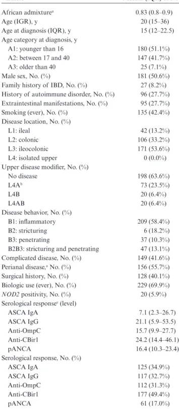
Data from 358 unrelated AA participants with CD were analyzed. The demographic and phenotypic characteristics of these participants are described in Table 1. The median age at diagnosis was 15 years (IQR, 12–22.5). Ileocolonic disease was observed in 53.6% of patients. Perianal disease was observed in 55.7%. Disease behavior was complicated in 41.6%. Biologic agents were used at some point in the disease course in 69.9% of patients. NOD2 positivity was observed in 5.9% of patients. More than 15% of data was missing for percent African admix-ture. The median serological levels and respective IQR for all serological markers are displayed in Table 1. The distribution of percent African admixture in the cohort is illustrated in Fig. 1: Percent admixture rates range from 0.31 to 1.0, with a median of 0.83 (IQR, 0.8–0.9).
Patient Demographic and Phenotypic
serological status are displayed in Table 2. On univariate ana-lysis, the following characteristics were associated with ASCA positivity: personal history of immune-mediated (P = 0.02), EIM (P = 0.02), complicated disease (P < 0.0001), ileal dis-ease (P < 0.0001), and disdis-ease requiring surgery (P = 0.002). Anti-OmpC positivity was significantly associated with age at enrollment (P = 0.005), complicated disease (P = 0.02), and disease requiring surgery (P < 0.0001). Anti-CBir1 posi-tivity was associated with age at enrollment (P = 0.001), age at diagnosis (P = 0.008), percent African admixture (P = 0.004), current smoke exposure (P = 0.004), and isolated ileal disease (P = 0.03). A negative ANCA was significantly associated with EIM (P = 0.01), complicated disease (P = 0.003), and ileal dis-ease (P = 0.01).
Patient Characteristics and Serological Levels
Associated With Complicated Disease
One-knot linear splines were set at 15 EU/mL, 20 EU/ mL, and 20 EU/mL for the variables ASCA IgG, anti-OmpC, and anti-CBir1, respectively (Supplemental Digital Content, Fig. S1). The final multivariable model (Table 3), after fast backwards selection of individual factors at the 0.05 signifi-cance level, showed that OmpC (OR, 2.23; 95% CI, 1.41–3.53), IgG ASCA (OR, 2.67; 95% CI, 1.67–4.28), and ileal disease (OR, 2.94; 95% CI, 5.26–8.77) were independently associated with complicated disease. There was no interaction between NOD2 positivity and any of the serological markers in the final model. Additionally, the interaction between percent African admixture and all covariates included in the initial and final model was assessed and found to be insignificant.
TABLE 1. Demographic and Phenotypic Characteristics
of Cohort
Variable Median (IQR) or No. (%)
African admixturea 0.83 (0.8–0.9)
Age (IGR), y 20 (15–36)
Age at diagnosis (IQR), y 15 (12–22.5) Age category at diagnosis, y
A1: younger than 16 180 (51.1%) A2: between 17 and 40 147 (41.7%) A3: older than 40 25 (7.1%) Male sex, No. (%) 181 (50.6%) Family history of IBD, No. (%) 27 (8.2%) History of autoimmune disorder, No. (%) 96 (27.7%) Extraintestinal manifestations, No. (%) 95 (27.7%) Smoking (ever), No. (%) 135 (42.4%) Disease location, No. (%)
L1: ileal 42 (13.2%)
L2: colonic 106 (33.2%)
L3: ileocolonic 171 (53.6%) L4: isolated upper 0 (0.0%) Upper disease modifier, No. (%)
No disease 198 (63.6%)
L4Ab 73 (23.5%)
L4B 20 (6.4%)
L4AB 20 (6.4%)
Disease behavior, No. (%)
B1: inflammatory 209 (58.4%)
B2: stricturing 6 (18.2%)
B3: penetrating 37 (10.3%) B2B3: stricturing and penetrating 47 (13.1%) Complicated disease, No. (%) 149 (41.6%) Perianal disease,a No. (%) 156 (55.7%)
Surgical history, No. (%) 128 (40.1%) Biologic use (ever), No. (%) 229 (69.9%)
NOD2 positivity, No. (%) 20 (5.9%) Serological responsec (level)
ASCA IgA 7.1 (2.3–26.7)
ASCA IgG 21.1 (5.9–53.5)
Anti-OmpC 15.7 (9.9–27.7)
Anti-CBir1 24.2 (14.4–46.1)
pANCA 16.4 (10.3–23.4)
Serological response, No. (%)
ASCA IgA 125 (34.9%)
ASCA IgG 117 (32.7%)
Anti-OmpC 112 (31.3%)
Anti-CBir1 177 (49.4%)
pANCA 61 (17.0%)
aMissing Greater than 15% of the data.
bL4A: disease proximal to the ligament of treitz; L4B: disease distall to the ligament
of treitz but proximal to the distal 1/3 of the ileum.
cResults are classified a s n egative o r p ositive a ccording t o t he m anufacturer’s
determined cutoffs. A negative response was defined as: ASCA IgA < 20 EU/mL;
ASCA IgG < 40 EU/mL; anti-OmpC < 23 EU/mL; anti-CBir1 < 25 EU/mL; pANCA < 17.46 EU/Ml.
anti-Cbir1 = flagellin-like bacterial antigen; Panca = perinuclear antinuclear cytoplas-mic antibody.
TABLE
2.
Demog
raphic and P
henot
ypic C
har
ac
teristics A
cc
or
ding t
o S
er
olog
ic S
ta
tus
ASCA a anti-OmpC anti-CBir1 pANCA Pr edictors P ositi ve (n = 155) Negati ve (n = 203) P P ositi ve (n = 112) Negati ve (n = 246) P P ositi ve (n = 177) Negati ve (n = 181) P P ositi ve (n = 61) Negati ve (n = 297) P Age (IGQ),b y
21 (16–35) 18 (15–37) 0.11 25 (16–40) 18 (15–33) 0.005 18 (15–28) 22 (16–44) 0.001 18 (16–39) 20 (15–36) 0.61 Age a t dia gno-sis , y 16 (13–22) 15 (11–24) 0.25 15 (12–23.5) 15 (11.5– 21.5) 0.44 15 (12–19) 16 (12–25) 0.008 16 (12–24) 15 (12–22) 0.31 Male se x, No . (%) 81 (52.3%) 100 (49.2%) 0.57 57 (50.9%) 124 (50.4%) 0.93 93 (53.5%) 88 (48.6%) 0.46 25 (41.0%) 156 (53.5%) 0.10 NOD2 positi ve , No . (%) 12 (8.7%) 8 (4.7%) 0.16 4 (4.2%) 16 (7.5%) 0.28 d 8 (5.4%) 12 (7.5%) 0.45 2 (3.9%) 18 (7.0%) 0.4 d
African admixtur
e b, c 0.80 (0.74– 0.88) 0.84 (0.76– 0.89) 0.41 0.84 (0.78– 0.90) 0.82 (0.62– 0.88) 0.06 0.84 (0.79– 0.90) 0.82 (0.71– 0.93) 0.004 0.83 (0.76– 0.89) 9.83 (0.76– 0.89) 0.80 F amil
y history of
IBD , No . (%) 11 (7.9%) 16 (8.5%) 0.83 7 (6.9%) 20 (8.8%) 0.56 12 (7.3%) 15 (9.1%) 0.55 4 (7.0%) 23 (8.4%) 0.72 d A utoimm une Disease , no . (%) 33 (21.5%) 63 (32.5%) 0.02 31 (28.4%) 65 (27.3%) 0.83 45 (26.3%) 51 (29.0%) 0.58 14 (24.6%) 82 (28.3%) 0.57 EIM, No . (%) 32 (21.5%) 63 (32.5%) 0.02 33 (31.4%) 62 (26.1%) 0.31 43 (25.2%) 52 (30.2%) 0.30 24 (40.7%) 71 (29.1%) 0.01 Curr ent smok e exposur e, No . (%) 35 (28.2%) b 49 (29.2%) 0.86 27 (29.0%) 57 (28.6%) 0.95 30 (20.6%) 54 (37.0%) 0.002 14 (27.5%) 70 (29.1%) 0.82 Upper disease , e No . (%) 50 (36.2%) 63 (26.4%) 0.97 32 (33.7%) 81 (37.5%) 0.52 56 (37.3%) 57 (35.4%) 0.72 18 (33.3%) 95 (37.0%) 0.61 P erianal disease , No . (%) 78 (61.4%) 78 (51.0%) 0.08 59 (64.8%) 97 (51.3%) 0.03 85 (57.4%) 71 (53.8%) 0.54 18 (42.9%) 138 (58.0%) 0.07 Outcomes Complica ted dis-ease ,
f No
. (%) 85 (54.8%) 64 (31.5%) <0.0001 57 (50.9%) 92 (37.4%) 0.02 82 (46.3%) 67 (37.0%) 0.07 15 (24.6%) 134 (45.1%) 0.003 Ileal disease ,
g No
. (%) 85 (54.8%) 103 (56.0%) <0.0001 68 (68.0%) 145 (66.2%) 0.75 109 (66.1%) 104 (67.5%) 0.78 28 (50.1%) 185 (70.1%) 0.01 Isola
ted ileal
dis-ease , No . (%) 35 (11.1%) 46 (14.7%) 0.35 24 (12.0%) 57 (13.7%) 0.68 27 (9.1%) 54 (17.5%) 0.03 12 (10.9%) 69 (13.6%) 0.59 Sur gery , No . (%) 72 (49.3%) 56 (32.4%) 0.002 56 (56.0%) 72 (32.9%) <0.0001 62 (38.5%) 66 (41.7%) 0.55 16 (28.6%) 112 (42.6%) 0.05
IgA ASCA w
as classified as nega
ti
ve (0.0–20.0 EU/mL) and positi
ve (≥20.0 EU/mL); IgG ASCA w
as classified as nega
ti
ve (0.0–40.0 EU/mL) and positi
ve (≥40.0 EU/mL); Anti-OmpC w
as classified as nega
ti ve (0.0–23.0 EU/mL) and positi ve (≥23.0 EU/mL); Anti-CBir1 w as classified as nega ti ve (0.0–25.0
EU/mL) and positi
ve
(≥25.0 EU/mL); pANCA w
as classified as nega ti ve (0.0–30.0 EU/mL) and positi ve (≥30.0 EU/mL).
aASCA positi
vity is defined as being either IgA or IgG ASCA positi
ve
.
bMedian (IQR). cMissing gr
ea
ter than 15% of
the da
ta.
dFisher e
xact test.
eUpper disease is defined as L4A, L4B
, or L4AB positi
vity
.
fComplica
ted disease is defined as B2 or B3 or B2B3 disease beha
vior
.
gIleal disease is defined as the (L1) isola
te ileal or (L3) ileocolonic disease
.
anti-Cbir1 = fla
gellin-lik
e bacterial antigen; pANCA = perin
uclear antin
uclear cytoplasmic antibod
Patient Characteristics and Serological Levels
Associated With Surgery
One-knot linear splines were set at 25 years, 40 EU/ mL, 20 EU/mL, and 20 EU/mL for the variables age at diag-nosis, IgA ASCA, IgG ASCA, and anti-OmpC, respectively (Supplemental Digital Content, Fig. S2). The final multivaria-ble model (Table 4), after fast backwards selection of individual factors at the 0.05 significance level, showed that disease requir-ing surgery was independently associated with NOD2 positivity (OR, 2.9; 95% CI, 1.01–8.33), IgG ASCA (OR, 2.51, 95% CI, 1.49–4.22), and anti-OmpC (OR, 3.57; 95% CI, 2.12–6.00). There was no interaction between NOD2 positivity and any of the serological markers in the final model. Additionally, inter-action between percent African admixture and all covariates included in the initial models and the final remaining model was assessed and found to be insignificant.
DISCUSSION
Our study was designed to describe the phenotypic be-havior of CD in AAs and to explore the relationship between antimicrobial serology and disease outcomes, while controlling for genetic ancestry. Our series comprises the largest cohort of AAs with CD and is the first study to evaluate OmpC, anti-CBir1, and ANCA and their association with phenotype and disease behavior.
We found that the majority of AAs with CD had in-flammatory, noncomplicated disease type (58.4%), mostly the colon (86.8%). Fifty-three percent of patients with colonic dis-ease also had disease affect the small bowel, and 55.7% of our patients had perianal disease at the time of enrollment. Our
observations regarding primary disease location and disease be-havior concur with the findings of both Mahid et al. and Hou et al., who published systematic reviews on the epidemiology and phenotypic presentation of IBD in AAs.7, 8 These findings
are also consistent with those found in European ancestry pop-ulations, which is contrary to earlier literature reporting that AAs have a more severe course with different disease distribu-tion.22, 23
However, we found a notably higher prevalence of peri-anal disease than previously reported. Mahid et al. reported the prevalence of perianal disease to be 26%, Hou et al. reported a prevalence of 25%, and Dassopoulos et al. reported a preva-lence of 34%. In other reports, the prevapreva-lence of perianal dis-ease ranged from 0% to 60%.24–31 The wide range appears to
be a function of (i) the sample size, (ii) the disease severity of the patient population, and (iii) the definition of perianal disease. The perianal disease modifier, as described in the Montreal Classification, includes skin tags, anal fissures, peri-anal abscesses, or periperi-anal fistulas, but many studies either did not clarify how perianal disease was defined or included only perianal abscesses and fistulas in their definition.12, 13
While numerous studies have reported the presence and magnitude of serological antibodies with complicated disease, ileal involvement, and earlier-onset disease in NE populations, less is known about these associations in AAs. Dassopoulos et al. conducted the first study to assess ASCA levels in AAs with CD. They reported that, similar to NEs, ASCA was independently associated with complicated behavior and surgery in AAs with CD.9 Our study expands upon this study by exploring the
associ-ation between ASCA, anti-OmpC, anti-CBir1, and ANCA with complicated disease and disease requiring surgery. We not only
TABLE 3. Adjusted Odds Ratio for Complicated Crohn’s Disease
Odds Ratioa 95% Confidence Interval
Anti-OmpCb Comparing 27.7 EU/mL with 17.7 EU/mL 2.23 1.41–3.53
IgG ASCAb Comparing 53.5 EU/mL with 5.9 EU/mL 2.67 1.67–4.28
Ileal disease Positive to negative 2.94 5.26–8.77
aORs were calculated by comparing the 75th percentile with the 25th percentile.
bOne-knot linear splines were created based on review of restricted cubic spline and subject matter knowledge.
TABLE 4. Adjusted Odds Ratio for Disease Requiring Surgery
Odds Ratioa 95% Confidence Interval
NOD2 positivity Positive to negative 2.9 1.01–8.33
IgG ASCAb Comparing 53.3 EU/mL with 5.9 EU/mL 2.51 1.49–4.22
Anti-OmpCb Comparing 27.7 EU/mL with 9.9 EU/mL 3.57 2.12–6.00
aORs were calculated by comparing the 75th percentile with the 25th percentile.
confirmed the association between IgG ASCA with complicated disease and surgeries but also identified an association between anti-OmpC with complicated disease and disease requiring sur-gery in AAs. We also found that IgG ASCA remained statis-tically significant after controlling for ileal disease, NOD2 positivity, and percent African Admixture. In contrast to prior reports, in which ASCAs were known to be associated with white populations, ileal involvement, and NOD2 status, the as-sociation between ASCA and complicated disease and disease requiring surgery is not the result of European admixture.9, 32
Our findings do concur with prior literature regarding the as-sociation between anti-OmpC with complicated disease and the need for surgery in NE populations.5, 33, 34
Dubinsky et al. were the first to tabulate antibody sum scores in NEs and found that the frequency of internal pene-trating disease, stricturing disease, and disease requiring surgery increased as the number of immune responses increased.5 In a
subanalysis, we calculated antibody sum scores according to the method described by Dubinsky et al. and observed similar results (Supplemental Digital Content, Fig. S3).5 We saw an
in-crease in complicated behavior types (B2, B3, and B2B3) with a higher sum score, and, conversely, inflammatory disease (B1) was least present in the highest-sum group.
These findings suggest that the phenotypes of CD in Caucasians and AAs are not as dissimilar as initially believed.22, 23
We hypothesized that the genetic differences between AAs and NEs would result in statistically significant, clinically mean-ingful differences between antimicrobial antibodies and their re-lationship with disease phenotype in CD. We also hypothesized that there would be an interaction between African admixture and serological levels, which was also not observed. Contrary to our initial hypotheses, we found that antimicrobial antibod-ies and their association with disease phenotype and behavior can be interpreted similarly between AAs and NEs. We postu-late that these similarities are more of a function of similari-ties among the intestinal microbiota than differences in genetic make-up, emphasizing the role of the environment and the limi-tation of genetics to diagnose IBD or predict disease behavior.1
These findings are further corroborated by twin concordance studies that have demonstrated that only a small percentage of the risk of developing inflammatory bowel disease is related to genetics.35
Limitations of our study include the substantial missing data from our participant population and the predominance of participants diagnosed younger than age 16 years. While there was no missing data related to serological levels, we were missing more than 15% of data in variables measuring the pres-ence of perianal disease and percent African admixture. Eleven additional confounding variables were missing values for any-where between 1.4% and 14.0% of the participants. As a result, our sample size decreased by more than 50% when we per-formed the multivariable analysis using our original model. We addressed this limitation by performing multiple imputations
by chained equations, a principled method of dealing with miss-ing data.20 We were also limited by the absence of demographic
or phenotypic information on those patients who chose not to participate. Additionally, our study population was based on a population with a pediatric majority. The majority of our patients were diagnosed younger than age 16 years, which is not reflective of the general population and likely suggests a population with more severe disease.
In contrast to the hypothesis that the diagnostic value of antimicrobial and autoimmune antibodies would vary among different ethnic and geographic populations due to differences in environmental and genetic influences, we found that with re-spect to ASCA and anti-OmpC, they behave similarly in AA and NE populations recruited from similar geographic areas.4
Our study also provides reassurance that the utility of sero-logical markers for prognosis of CD applies equally to AA populations.
ACKNOWLEDGMENTS
Author contributions: Madeline Bertha helped concep-tualize the paper, contributed to data acquisition, performed statistical analysis, wrote the manuscript, and reviewed and approved the final manuscript. Arthi Vasantharoopan contrib-uted to statistical analysis, contribcontrib-uted to the manuscript, and reviewed and approved the final manuscript. Archana Kumar helped conceptualize the paper, contributed to data acqui-sition, contributed to statistical analysis, contributed to the manuscript, and reviewed and approved the final manuscript. Beau Bruce helped conceptualize the paper, performed statis-tical analysis, contributed to the manuscript, and reviewed and approved the final manuscript. Tatyana Hofmekler contributed to the manuscript and reviewed and approved the final manu-script. Jarod Prince contributed to data acquisition, contrib-uted to the manuscript, and reviewed and approved the final manuscript. Cary Sauer helped contribute to the manuscript and reviewed and approved the final manuscript. Dermot McGovern conceptualized the manuscript, performed the assays, helped contributed to the manuscript, and reviewed and approved the final manuscript. Subra Kugathasan is the prin-ciple investigator of this project and responsible for the overall conduct, results, and conclusions of the paper. He conceptual-ized the paper, contributed to the manuscript, and reviewed and approved the final manuscript. All other authors contributed to the acquisition of data.
REFERENCES
1. Gerich ME, McGovern DP. Towards personalized care in ibd. Nat Rev
Gastroenterol Hepatol. 2014;11:287–299.
2. Mow WS, Landers CJ, Steinhart AH, et al. High-level serum antibodies to bac-terial antigens are associated with antibiotic-induced clinical remission in Crohn’s disease: a pilot study. Dig Dis Sci. 2004;49:1280–1286.
4. Prideaux L, De Cruz P, Ng SC, et al. Serological antibodies in inflammatory bowel disease: a systematic review. Inflamm Bowel Dis. 2012;18:1340–1355. 5. Dubinsky MC, Kugathasan S, Mei L, et al; Western Regional Pediatric IBD
Research Alliance; Pediatric IBD Collaborative Research Group; Wisconsin Pediatric IBD Alliance. Increased immune reactivity predicts aggressive compli-cating Crohn’s disease in children. Clin Gastroenterol Hepatol. 2008;6:1105–1111. 6. Straus WL, Eisen GM, Sandler RS, et al. Crohn’s disease: does race matter? The Mid-Atlantic Crohn’s Disease Study Group. Am j Gastroenterol. 2000;95:479–483. 7. Hou JK, El-Serag H, Thirumurthi S. Distribution and manifestations of inflam-matory bowel disease in Asians, Hispanics, and African Americans: a systematic review. Am j Gastroenterol. 2009;104:2100–2109.
8. Mahid SS, Mulhall AM, Gholson RD, et al. Inflammatory bowel disease and African Americans: a systematic review. Inflamm Bowel Dis. 2008;14:960–967. 9. Dassopoulos T, Nguyen GC, Talor MV, et al; NIDDK IBD Genetics Consortium.
nod2 mutations and anti-saccharomyces cerevisiae antibodies are risk factors for Crohn’s disease in African Americans. Am j Gastroenterol. 2010;105:378–386. 10. Tishkoff SA, Reed FA, Friedlaender FR, et al. The genetic structure and history
of Africans and African Americans. Science. 2009;324:1035–1044.
11. Bernstein CN, Fried M, Krabshuis JH, et al. World Gastroenterology Organization practice guidelines for the diagnosis and management of ibd in 2010. Inflamm Bowel Dis. 2010;16:112–124.
12. Satsangi J, Silverberg MS, Vermeire S, et al. The montreal classification of inflamma-tory bowel disease: controversies, consensus, and implications. Gut. 2006;55:749–753. 13. Levine A, Griffiths A, Markowitz J, et al. Pediatric modification of the Montreal
classification for inflammatory bowel disease: the Paris classification. Inflamm
Bowel Dis. 2011;17:1314–1321.
14. Landers CJ, Cohavy O, Misra R, et al. Selected loss of tolerance evidenced by Crohn’s disease-associated immune responses to auto- and microbial antigens.
Gastroenterology. 2002;123:689–699.
15. Price AL, Helgason A, Palsson S, et al. The impact of divergence time on the nature of population structure: an example from Iceland. Plos Genet. 2009;5:e1000505. 16. Pasaniuc B, Sankararaman S, Kimmel G, et al. Inference of locus-specific
an-cestry in closely related populations. Bioinformatics. 2009;25:i213–i221. 17. Seldin MF, Pasaniuc B, Price AL. New approaches to disease mapping in admixed
populations. Nat Rev Genet. 2011;12:523–528.
18. Alexander DH, Novembre J, Lange K. Fast model-based estimation of ancestry in unrelated individuals. Genome Res. 2009;19:1655–1664.
19. Harrell F. Regression Modeling Strategies: With Applications to Linear Models,
Logistic Regression, and Survival Analysis. New York: Springer-Verlag, Inc.; 2001.
20. Azur MJ, Stuart EA, Frangakis C, Leaf PJ. Multiple imputation by chained equa-tions: what is it and how does it work? Int j Methods Psychiatr Res. 2011;20:40–49. 21. Lawless JF, Singhal K. Efficient screening of non-normal regression models.
Biometrics. 1978;34:318–327.
22. Agrez MV, Valente RM, Pierce W, et al. Surgical history of Crohn’s disease in a well-defined population. Mayo Clin Proc. 1982;57:747–752.
23. Sonnenberg A, McCarty DJ, Jacobsen SJ. Geographic variation of inflammatory bowel disease within the United States. Gastroenterology. 1991;100:143–149. 24. Basu D, Lopez I, Kulkarni A, et al. Impact of race and ethnicity on inflammatory
bowel disease. Am j Gastroenterol. 2005;100:2254–2261.
25. Cross RK, Jung C, Wasan S, et al. Racial differences in disease phenotypes in patients with Crohn’s disease. Inflamm Bowel Dis. 2006;12:192–198.
26. Deveaux PG, Kimberling J, Galandiuk S. Crohn’s disease: presentation and se-verity compared between black patients and white patients. Dis Colon Rectum. 2005;48:1404–1409.
27. Goldman CD, Kodner IJ, Fry RD, et al. Clinical and operative experience with non-Caucasian patients with Crohn’s disease. Dis Colon Rectum. 1986;29:317–321. 28. Mahid SS, Minor KS, Stromberg AJ, et al. Active and passive smoking in child-hood is related to the development of inflammatory bowel disease. Inflamm Bowel Dis. 2007;13:431–438.
29. Nguyen GC, Torres EA, Regueiro M, et al. Inflammatory bowel disease character-istics among African Americans, Hispanics, and non-Hispanic whites: characteriza-tion of a large North American cohort. Am j Gastroenterol. 2006;101:1012–1023. 30. Ogunbi SO, Ransom JA, Sullivan K, et al. Inflammatory bowel disease in
African-American children living in Georgia. j Pediatr. 1998;133:103–107. 31. Simsek H, Schuman BM. Inflammatory bowel disease in 64 black patients:
ana-lysis of course, complications, and surgery. j Clin Gastroenterol. 1989;11:294–298. 32. Adeyanju O, Okou DT, Huang C, et al. Common nod2 risk variants in African Americans with Crohn’s disease are due exclusively to recent Caucasian admix-ture. Inflamm Bowel Dis. 2012;18:2357–2359.
33. Ferrante M, Henckaerts L, Joossens M, et al. New serological markers in inflam-matory bowel disease are associated with complicated disease behaviour. Gut. 2007;56:1394–1403.
34. Mow WS, Vasiliauskas EA, Lin YC, et al. Association of antibody responses to microbial antigens and complications of small bowel Crohn’s disease.
Gastroenterology. 2004;126:414–424.
35. Halfvarson J, Bodin L, Tysk C, et al. Inflammatory bowel disease in a Swedish twin cohort: a long-term follow-up of concordance and clinical characteristics.