Am. J. Respir. Cell Mol. Biol. Vol. 24, pp. 662–670, 2001 Internet address: www.atsjournals.org
Evidence for Stem-Cell Niches in the Tracheal Epithelium
Duncan W. Borthwick, Mariam Shahbazian, Q. Todd Krantz, Julia R. Dorin, and Scott H. Randell
Medical Research Council Human Genetics Unit, Edinburgh, United Kingdom; U.S. Environmental Protection Agency, Research Triangle Park; and Cystic Fibrosis/Pulmonary Research and Treatment Center, The University of North Carolina, Chapel Hill, North Carolina
It is generally important to elucidate airway epithelial cell lin-eages and to identify multipotent progenitors as targets for gene therapy. Stem (S) cells are typically present in specialized compartments spatially proximal to their differentiated prog-eny, but an equivalent paradigm has not been demonstrated in the airway. We discovered a distinct population of cells dis-playing high levels of keratin expression in murine tracheal submucosal gland ducts, and tested the hypothesis that bro-modeoxyuridine (BrdU) label–retaining cells (LRCs), thought to represent the S-cells, were present in this compartment. Mice received weekly epithelial damage by intratracheal de-tergent or SO2 inhalation for 4 wk and received intraperito-neal injections of BrdU every 48 h during the injury and repair period. At 3 and 6 d after injury, BrdU-positive epithelial cells were noted along the entire tracheal length in both basal and lumenal cell positions. At later time points (20 and 95 d) LRCs were localized to gland ducts in the upper trachea and to sys-tematically arrayed foci in the lower trachea, typically near the cartilage–intercartilage junction. LRCs were not pulmonary neuroendocrine cells. Heterotopic tracheal grafts after surface epithelial removal demonstrated reconstitution of a surface-like epithelium from gland remnants. These results suggest that airway epithelial S cells are localized to specific niches.
Airway diseases such as asthma, chronic bronchitis, and cystic fibrosis are characterized by bronchial epithelial hy-perplasia and metaplasia that likely contribute to the down-ward spiral of physiologic impairment. Elucidation of epi-thelial cell lineage is fundamental toward understanding mechanisms that alter epithelial phenotype. It is also im-portant to identify cells with extensive progenitorial ca-pacity as potential targets for gene therapy. Despite many studies directed at understanding the growth properties of specific subsets of cells constituting the pseudostratified airway epithelium, a definitive model establishing progeni-tor–progeny relationships in the normal steady state or in response to injury has not yet been established (1).
Stem (S)-cell theory divides epithelial cell types in re-newing tissues into three major compartments according
to proliferation capacity and differentiation potential (re-cently reviewed in Refs. 2 and 3). S cells are pluripotent to generate all cell types in the tissue compartment and usu-ally have adequate growth capacity for the life of the ani-mal. In general, S cells turn over slowly and display mini-mal physiologic differentiation. As early descendents of S cells, transiently amplifying (TA) cells retain significant growth capacity while acquiring differentiated functions. TA cells eventually become incapable of proliferation and enter the terminally differentiated (TD) compartment. To conserve growth potential and to prevent genetic injury while vulnerable during mitosis, S cells are thought to cy-cle slowly and are recruited only as demanded by tissue turnover. Thus, much of the increase in cell number in the steady state occurs in the TA population. One conse-quence is that a pulse label of [3H]thymidine or
bromode-oxyuridine (BrdU) will label mostly TA cells. Long-term [3H]thymidine or BrdU labeling will mark S cells that
re-tain the label for an extended period due to slow turnover. Thus, an adequate labeling intensity and a suitable wash-out period of the TA and TD compartments will result in so-called label-retaining cells (LRCs) thought to represent the S-cell compartment.
In extensively studied tissues such as the epidermis, in-testinal epithelium, and cornea, label-retaining S cells re-side in specialized and generally well protected niches spa-tially proximal to their more differentiated TA and TD progeny. However, S-cell niches have not been demon-strated in the psuedostratified airway epithelium.
In studies aiming to isolate specific subpopulations of murine tracheal epithelial cells, we discovered a distinct population of cells displaying high levels of keratin gene and protein expression in submucosal gland ducts. Intrigu-ingly, high keratin protein content in a subset of corneal epithelial cells (4) ultimately led to the discovery that LRCs of the cornea reside in the limbus (5). Further, ex-pression of a specific keratin has been reported to be a marker for hair-follicle S cells (6). To determine whether the keratin-rich cells in the murine trachea were LRCs, we performed long-term BrdU labeling and washout stud-ies of purposefully injured murine tracheas. Injury was required to stimulate cell division in the normally quies-cent tracheal epithelium. Pulmonary neuroendocrine cells (PNECs) were localized to assess their role in S-cell pat-terning. Heterotopic tracheal grafts denuded of their sur-face epithelium were used to determine whether gland and gland duct remnants could reconstitute a surface-like epi-thelium.
Materials and Methods Transgenic Mice
All animals were handled under Institutional Animal Care and
Use Committee–approved protocols. The K5gal6000 transgenic
(Received in original form April 18, 2000 and in revised form December 12,
2000)
EPA Disclaimer: This report has been reviewed by the National Health
and Environmental Effects Research Laboratory, U.S. Environmental Protection Agency, and approved for publication. Approval does not sig-nify that the contents necessarily reflect the views and policies of the agency, nor does mention of trade names and commercial products consti-tute endorsement or recommendation for use.
Address correspondence to: Scott H. Randell, Ph.D., UNC CF Center, CB
7248, Rm. 4011 Thurston-Bowles, Chapel Hill, NC 27599. E-mail: randell@ med.unc.edu
Abbreviations: -galactosidase, gal; bromodeoxyuridine, BrdU; calcitonin
gene–related peptide, CGRP; Griffonia simplicifolia isolectin B4, GSIB4;
Borthwick, Shahbazian, Krantz, et al.: Airway Epithelial Stem Cells 663
mouse strain in which 6,000 base pairs (bp) of the basal cell–spe-cific human keratin 5 promoter drives expression of the bacterial LacZ gene (7, 8) was a kind gift from Dr. Elaine Fuchs
(Univer-sity of Chicago, Chicago, IL). -Galactosidase (gal) activity in
excised tissues was detected using X-gal substrate as previously described (9). Whole-mount images were obtained and tissues were processed for paraffin embedding. Sections were counter-stained with nuclear fast red.
Lectin and Keratin Staining
Griffonia simplicifolia isolectin B4 (GSIB4) binding sites in
mouse trachea were detected using biotinylated lectin as previ-ously described (10). Polyclonal antibodies against the mouse ho-mologues of human cytokeratin 14 and 18 were generated by immunizing rabbits with peptides CGKVVSTHEQVLRTKN-COOH and CGRVVSETNDTRVLRH-CGKVVSTHEQVLRTKN-COOH, respectively, con-jugated to maleimide-activated ovalbumin or bovine serum albu-min (Pierce, Rockford, IL). Antibodies were affinity-purified on peptide linked to maleimide-activated Sepharose (Pierce). Im-munostaining was performed on formalin-fixed paraffin sections using Texas Red–labeled antirabbit antibody (Jackson Immu-noresearch, West Grove, PA) and confocal microscopy.
Induction of Epithelial Damage by Polidocanol or SO2 Because the normal rodent tracheobronchial epithelium is
mitot-ically quiescent, with 2% of the cells being labeled by a pulse
of [3H]thymidine or BrdU (11, 12), it is necessary to recruit S
cells into the actively dividing pool to possibly visualize LRCs. Two methods of tracheal epithelial damage were used: intratra-cheal instillation of polidocanol (Sigma, St. Louis, MO), a deter-gent clinically used as a sclerosing adeter-gent and to facilitate gene
transfer to airways (13); and SO2 inhalation, a well-studied model
of pulmonary epithelial injury (14, 15).
Ten l of 2% polidocanol in phosphate-buffered saline (PBS)
was directly instilled into the trachea just below the pharynx in anesthetized (intraperitoneal ketamine/xylazine mixture) CD1 mice (4- to 6-wk-old males; Charles River, Raleigh, NC). To assess the damage qualitatively, three animals were killed by barbiturate overdose at 10 min or 2 or 24 h after instillation and their tracheas were removed for conventional histology. Nine mice were ex-posed to an ambient environment of 500 parts per million (ppm)
SO2 for 3 h in monitored chambers at the U.S. Environmental
Protection Agency facility in Research Triangle Park, NC, and tracheal epithelial damage was assessed histologically at 2 or 24 h.
Repeated Damage and BrdU Labeling
Male CD1 mice (6 wk old, 25 g) received weekly tracheal
dam-age by instillation of 10 l of 2% polidocanol or by SO2
inhala-tion (four escalating doses: 500 ppm for 3, 3.5, 4, or 4.5 h) as de-scribed earlier. In addition, the mice received intraperitoneal injections of BrdU (2 mg) every 48 h beginning 2 h after the first injury and extending until 24 h after the final injury. Groups of
mice (n 4–9) were killed at different time points (see Table 1)
and their tracheas were examined for BrdU incorporation by im-munohistochemistry as described later. Controls included groups of mice receiving BrdU but without tracheal damage, and dam-aged mice receiving no BrdU.
BrdU Immunohistochemistry
BrdU incorporation was detected in sections of trachea and in-testine using a staining procedure modified from previously re-ported methods (16). Fixation with Omnifix (FR Chemical, Mt. Vernon, NY) resulted in much stronger staining than with forma-lin and we also avoided antimouse immunoglobuforma-lin (Ig) G sec-ondary antibodies to prevent high background. We used a fluo-rescein isothiocyanate (FITC)–labeled anti-BrdU monoclonal
antibody (Caltag, Burlingame, CA) followed by rabbit anti-FITC IgG (Molecular Probes, Eugene, OR) which was detected with peroxidase-conjugated goat antirabbit IgG (Jackson Immunore-search). Optimal antibody dilutions were determined empirically. A duplicate section was included on each slide in which an equal concentration of nonspecific FITC-labeled mouse IgG1 replaced the primary antibody. A positive control slide of a BrdU pulse– labeled (2 h) mouse intestine was included in every staining ses-sion. This staining method was very sensitive, resulting in uni-form, densely stained nuclei in crypt enterocytes after 2-h pulse labeling. As well as densely stained cells, we also observed fainter and heterogeneously labeled nuclei (small “dots” within the nu-clei instead of uniformly brown nunu-clei) in the tracheas of animals chronically exposed to BrdU, possibly daughter cells in which BrdU was diluted.
Measurement of BrdU Labeling
Approximately 600 cells from three different regions (200/region) of every longitudinally sectioned trachea were examined for cell type and BrdU incorporation. The mouse trachea contains ap-proximately 14 cartilage rings and, in the CD1 strain, tracheal glands are typically present only within the uppermost four rings (17). Region 1 began just above the most cephalad cartilage ring, with regions 2 and 3 beginning at rings 5 and 9 distal to the phar-ynx, respectively. A total of 100 consecutive cells beginning at each level on both sides of the trachea were scored. Staining was considered positive if the signal was nuclear and clearly above that detected as background on the adjacent negative control sec-tion, including faintly and heterogeneously stained nuclei. Cells were categorized as basal if they were small, cuboidal to squam-oid in shape, and closely adherent to the basal lamina, and the nucleus was in the bottom third of the epithelial layer. All other cells were categorized as columnar. Several other studies have discriminated between specific cell types, but these investigations
generally relied upon the higher resolution afforded by [3
H]thy-midine autoradiography on thin plastic or epoxy sections. Using Omnifix and paraffin embedding, and after acid and trypsin treatment needed for BrdU localization, we were confident dis-criminating only between basal and columnar cells. Areas of ob-vious artifact due to tissue processing were excluded. We noted densely stained BrdU-positive nuclei in the tracheas of mice chronically labeled with BrdU. Because these cells are of particu-lar interest we categorized the specific location of each densely stained nucleus.
Localization of PNECs
PNECs are proposed to play important roles in pulmonary devel-opment and epithelial cell growth (18–22). We immunostained for calcitonin gene–related peptide (CGRP) to localize PNECs in the Day 95 experimental groups. Anti-CGRP (Sigma) was used
TABLE 1
Schedule for airway injury, BrdU injections, and tissue analysis
Day 0 7 14 21 22
Time Point +3 d +6 d +21 d +95 d
Damage† * * * *
BrdU‡
---n 4 4 4 9
†Injury was induced once per week as indicated (*) by inhalation of 500 ppm
SO2 or intratracheal instillation of 10 l 2% polidocanol solution.
‡2 mg of BrdU was administered intraperitoneally every 48 h beginning 24 h
664 AMERICAN JOURNAL OF RESPIRATORY CELL AND MOLECULAR BIOLOGY VOL. 24 2001
as the primary antibody followed by biotinylated secondary anti-body and streptavidin-peroxidase.
Tracheal Grafts
We studied heterotopic tracheal grafts to examine whether tra-cheal gland duct and/or acinar remnants could reconstitute a sur-face-like epithelium in denuded tracheas. Tracheas were re-moved from donor CD1 mice and the surface cells were rere-moved
by overnight incubation at 4C in 0.1% Protease XIV (Sigma) in
F-12 medium on a tilting platform. Histologic analysis demon-strated complete removal of the surface epithelium (illudemon-strated in Figure 6). After surface denudation, Protease XIV action was terminated by addition of fetal bovine serum to 10% and the grafts were washed with three changes of F-12 medium. The pha-ryngeal opening and the trachea distal to ring 5 were closed with ligating clips. The upper, gland-containing tracheal portion was transplanted to the subcutaneous space of Nu/Nu mice and al-lowed to grow for up to 42 d before removal and routine histo-logic analysis.
Results and Discussion
Detection of a Keratin-Rich Compartment in the Murine Tracheal Epithelium
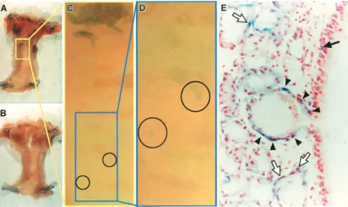
In initial studies directed toward isolation of specific air-way epithelial cell types, we localized gal activity in the trachea of transgenic mice in which 6,000 bp of the basal cell–specific human keratin 5 promoter drives expression of the bacterial LacZ gene. These mice express gal in a pattern faithful to endogenous expression in several or-gans examined but with some notable exceptions (7), and high-level activity appears to correlate with growth poten-tial in developing epidermis (8). Unexpectedly, gal activ-ity was visible only in a circumferential band in the upper trachea, with a few small patches along a descending pos-terior stripe (Figures 1A, 1C, and 1D), a pattern most con-sistent with the localization of tracheal glands. No blue cells were visible in the lower trachea. Cross sections re-vealed scattered positive basal cells in the upper tracheal surface epithelium, prominently stained small groups of cells in the ducts of submucosal glands, and positive gland myoepithelial cells (Figure 1E). This was not due to an
ab-sence of tracheal basal cells, which were clearly visible at all tracheal levels when stained with GSIB4 lectin (Figure
2A). As shown in Figures 2B and 2C, cellular gal activity correlated with high-level keratin protein expression as detected with antibodies against mouse keratins 14 and 18. Keratin 14 is usually coexpressed with keratin 5 (23, 24) and is present in surface basal cells and gland
myoepithe-Figure 1. High levels of keratin 5 promoter activity in tracheal gland duct cells.
Whole-mount photographs of gal-stained tracheas
from heterozygous K5gal6000 mice (A,
C,and D) or negative control littermates
(B) and a histologic section through a
pos-itive trachea at the level of a gland duct
opening (E). Squamous epithelial cells along
the pharyngeal floor are positive (
upper-most blue cells in A), as are cells in and around tracheal glands. Groups of faintly positive cells are visible in a descending posterior stripe (circled in C and D). (E)
Scattered surface basal cells (solid arrow),
gland duct cells (arrowheads), and gland
myoepithelial cells (open arrows) are stained.
Original magnifications: A and B, 8; C
and D, 64; E, 400.
Figure 2. Keratin-rich cells in tracheal gland ducts. GSIB4 lectin
(A), keratin 14 (B), keratin 18 (C), and rabbit IgG control (D)
staining of mouse upper trachea. The surface epithelium has a continuous lining of basal cells that extend into the gland duct (arrows in A). Clusters of cells in the gland duct and surrounding surface epithelium as well as gland myoepithelial cells are keratin
14–positive (arrows and arrowheads in B, respectively). Gland
duct cells, surface epithelial cells, and columnar acinar cells are
keratin 18–positive (arrows, arrowheads, and asterisk in C,
Borthwick, Shahbazian, Krantz, et al.: Airway Epithelial Stem Cells 665
lial cells. Keratin 18 is present in suprabasal, columnar sur-face cells and gland acinar cells. Interestingly, basal cells in gland ducts were positive for both keratins. These studies demonstrate the presence of a compartment of cells likely demonstrating very high keratin 5 promoter activity rela-tive to other airway cell types. These cells abundantly con-tain both basal and nonbasal cell type–specific keratin pro-teins. This result is intriguing in that relatively high keratin content has been viewed as a biochemically primitive phe-notype (4) and this observation led to formal demonstra-tion that corneal epithelial S cells reside in the limbus (5).
Damage Models to Enable Localization of LRCs in the Murine Tracheal Epithelium
Prolonged metabolic labeling with [3H]thymidine or BrdU
will mark epithelial S cells which, by virtue of their slow cycle time, will retain the label for an extended period. Rapidly renewing systems such as the epidermis (25–27) are amenable to localization of LRCs without additional recruitment of S cells, as was necessary for the cornea (5). Because the basal rate of mitosis in healthy airways is
exceedingly low (11) it was necessary to injure the epi-thelium to cause cell proliferation. Two types of injury were used: intratracheal instillation of the detergent poli-docanol, and SO2 inhalation. As shown in Figure 3, the
direct instillation of 10 l of 2% polidocanol in PBS caused widespread removal of the epithelium. At first the superficial columnar layer was lost, and by 24 h patches of either epithelial remnants or denuded basal lamina were visible. Heterogeneity was possibly due to mechanical damage by the cannula during instillation and/or inhomoge-neous physical dispersion of the detergent solution. At 2 h after inhalation of 500 ppm SO2, the superficial columnar
epithelial cells uniformly displayed cytopathic changes indicative of degeneration. Gland acini appeared rela-tively empty and gland ducts were dilated with mucin. At 24 h, the basal cell layer was largely retained. In both cases, eosinophilic material constituting a hyaline membrane was frequently observed. Within 3 d after both injuries, repair was well underway and a columnar layer was present but the percentage of the epithelial surface covered by cilia was still depressed. By 7 d, a normal-appearing
epi-Figure 3. Characterization of tracheal epithelial injuries and the subacute proliferative response. Histologic sections of upper (A–C and
G–I) or lower (D–F) trachea from control (A, D,and G), polidocanol-treated (B, E,and H), or SO2-exposed (C, F,and I) mice at 2 (A–
C) or 24 h (D–I) after treatment. At 2 h after both injuries, cells closely adherent to the basal lamina persisted; eosinophilic material
covered the epithelial surface 2 h after SO2 (arrows in C). By 24 h in polidocanol animals, there were patches of denuded basal lamina
with an eosinophilic covering (arrows in E). However, 24 h after SO2, a uniform epithelial monolayer was present (F). A 1-h pulse label
of BrdU revealed a 1.3% labeling index in untreated controls (G) but very high labeling of remaining epithelial cells 24 h after
poli-docanol (H) or SO2 (I). A–F: hematoxylin and eosin stain, original magnification 200; G–I: BrdU immunostaining, original
666 AMERICAN JOURNAL OF RESPIRATORY CELL AND MOLECULAR BIOLOGY VOL. 24 2001
thelium was re-established. Cell proliferation was assessed with a 1-h pulse label of BrdU 24 h after injury. Nuclear staining was negligible in isotype-matched antibody con-trols or in animals not receiving BrdU. An average tracheal BrdU labeling index of 1.3 0.3% (mean standard er-ror of the mean [SEM], n 3) was obtained from normal untreated animals. At 24 h after both injuries, the BrdU labeling index of viable surface epithelial cells was greater than 50%.
Identification of LRCs in Repetitively Injured Tracheas Chronically Labeled with BrdU
Mouse tracheas were injured with polidocanol or SO2
once per week for 4 wk and the mice received BrdU injec-tions every other day from the day of the first injury to 1 d after the last injury. There were no treatment-related mouse deaths in our studies, and groups of mice were killed at 3, 6, 21, and 95 d after the final injury. Trachea and duodenum were processed for BrdU immunostaining. Representative photomicrographs and morphometry re-sults are shown in Figures 4 and 5, respectively. At the 3-d time point, BrdU-positive duodenal enterocytes were lo-calized to the lower half and middle of the villus, whereas by 6 d only scattered positive cells were present at the vil-lus tip, consistent with the known cell migration pattern in the crypt–villus unit (not shown). BrdU-positive entero-cytes were no longer detectable at 21 and 95 d. Positive nu-clei were absent in animals that were injured but did not receive BrdU.
Figure 4. BrdU LRCs localize to upper tracheal gland ducts and
to systematically arrayed foci in the lower trachea. Panels A–D
are sections of upper trachea from mice 3, 6, 22, or 95 d,
respec-tively, after repeated SO2 injury and BrdU administration. Panel
E is from a polidocanol-treated mouse at 95 d. Densely stained
LRCs are clearly visible in gland ducts (arrows in D and E).
Pan-els F–J are from the distal trachea. Panels F and G demonstrate a
high labeling index in both basal and columnar cells 3 d after SO2
or polidocanol, respectively. Panel H shows two foci of epithelial
LRCs in the distal trachea 95 d after polidocanol treatment (
ar-rows). Panels I and J are low- and high-power views, respectively,
of distal trachea 95 d after SO2 treatment. Epithelial LRC
clus-ters are systematically arrayed and tend to correspond to the
lo-cation of the cartilage–intercartilage junction (arrows). All
pan-els were immunostained for BrdU. Original magnifications: A–H
and J,400; I, 100.
Figure 5. BrdU labeling index and locations of LRCs. (A) The mean tracheal surface epithelial BrdU labeling index including both faintly and strongly labeled cells. There was a very
signifi-cant overall time effect by analysis of variance (ANOVA) and P
0.05 for all comparisons with the Day 3 groups (two-tailed t test).
(B) The ratio of BrdU-positive basal to columnar cells. There
was a significant time effect by ANOVA. In the SO2 group, Day
6 is significantly different than all other time points, whereas in the polidocanol group Day 3 is significantly different than all
other time points (P 0.05, two-tailed t test). In A and B, values
from the upper, middle, and lower trachea were not different and
were averaged. (C–F) The locations of strongly labeled cells in
the upper and lower trachea 95 d after SO2 or polidocanol
treat-ment expressed as percentages of the total strongly labeled cells.
P values for the comparison with gland duct in C and D, and
co-lumnar versus basal in E and F, are given in parentheses
(two-tailed t test). Four or five animals were studied per time point.
Borthwick, Shahbazian, Krantz, et al.: Airway Epithelial Stem Cells 667
Staining results in tracheas repetitively injured with polidocanol or SO2 and chronically labeled with BrdU
were similar. At 3 d after the final injury (31 and 34 d after the first BrdU injections, respectively), the surface epithe-lial BrdU labeling index was approximately 65%. BrdU-positive epithelial cells were noted in both luminal and basal positions along the surface of the entire trachea. In the tra-cheal gland system, duct, acinar, and myoepithelial cells were labeled. A heterogeneous pattern of nuclear BrdU staining was observed in both the surface epithelium and in glands. Many cells had focally positive “dots” within the nucleus and faint background staining of nuclear matrix, whereas other cells had more uniformly densely stained nuclei. Presumably, the fainter staining was due to dilution of the BrdU label, which occurred as cells proliferated in the absence of BrdU. Even at 3 and 6 d, densely stained cells were preferentially localized to gland ducts and in scattered surface foci in both the upper and lower trachea. By 21 and 95 d after the last injury the overall BrdU stain-ing was much weaker, although numerous faintly stained BrdU-positive cells were still visible. The labeling index, including faintly stained cells, still exceeded 30%. Densely stained cells were preferentially localized to submucosal gland ducts in the upper trachea. Clusters of cells with strong BrdU staining were also systematically distributed along the surface epithelium in the glandless lower trachea and tended to correspond to the position of the cartilage– intercartilage junction. The ratio of basal to columnar LRCs in the distal trachea was approximately 2 to 1.
LRCs Are Not PNECs
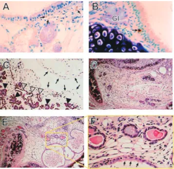
PNECs are proposed to play important roles in lung devel-opment and as regulators of epithelial cell growth. The unique distribution of LRCs suggested a possible associa-tion with PNECs, and we used CGRP staining to assess the relationship. As illustrated in Figures 6A and 6B, gland duct openings in the upper trachea were innervated with CGRP-positive fibers but PNECs were rare in this loca-tion. Because several LRCs were visible in each section from Day 95 animals containing gland ducts, it is clear that upper tracheal LRCs were not CGRP-positive PNECs. In the lower trachea, PNECs were frequently located near the cartilage–intercartilage junction where gland rudiments were occasionally found and where foci of LRCs were typ-ically present. However, there were many more LRCs than PNECs. Thus, in the lower trachea, PNECs were systemat-ically distributed along with LRCs, which may reflect their contribution to an airway trophic unit.
Gland and Gland Duct Remnants Can Regenerate a Surface-like Epithelium
Heterotopic tracheal grafts were used to test the hypothe-sis that gland and/or gland duct cells could contribute to surface epithelial regeneration. In this experiment, tra-cheas were harvested and the surface epithelium removed by exposure to protease. No remaining surface epithelial cells were seen in eight of eight protease-treated tracheas when viewed histologically (Figure 6C). Gland duct and acinar cells were present but were apparently “loosened” from the basal lamina by the protease. During the early stages after transplantation into Nu/Nu mice the glands
re-gressed, but by 28 d they had increased in size. Also at 28 d, cystic spaces lined by a cuboidal epithelium containing cil-iated cells appeared. Because of a lack of specific markers for gland ducts, it is impossible to determine whether this was a dilated gland duct structure or a new surface epithe-lium. However, the cells resembled the low cuboidal epi-thelium typically present in mouse bronchi. Although not conclusive, this experiment supports the idea that gland or gland duct cells can reconstitute a surface-like epithelium.
Are Airway Epithelial Stem Cells Present in Distinct Morphologic Compartments?
There has been a long-standing debate regarding the iden-tity of airway epithelial S cells. Several studies suggest that basal cells are progenitors (28–32), but a strong prolifera-tive response in secretory cells following injury (33), the late developmental appearance of basal cells (34–36), and studies with isolated cell populations (37) make a case for small-granule secretory cells or an undifferentiated colum-nar cell. There is evidence for great plasticity in growth and differentiation potential of airway epithelial cells (38) and our earlier studies showing that both basal and
non-Figure 6. LRCs are not pulmonary neuroendocrine cells and
gland remnants regenerate a surface-like epithelium. (A and B)
Immunostaining for CGRP in upper and middle trachea,
respec-tively, 95 d after SO2 exposure. Positive nerve fibers are visible in
the lamina propria (arrows in A) but PNECs are not present in
the epithelium that is predicted to have several LRCs. A solitary
PNEC is visible in the surface epithelium (arrow in B) near a
small gland rudiment (Gl) at the cartilage–intercartilage junction. Panel C is from a protease-treated trachea before transplantation
and shows complete removal of the surface epithelium (arrows)
and “loosening” of gland duct (open arrowheads) and acinar (
ar-rowheads) cells. Panel D shows small gland remnants 14 d after transplantation. Low- and high-power views of tracheal grafts 28 d
after transplantation (E and F, respectively) show an increase in
gland size and the presence of cystic spaces lined by a ciliated
ep-ithelium resembling the bronchial surface (arrows in F). Original
668 AMERICAN JOURNAL OF RESPIRATORY CELL AND MOLECULAR BIOLOGY VOL. 24 2001
basal cells could regenerate a complete mucociliary epi-thelium in tracheal grafts support this notion (16, 39). How-ever, detailed analysis of gland neogenesis in xenografts suggests that only a subset of human airway basal cells are pluripotent, at least to form glands (40). A critical factor in this debate is the specificity of the assay for “stemness.” Within the context of a three-compartment model of cell lineages, DNA precursor LRCs have been considered syn-onymous with S cells in several renewing tissues (5, 27, 41). To our knowledge, the present studies are the first attempt to localize LRCs in the respiratory tract. Because epithe-lial proliferation in the normal airway is too slow for suffi-cient S-cell labeling, we chose two different tolerable inju-ries. These treatments may not have been ideal in that both tended to selectively remove superficial cells. Loss of columnar cells and re-establishment of barrier function by basal cells appears to be a basic property of the pseudo-stratified epithelium (42). Selective removal of columnar cells may have obscured their potential contribution. Fur-ther studies, such as mechanical injury in the rat that cleanly denudes a portion of the epithelium, leaving both columnar and basal cells in the remaining portion (43), would be useful but this was not feasible in mice. Although [3H]thymidine labeling and autoradiography on thin
plas-tic or epoxy sections would have enabled more accurate identification and quantification of labeled cells, we ruled out the use of isotope on the basis of expense, environ-mental impact, and the availability of BrdU as an alterna-tive. Further, we had to decide on the route, dose, and fre-quency of BrdU administration. Preliminary experiments (S.H.R.’s unpublished results) with slow-release pellets that may have allowed for continuous labeling demonstrated unacceptable toxicity, and suitable implantable minipumps have only a 7-d duration. There was no adverse effect on body weight with the selected regimen of 2 mg BrdU every other day. Although this dose strongly labeled dividing cells within 1 h, low BrdU plasma levels between injections probably resulted in labeling of only a subset of prolifer-ating cells. Despite technical limitations due to injury type and incomplete cell labeling, we were nonetheless able to demonstrate clearly the preferential localization of LRCs to specific niches in the mouse trachea, namely, gland ducts in the upper trachea and systematically arrayed foci associated with the cartilage–intercartilage junction in the lower trachea. Perhaps these niches are analogous to “S cell clusters” present in the epidermis (44). Although these observations do not rule out a degree of progenitorial ca-pacity among various tracheal-cell populations, they are most consistent with localization of airway epithelial S cells to distinct morphologic compartments. It is important to note that LRCs may be either slow-cycling S cells or re-sidual TD cells. The latest time point that we studied was 95 d, which we thought was a good compromise between the probable epithelial cell life span (which, to our knowl-edge, is not precisely known for the various cells of the mouse trachea) and loss of BrdU label. It is likely that high levels of proliferation occurred in locations between clus-ters of LRCs. Some of the LRCs we noted may have been residual TD cells, but minimal labeling of ciliated cells (the one airway epithelial cell type generally considered to have limited proliferation capacity) suggests that many of
the LRCs were slowly cycling S cells. More studies are necessary to define tracheal cell life spans, to precisely map LRCs in three dimensions, and to illustrate patterns of cell proliferation and migration in relation to putative S cell niches.
A “Zonal Model” for Airway Epithelial Stem Cells
One may envision the respiratory tract as comprised of distinct “zones” with different cell lineage systems. Epi-thelial cell composition and zone boundaries depend on both the species and the individual animal history. In nor-mal mice, a renewing cell system encompassing a gland-containing, pseudostratified epithelium with Clara cells and few goblet cells is present in the upper trachea. In rats, a similar system, but with more goblet cells and no Clara cells, is present in the entire trachea; whereas in humans this zone penetrates many bronchial generations. Distally, the airway epithelium becomes glandless, cuboidal, and dominated by a Clara cell–based lineage system (45) be-fore transitioning to a type II cell–based system (46) in the alveoli. After injury or infection, cell composition and/or positional boundaries may change. For example, glands may form more distally, accompanied by goblet-cell meta-plasia in small airways or bronchiolarized metameta-plasia of the proximal alveolar region. Our results showing localiza-tion of LRCs to upper tracheal gland ducts and to distinct foci in the glandless distal trachea illustrate the presence of related, but likely different, lineage systems in these dif-ferent airway zones.
“Trophic Units” in the Airway Epithelium
Borthwick, Shahbazian, Krantz, et al.: Airway Epithelial Stem Cells 669
may help to resolve this issue. The relationship of LRCs in gland ducts to oncocytes (53) or cystic fibrosis transmem-brane conductance regulator–rich “flasklike cells” (54) re-mains to be determined.
Conclusions
This report suggests the existence of S-cell niches in the pseudostratified airway epithelium. Potential S-cell targets for gene therapy likely reside within gland ducts and in foci systematically arrayed along the surface of glandless airways. Further studies are needed to discover unique markers of airway epithelial S-cells, to better understand the niche microenvironment, and to isolate and test puta-tive airway epithelial S cells.
Acknowledgments: The authors gratefully acknowledge expert technical sup-port by Tracy Bartollotta and Kimberlie Burns of the UNC CF Center Histol-ogy Core, and thank Dr. Euan Slorach for critical reading of the manuscript.
The authors thank Dr. Elaine Fuchs for providing the K5βgal6000 transgenic
mice. This work was supported by NIH grant HL58345 to one author (S.H.R.) and by support to one author (J.R.D.) from the MRC.
References
1. Mason, R. J., M. C. Williams, H. L. Moses, S. Mohla, and M. A. Berberich. 1997. Stem cells in lung development, disease, and therapy. Am. J. Respir. Cell Mol. Biol. 16:355–363.
2. Watt, F. M., and B. L. Hogan. 2000. Out of eden: stem cells and their niches. Science 287:1427–1430.
3. Slack, J. M. 2000. Stem cells in epithelial tissues. Science 287:1431–1433. 4. Schermer, A., S. Galvin, and T. T. Sun. 1986. Differentiation-related
expres-sion of a major 64K corneal keratin in vivo and in culture suggests limbal location of corneal epithelial stem cells. J. Cell Biol. 103:49–62.
5. Cotsarelis, G., S. Z. Cheng, G. Dong, T. T. Sun, and R. M. Lavker. 1989. Ex-istence of slow-cycling limbal epithelial basal cells that can be preferen-tially stimulated to proliferate: implications on epithelial stem cells. Cell 57:201–209.
6. Lyle, S., M. Christofidou-Solomidou, Y. Liu, D. E. Elder, S. Albelda, and G. Cotsarelis. 1998. The C8/144B monoclonal antibody recognizes cytokera-tin 15 and defines the location of human hair follicle stem cells. J. Cell Sci. 111:3179–3188.
7. Byrne, C., and E. Fuchs. 1993. Probing keratinocyte and differentiation specificity of the human K5 promoter in vitro and in transgenic mice. Mol. Cell. Biol. 13:3176–3190.
8. Byrne, C., M. Tainsky, and E. Fuchs. 1994. Programming gene expression in
developing epidermis. Development 120:2369–2383.
9. Cepko, C., E. F. Ryder, C. P. Austin, C. Walsh, and D. M. Fekete. 1995. Lin-eage analysis using retrovirus vectors. Methods Enzymol. 254:387–419. 10. Shimizu, T., P. Nettesheim, J. F. Mahler, and S. H. Randell. 1991. Cell
type-specific lectin staining of the tracheobronchial epithelium of the rat: quan-titative studies with Griffonia simplicifolia I isolectin B4. J. Histochem. Cy-tochem. 39:7–14.
11. Evans, M. J., S. S. 1989. Lung Cell Kinetics. In Lung Cell Biology. D. Massar, editor. Marcel Dekker, New York. 1–36.
12. Taya, A., A. Morgan, S. T. Baker, J. A. Humphreys, M. Bisson, and C. G. Collier. 1994. Changes in the rat lung after exposure to radon and its prog-eny: effects on incorporation of bromodeoxyuridine in epithelial cells and on the incidence of nuclear aberrations in alveolar macrophages. Radiat. Res. 139:170–177.
13. Parsons, D. W., B. R. Grubb, L. G. Johnson, and R. C. Boucher. 1998. En-hanced in vivo airway gene transfer via transient modification of host bar-rier properties with a surface-active agent. Hum. Gene Ther. 9:2661–2672. 14. Kavet, R. I., and J. D. Brain. 1974. Reaction of the lung to air pollutant
ex-posure. Life Sci. 15:849–861.
15. Lamb, D., and L. Reid. 1968. Mitotic rates, goblet cell increase and his-tochemical changes in mucus in rat bronchial epithelium during exposure to sulphur dioxide. J. Pathol. Bacteriol. 96:97–111.
16. Liu, J. Y., P. Nettesheim, and S. H. Randell. 1994. Growth and differentia-tion of tracheal epithelial progenitor cells. Am. J. Physiol. 266:L296–L307. 17. Borthwick, D. W., J. D. West, M. A. Keighren, J. H. Flockhart, B. A. Innes, and J. R. Dorin. 1999. Murine submucosal glands are clonally derived and show a cystic fibrosis gene-dependent distribution pattern. Am. J. Respir. Cell Mol. Biol. 20:1181–1189.
18. Peake, J. L., S. D. Reynolds, B. R. Stripp, K. E. Stephens, and K. E. Pinker-ton. 2000. Alteration of pulmonary neuroendocrine cells during epithelial repair of naphthalene-induced airway injury. Am. J. Pathol. 156:279–286.
19. Reynolds, S. D., A. Giangreco, J. H. Power, and B. R. Stripp. 2000. Neu-roepithelial bodies of pulmonary airways serve as a reservoir of progenitor cells capable of epithelial regeneration. Am. J. Pathol. 156:269–278. 20. Emanuel, R. L., J. S. Torday, Q. Mu, N. Asokananthan, K. A. Sikorski, and
M. E. Sunday. 1999. Bombesin-like peptides and receptors in normal fetal
baboon lung: roles in lung growth and maturation. Am. J. Physiol. 277:
L1003–L1017.
21. Hoyt, R. F., Jr., N. A. McNelly, E. M. McDowell, and S. P. Sorokin. 1991. Neuroepithelial bodies stimulate proliferation of airway epithelium in fe-tal hamster lung. Am. J. Physiol. 260:L234–L240.
22. Hoyt, R. F., Jr., S. P. Sorokin, E. M. McDowell, and N. A. McNelly. 1993. Neuroepithelial bodies and growth of the airway epithelium in developing hamster lung. Anat. Rec. 236:15–22; discussion 22–14.
23. Fuchs, E., and C. Byrne. 1994. The epidermis: rising to the surface. Curr. Opin. Genet. Dev. 4:725–736.
24. Fuchs, E. 1995. Keratins and the skin. Annu. Rev. Cell Dev. Biol. 11:123–153. 25. Lavker, R. M., and T. T. Sun. 1983. Epidermal stem cells. J. Invest.
Derma-tol. 81:121s–127s.
26. Bickenbach, J. R., and I. C. Mackenzie. 1984. Identification and localization of label-retaining cells in hamster epithelia. J. Invest. Dermatol. 82:618–622. 27. Mackenzie, I. C., and J. R. Bickenbach. 1985. Label-retaining keratinocytes
and Langerhans cells in mouse epithelia. Cell Tissue Res. 242:551–556. 28. Donnelly, G. M., D. G. Haack, and C. S. Heird. 1982. Tracheal epithelium:
cell kinetics and differentiation in normal rat tissue. Cell Tissue Kinet. 15:119–130.
29. Breuer, R., G. Zajicek, T. G. Christensen, E. C. Lucey, and G. L. Snider. 1990. Cell kinetics of normal adult hamster bronchial epithelium in the steady state. Am. J. Respir. Cell Mol. Biol. 2:51–58.
30. Ford, J. R., and M. Terzaghi-Howe. 1992. Characteristics of magnetically separated rat tracheal epithelial cell populations. Am. J. Physiol. 263:L568– L574.
31. Ford, J. R., and M. Terzaghi-Howe. 1992. Basal cells are the progenitors of primary tracheal epithelial cell cultures. Exp. Cell Res. 198:69–77. 32. Boers, J. E., A. W. Ambergen, and F. B. Thunnissen. 1998. Number and
proliferation of basal and parabasal cells in normal human airway epithe-lium. Am. J. Respir. Crit. Care Med. 157:2000–2006.
33. McDowell, E. M., J. W. Combs, and C. Newkirk. 1983. Changes in secretory cells of hamster tracheal epithelium in response to acute sublethal injury: a quantitative study. Exp. Lung Res. 4:227–243.
34. Plopper, C. G., J. L. Alley, and A. J. Weir. 1986. Differentiation of tracheal epithelium during fetal lung maturation in the rhesus monkey Macaca mu-latta. Am. J. Anat. 175:59–71.
35. Broers, J. L., L. de Leij, M. K. Rot, A. ter Haar, E. B. Lane, I. M. Leigh, S. S. Wagenaar, G. P. Vooijs, and F. C. Ramaekers. 1989. Expression of in-termediate filament proteins in fetal and adult human lung tissues. Differ-entiation 40:119–128.
36. McDowell, E. M., C. Newkirk, and B. Coleman. 1985. Development of hamster tracheal epithelium: II. Cell proliferation in the fetus. Anat. Rec. 213:448–456.
37. Johnson, N. F., and A. F. Hubbs. 1990. Epithelial progenitor cells in the rat trachea. Am. J. Respir. Cell Mol. Biol. 3:579–585.
38. Basbaum, C., and B. Jany. 1990. Plasticity in the airway epithelium. Am. J. Physiol. 259:L38–L46.
39. Randell, S. H., C. E. Comment, F. C. Ramaekers, and P. Nettesheim. 1991. Properties of rat tracheal epithelial cells separated based on expression of cell surface alpha-galactosyl end groups. Am. J. Respir. Cell Mol. Biol. 4: 544–554.
40. Engelhardt, J. F., H. Schlossberg, J. R. Yankaskas, and L. Dudus. 1995. Pro-genitor cells of the adult human airway involved in submucosal gland
de-velopment. Development 121:2031–2046.
41. Cotsarelis, G., T. T. Sun, and R. M. Lavker. 1990. Label-retaining cells re-side in the bulge area of pilosebaceous unit: implications for follicular stem cells, hair cycle, and skin carcinogenesis. Cell 61:1329–1337.
42. Erjefalt, J. S., F. Sundler, and C. G. Persson. 1997. Epithelial barrier forma-tion by airway basal cells. Thorax 52:213–217.
43. Shimizu, T., M. Nishihara, S. Kawaguchi, and Y. Sakakura. 1994. Expres-sion of phenotypic markers during regeneration of rat tracheal epithelium following mechanical injury. Am. J. Respir. Cell Mol. Biol. 11:85–94. 44. Jensen, U. B., S. Lowell, and F. M. Watt. 1999. The spatial relationship
be-tween stem cells and their progeny in the basal layer of human epidermis: a new view based on whole-mount labelling and lineage analysis.
Develop-ment 126:2409–2418.
45. Evans, M. J., L. V. Johnson, R. J. Stephens, and G. Freeman. 1976. Renewal of the terminal bronchiolar epithelium in the rat following exposure to NO2 or O3. Lab. Invest. 35:246–257.
46. Evans, M. J., L. J. Cabral, R. J. Stephens, and G. Freeman. 1975. Transfor-mation of alveolar type 2 cells to type 1 cells following exposure to NO2. Exp. Mol. Pathol. 22:142–150.
47. Baker, D. G., D. M. McDonald, C. B. Basbaum, and R. A. Mitchell. 1986. The architecture of nerves and ganglia of the ferret trachea as revealed by acetylcholinesterase histochemistry. J. Comp. Neurol. 246:513–526. 48. McDonald, D. M. 1988. Neurogenic inflammation in the rat trachea: I.
670 AMERICAN JOURNAL OF RESPIRATORY CELL AND MOLECULAR BIOLOGY VOL. 24 2001
49. Hogan, B. L. 1999. Morphogenesis. Cell 96:225–233.
50. Hogan, B. L., and J. M. Yingling. 1998. Epithelial/mesenchymal interactions and branching morphogenesis of the lung. Curr. Opin. Genet. Dev. 8:481–486. 51. Duan, D., Y. Yue, W. Zhou, B. Labed, T. C. Ritchie, R. Grosschedl, and J. F. Engelhardt. 1999. Submucosal gland development in the airway is
controlled by lymphoid enhancer binding factor 1 (LEF1). Development
126:4441–4453.
52. Franklin, W. A., A. F. Gazdar, J. Haney, Wistuba, II, F. G. La Rosa, T.
Kennedy, D. M. Ritchey, and Y. E. Miller. 1997. Widely dispersed p53 muta-tion in respiratory epithelium. A novel mechanism for field carcinogenesis. J. Clin. Invest. 100:2133–2137. [published erratum J. Clin. Invest. 1997 100:2639] 53. Matsuba, K., T. Takizawa, and W. M. Thurlbeck. 1972. Oncocytes in human
bronchial mucous glands. Thorax 27:181–184.