Original Research Article
Assessment of chronic suppurative otitis media patients with
cholesteatoma on the basis of gadolinium enhanced T1-T2
weighted images of MRI
Sumeer Verma
1, Anshu Arora
2*, Ajay Kumar Jain
3INTRODUCTION
Chronic suppurative otitis media (CSOM) is a chronic middle ear infection with or without discharge with a permanent perforation in the tympanic membrane.1 Incidence is higher in the developing country because of overcrowding, inadequate health care, poor hygiene, recurrent upper respiratory tract infections, poor nutrition, and pollution.2
CSOM is usually classified into two main groups: tubotympanic and atticoantral disease.1 Both types of CSOM, tubotympanic which is considered safe, as well as atticoantral which is considered as unsafe, may lead to erosion of the ossicular chain. This propensity for ossicular destruction is much greater in case of unsafe CSOM due to presence of cholesteatoma and/or granulations.3 Cholesteatoma is a lesion lined with keratin producing squamous epithelium and filled with desquamation debris. It can be located in the external ABSTRACT
Background: The propensity for ossicular destruction is much greater in case of unsafe CSOM due to presence of cholesteatoma and/or granulations. Partial or total destruction of ossicles is seen in approximately 80% of patients with cholesteatoma, whereas in chronic otitis media without cholesteatom, ossicular chain erosion can be seen in approximately 20% cases. The present study aims to evaluate the clinical profile of patients of unsafe chronic suppurative otitis media with cholesteatoma and assesses patients on the basis of gadolinium enhanced T1-T2
weighted images of MRI.
Methods: The study was conducted among patients who were fulfilling the criteria for unsafe CSOM i.e., retraction pocket in pars tensa, marginal perforation, perforation in pars flaccida, presence of granulation tissue, presence of polyp, blood stained discharge etc. were selected for the study. MRI was performed in all cases by using gadolinium enhanced T1-T2 sequences for diagnosis of cholesteatoma. Mastoidectomy was done to confirm the findings of MRI.
Results: In maximum number of cases perforation was found in attic region. Most common complication of disease is the ossicular chain erosion. In present study sensitivity was 84%, specificity was 100% and positive predictive value and negative predictive value were 100% and 66% respectively.
Conclusions: It can be concluded that MRI can differentiate cholesteatoma from other inflammatory etiology. By using MRI with 1.5 or 3T unit a small cholesteatoma (even 2-3 mm) can be easily detected at its early stage and further complications can be prevented.
Keywords: Chronic suppurative otitis media, Cholesteatoma, MRI
1Department of ENT, R.D. Gardi Medical College, 2Medical Officer, Civil Hospital, Ujjain, Madhya Pradesh, India 3
Department of ENT, Gajra Raja Medical College, Gwalior, Madhya Pradesh, India
Received: 20 January 2019
Revised: 28 March 2019
Accepted: 01 April 2019
*Correspondence:
Dr. Anshu Arora,
E-mail: doctoraroraanshu@gmail.com
Copyright: © the author(s), publisher and licensee Medip Academy. This is an open-access article distributed under the terms of the Creative Commons Attribution Non-Commercial License, which permits unrestricted non-commercial use, distribution, and reproduction in any medium, provided the original work is properly cited.
auditory canal, mastoid process of temporal bone, middle ear cavity, or within the petrous apex. Due to it’s locally aggressive nature and insidious clinical course cholesteatomas are potentially dangerous, surgery being the primary universally accepted treatment for this condition.4 Partial or total destruction of ossicles is seen in approximately 80% of patients with cholesteatoma, whereas in chronic otitis media without cholesteatom, ossicular chain erosion can be seen in approximately 20% cases.3
Imaging plays an important role, especially in complicated and recurrent conditions imaging findings may fundamentally influence the treatment. Also, in non-inflammatory conditions of external and middle ear computed tomography (CT) or magnetic resonance imaging (MRI) would provide a diagnosis and/or necessary information for surgery in a significant number of cases.5 The additional value of MRI in primary acquired cholesteatoma is due mainly to its capacity to unequivocally confirm the diagnosis of cholesteatoma in cases of clinical doubt; in its capacity to distinguish cholesteatoma from other soft tissues, such as fibrosis, granulation tissue and cholesterol granuloma; and it’s potential to document invasion of the labyrinth and of the intracranial space.6 The present study aims to evaluate the clinical profile of patients of unsafe chronic suppurative otitis media with cholesteatoma and assesses patients on the basis of gadolinium enhanced T1-T2 weighted images
of MRI. METHODS
The study was conducted in the Department of Otorhinolaryngology, tertiary care center over a period of one year October 2010 to October 2011. 25 cases from all age, sex, religion and different socio-economic status were selected.
A detailed history regarding the illness of the patient followed by a thorough clinical examination was carried out on the patients in a systemic manner as per attached proforma. All patients who were fulfilling the criteria for unsafe CSOM i.e., retraction pocket in pars tensa, marginal perforation, perforation in pars flaccida, presence of granulation tissue, presence of polyp, blood stained discharge etc. were selected for the study. MRI was performed in all cases by using gadolinium enhanced T1-T2 sequences for diagnosis of cholesteatoma.
Mastoidectomy was done to confirm the findings of MRI. Detailed clinical history was taken regarding ear discharge (colour, amount, odour, laterality, duration) and ear ache. Hearing impairment and other associated symptoms like - vertigo, tinnitus, headache, vomiting, fever, facial nerve, involvement, swelling around the ear, convulsions etc. Examination of ear was carried out by using otoscope - the appearance of pinna, external auditory canal noted. The presence of any discharge, its colour odour and character were noted. Tympanic
membrane's condition with regards to perforation, retraction pocket was noted.
External auditory canal was also examined for presence of polypoidal mass. Preauricular and postauricular region was also inspected for swelling or any other abnormality. Otoscopic findings were confirmed by examination done under microscope in which tympanic membrane's status, ossicuar chain condition, middle ear mucosa were examined. Bedside routine blood and urine examination were done. Hearing tests with tuning fork (256, 512, 1024 Hz) were done in all cases. Radiological examination i.e., a skigram of both mastoid region using schuller's view was taken to see for extent of pneumatization. Finally MRI was performed by using gadolinium enhanced T1-T2
sequences for diagnosis of cholesteatoma and confirmation of findings was done by doing mastoidectomy. The collected data were analyzed using SPSS software.
RESULTS
Study was done on twenty five patients of diagnose cholesteatoma by MRI using gadolinium enhanced T1-T2
weighted sequences.
Maximum number of cases were in age group of 5-15 yrs. Youngest of them was of 7 yrs and eldest was of 38 yrs. Majority of cases 52% were in age group of 5-15 yrs. Male predominance (68%) was seen over females (32%) (Table 1).
Table 1: Age and gender wise distribution.
Parameters No. of cases
Age (in years)
5-15 13
16-25 7
26-35 3
36-45 2
Sex Male 17
Female 8
Table 2: Distribution of cases according to involvement of ear and symptoms.
According to involvement of ear and symptoms
No. of cases
Affected ear
Right ear 5 Left ear 9 Bilateral 11
Symptoms
Otorrhoea 21 Hearing impairment 16 Ear ache 12 Swelling around ear 5
Vertigo 5
Tinnitus 3 Headache 8
Table 3: Distribution of cases according to character and odour of discharge.
Character and odour of discharge No. of cases
Character of discharge Mucopurulent 11 Purulent 6 Blood stained 4 No discharge 4
Odour of discharge Oudourless 6 Foetid 15 Table 4: Distribution of cases based on findings of
EUM (examination under microscope).
Parameters No. of cases
External auditory canal findings
Presence of polyp 3 Presence of
granulations 6 Normal 16
Site of perforation
Attic perforation 12 Marginal
perforation 5 Central perforation 6
Positive findings
Ossicular chain
erosion 14 Cholesteatoma
flakes 8
Status of tympanic membrane
Perforation
Right 8 Left 5 Bilateral 5 Retraction pocket
Right 2 Left 9 Bilateral 0 Table 5: Distribution of cases according to type of
hearing impairment.
Parameters No. of
cases
Hearing impairment
Conductive 9 Sensorineural hearing
loss 2
Mixed 5
Findings of x-ray mastoid
Completely sclerosed 15 Partially sclerosed 14
Positive finding on MRI
Presence of
cholesteatoma 16 Presence of
inflammatory etiology 9 Presence of intracranial extension 3 Among all cases in our study majority showed bilateral ear involvement (44%) results of study not affected by laterality (Table 2). Discharge was the most common symptoms (84%) presented by maximum number of patients followed by hearing impairment, ear ache, swelling around the ear and other symptoms (Table 2).
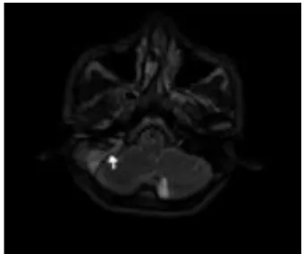
Figure 1: Axial image showing bright signals in right sigmoid sinus region (sigmoid sinus thrombosis).
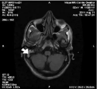
Figure 2: T2 weighted image showing oedema and collection in right petrous region (horizontal arrow) and right sided extradural collection (vertical arrow).
Character of discharge was mucopurulent in majority of patients (44%) in our study, followed by purulent discharge in 24% cases and blood stained discharge in 16% cases (Table 3). In majority of cases (71%) discharge was foul smelling (Table 3).
cases. Among all cases in our study 12 cases showed perforation in attic region, in 5 cases it was marginal and in 6 cases it was central perforation. All cases were examined and ossicular chain erosion was seen in 56% cases and presence of cholesteatoma flakes were seen in 32% cases.
Figure 3: T1 Weighted axial image showing cholesteatoma in right petromastoid region.
Figure 4: T2 weighted axial image showing small lesion in left middle ear with hyperintense component
suggestive of cholesteatoma.
Among all cases hearing impairment was seen in 64% patients, out of which 56% cases were of conductive type, sensorineural hearing loss was present in 12% cases and mixed type were seen in 31% cases (Table 5). Among all patients complete sclerosis on X-ray was seen in 15 cases and partial sclerosis was present in 14 cases. Among all patients, 16 patients showed cholesteatoma in MRI and rest 9 showed inflammatory lesion including granulation tissue, abscess cavity and fluid in middle ear etc. Intracranial complications were also observed in three cases which were in form of extradural abscess, sigmoid sinus thrombosis and abscess in temporal lobe region (Table 5).
DISCUSSION
Now-a-days various studies are going on to assess the role of magnetic resonance imaging in diagnosis of cholesteatoma. Present study also deals with role of MRI using gadolinium enhanced sequences in diagnosis of cholesteatoma.
In present study maximum number of cases (52%) were in 5-15 yrs age group followed by in 16-25 yrs (28%). The incidence in males (68%) was higher than females (32%). Sade et al found peak incidence of disease in 21-30 yrs age group.7 The high incidence in low age group in due to shortness and greater width of Eustachian tube in children by which infection can easily travel from nasopharynx to middle ear, blockage of eustachian tube due to peritubal lymphoid tissue and poor nutrition and lack of personal hygiene of children. Hossain et al analyzed the different types of ossicular chain defect in chronic suppurative otitis media with cholesteatoma on mastoid exploration and observed that cholesteatoma was more common in male (60%) than female (40%) and 53.33% patients were in young age group (21-35 years). 90% cases showed ossicular erosion while only 10% cases showed intact ossicles.3
In present study maximum number of patients presented with complaints of discharging ear (84%) followed by hearing impairment (64%) and then followed by ear ache (48%), headache (32%) swelling around the ear (20%), fever (24%) and tinnitus in (12%) cases. Sade et al also found in their series that otorrhoea was the most common symptom in 62% cases followed by hypoacuris (11%) cases.8
In present study, ossicular erosion was seen in 14 cases (56% cases) and cholesteatoma flakes found in 8 cases (32%). Varshney et al determined the status of the
ossicles in cases of chronic suppurative otitis
media (CSOM) and ossicular erosion was found to be much more common in unsafe CSOM than in safe
CSOM.9
In present study out of 25 cases, 9 cases showed conductive deafness, 2 showed sensorineural hearing loss and 5 showed mixed type of hearing impairment percentage were respectively - 36%, 8% and 20%. All cases under the study were undergone X-ray mastoid bilateral Schuller's view and complete sclerosis was revealed in 15 ears and partial sclerosis was seen in 14 cases. All cases under 2T-unit the study undergone MRI with using gadolinium enhanced T1-T2 sequences.
positive predictive value was 100% and negative predictive value was 66%. Khater et al evaluated the value MRI in diagnosis of middle ear pathologies following operation for chronic otitis media and reported
MRI sensitivity and specificity was 95.23% and 99.16% respectively.10Vaid et al evaluated the role of magnetic resonance imaging (MRI) in diagnosis of cholesteatoma and correlated imaging findings with intraoperative findings, based on specific MRI findings, presence of cholesteatoma was reported in 17 out of 31 patients and all 31 patients underwent surgery and 19 patients had confirmed intraoperative cholesteatoma and similar to present study, this study also showed high sensitivity of a specific sequence based MRI examination in detection of cholesteatoma and in differentiating cholesteatoma from postoperative inflammatory/granulation tissue.4
Martin et al studied with pre- and post-contrast magnetic resonance (MR) images to assess the role of MR imaging in the recognition of middle ear tissue abnormalities and reported that granulation tissue constantly appeared enhanced on studies done with gadolinium diethylene-triaminepentaacetic acid (DTPA), unlike cholesteatoma, cholesterol granuloma, or brain herniation into the middle ear cavities, moreover, when granulation tissue was associated with other soft-tissue masses, Gd-DTPA-enhanced MR images allowed accurate definition of the site and the extension of each lesion.11
The limitation of this study is by using MRI we can't get any information regarding bony structures (i.e., ossicular chain, scutum, tegmen, sinus plate etc.) which can be easily involved in case of cholesteatoma.
CONCLUSION
It can be concluded that MRI is an emerging diagnostic technique which can differentiate cholesteatoma from other inflammatory etiology. In present study sensitivity was 84%, specificity was 100% and positive predictive value and negative predictive value were 100% and 66% respectively. By using MRI with 1.5 or 3T unit a small cholesteatoma (even 2-3 mm) can be easily detected at its early stage and further complications can be prevented.
Funding: No funding sources Conflict of interest: None declared
Ethical approval: The study was approved by the Institutional Ethics Committee
REFERENCES
1. Rout MR, Mohanty D, Vijaylaxmi Y, Kamalesh B, Chakradhar M. Prevalence of cholesteatoma in chronic suppurative otitis media with central perforation. Indian J Otol. 2012;18(1):7.
2. Shrikrishna BH. Ossicular defects in patients with non cholesteatomatous CSOM, OJ Otol HNS. 2010;4:14-7.
3. Hossain MD, Ahamed MN, Sumon MM, Shoyeb BA. Status of ossicles in cholesteatoma, Bangladesh J Otorhinolaryngol . 2015;21(2):97-101.
4. Vaid S, Kamble Y, Vaid N, Bhatti S, Rawat S, Nanivadekar A, et al. Role of magnetic resonance imaging in cholesteatoma: the Indian experience. Indian J Otolaryngol Head Neck Surg. 2011;65(3):485-92.
5. Trojanowska A, Drop A, Trojanowski P, Rosińska-Bogusiewicz K, Klatka J, Bobek-Billewicz B. External and middle ear diseases: radiological diagnosis based on clinical signs and symptoms. Insights Imaging. 2011;3(1):33-48.
6. Vercruysse JP, De Foer B, Somers T, Casselman J, Offeciers E. Magnetic resonance imaging of cholesteatoma: an update. B-ENT. 2009;5(4):233-40.
7. Sade J, Fuchs C. Secretory otitis media in adults: I. The role of mastoid pneumatization as a risk factor. Ann Otol Rhinol Laryngol. 1996;105(8):643-7. 8. Sadé J, Fuchs C. Secretory otitis media in adults: II.
The role of mastoid pneumatization as a prognostic factor. Ann Otol Rhinol Laryngol. 1997;106(1):37-40.
9. Varshney S, Nangia A, Bist SS, Singh RK, Gupta N, Bhagat S. Ossicular chain status in chronic suppurative otitis media in adults. Indian J Otolaryngol Head Neck Surg. 2011;62(4):421-6. 10. Khater NH, Fahmy HS, Shahat HM, Khater AM.
Chronic inflammatory middle ear disease: postoperative CT and MRI findings. The Egyptian J Radiol Nuclear Med. 2015;46(3):629-38.
11. Martin N, Sterkers O, Nahum H. Chronic inflammatory disease of the middle ear cavities: Gd-DTPA-enhanced MR imaging. Radiology. 1990;176(2):399-405.