Original Research Article
Predicting and grading the degree of difficulty of cochlear implant
surgery by evaluating temporal bone using high resolution computed
tomography and magnetic resonance imaging
Vipul V. Chemburkar
1*, Gagandeep S. Saluja
1, Devdas S. Shetty
1, Ruchi R. Agrawal
2INTRODUCTION
The incidence of sensorineural hearing loss (SNHL) occurs in about 1.4 to 3 people per 1000 live births annually in the world.1,2 Hence, it is necessary to identify
a proper diagnostic method to treat this disorder in early stages. About 20% of the children with SNHL are impaired in the anatomy of labyrinth of the ear.3 Cochlear
implant is the important intervention procedure done in the children with SNHL.4,5 But before the cochlear
implant, it is necessary to undergo radiological assessments to diagnose or to identify the congenital abnormalities, cochlear nerve anomalies and/or to detect temporal bone abnormalities.6
High resolution computed tomography (HRCT) scans and magnetic resonance imaging (MRI) are important radiological techniques used to evaluate bony structures and provide accurate information related to the temporal bone diseases. HRCT of the temporal bone is helpful in
1Department of Radiology, T. N. Medical College and B. Y. L. Nair Hospital, Mumbai Central, Maharashtra, India 2Department of Radiology, Zen Hospital, Chembur, Mumbai, Maharashtra, India
Received: 14 September 2019
Accepted: 09 October 2019
*Correspondence:
Dr. Vipul V Chemburkar, E-mail: [email protected]
Copyright: © the author(s), publisher and licensee Medip Academy. This is an open-access article distributed under the terms of the Creative Commons Attribution Non-Commercial License, which permits unrestricted non-commercial use, distribution, and reproduction in any medium, provided the original work is properly cited.
ABSTRACT
Background: The current study was designed to assess the challenges that arise during cochlear implantation. Hence imaging based grading system, using a structured, 12-point scoring chart was developed with an aim to assess various anatomical factors of temporal bone helpful in contemplating complications involved in surgery and to assess various congenital and acquired abnormalities if detected during scan which can affect cochlear implant surgery.
Methods: This was a descriptive study done on 60 patients with sensorineural hearing loss. They were evaluated preoperatively by using HRCT and MRI findings and subsequently underwent cochlear implantation. A 12-point scoring chart was developed based on imaging findings. Surgical times were noted in each case and each imaging point on the scoring chart was correlated with the surgical times.
Results: Eleven out of 12 points in the scoring chart proved to be statistically significant in predicting the degree of difficulty of the surgical procedure. One point was not correlating with the surgical timings. Based on the grading system, in the present study, there were 37 patients (61.66%) classified as Grade 1, 16 patients (26.67%) classified as Grade 2 and 7 patients (11.67%) classified as Grade 3.
Conclusions: These radiological image findings and its related grading system are relatively easy and quick to assess on readily available pre-operative temporal bone CT scan and MRI. They can form a pre-operative checklist that provides a formalized approach for the surgeons and, in particular surgical trainees, predict and, thus prepare for, potentially challenging cochlear implant cases.
Keywords: Cochlear implant, High resolution computed tomography, Magnetic resonance imaging, Sensorineural hearing loss
determining variants of anatomy of temporal bone. MRI temporal bone is helpful in determining cochlear nerve and facial nerve anatomy.7 The current study was
designed using a grading system to assess the challenges
that arise during cochlear implant based on HRCT and MRI findings. The imaging based grading system, using a structured, 12-point scoring chart, would be useful to a trainee and experienced implant surgeon.
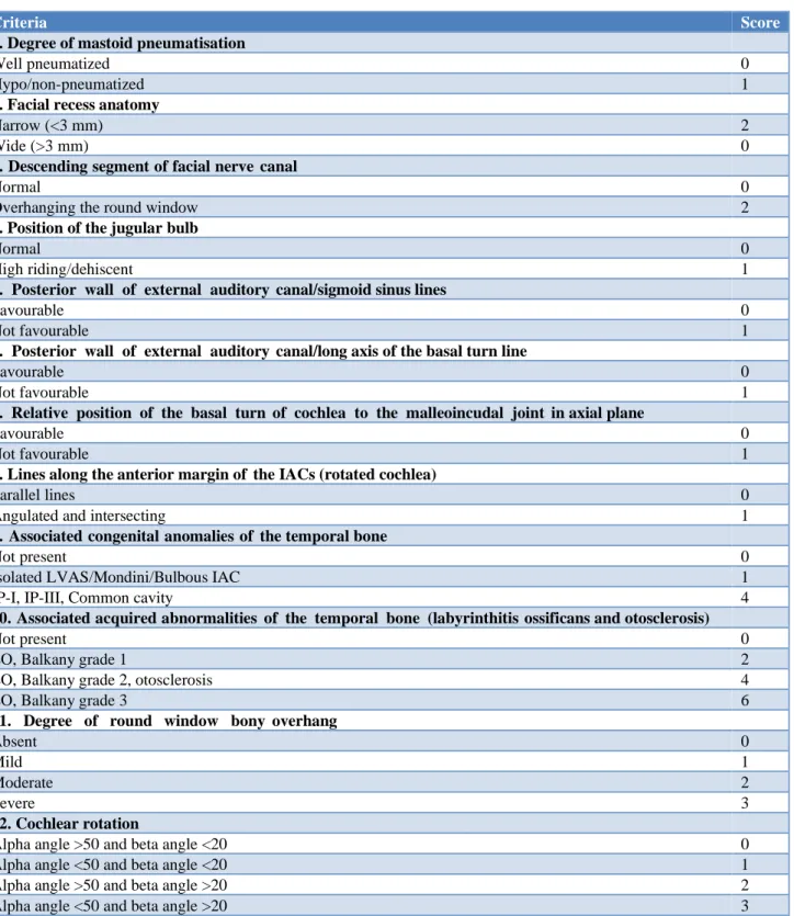
Table 1: Potential difficulty scoring system.
Criteria Score
1. Degree of mastoid pneumatisation
Well pneumatized 0
Hypo/non-pneumatized 1
2. Facial recess anatomy
Narrow (<3 mm) 2
Wide (>3 mm) 0
3. Descending segment of facial nerve canal
Normal 0
Overhanging the round window 2
4. Position of the jugular bulb
Normal 0
High riding/dehiscent 1
5. Posterior wall of external auditory canal/sigmoid sinus lines
Favourable 0
Not favourable 1
6. Posterior wall of external auditory canal/long axis of the basal turn line
Favourable 0
Not favourable 1
7. Relative position of the basal turn of cochlea to the malleoincudal joint in axial plane
Favourable 0
Not favourable 1
8. Lines along the anterior margin of the IACs (rotated cochlea)
Parallel lines 0
Angulated and intersecting 1
9. Associated congenital anomalies of the temporal bone
Not present 0
Isolated LVAS/Mondini/Bulbous IAC 1
IP-I, IP-III, Common cavity 4
10. Associated acquired abnormalities of the temporal bone (labyrinthitis ossificans and otosclerosis)
Not present 0
LO, Balkany grade 1 2
LO, Balkany grade 2, otosclerosis 4
LO, Balkany grade 3 6
11. Degree of round window bony overhang
Absent 0
Mild 1
Moderate 2
Severe 3
12. Cochlear rotation
Alpha angle >50 and beta angle <20 0
Alpha angle <50 and beta angle <20 1
Alpha angle >50 and beta angle >20 2
Alpha angle <50 and beta angle >20 3
contemplating complications involved in surgery and to assess various congenital and acquired abnormalities if detected during scan which can affect cochlear implant surgery.
METHODS
After obtaining approval from institutional ethics committee, the current descriptive study was done on 60 patients during the study period September 2017 to October 2018. All the patients were evaluated for pre-cochlear implant imaging by using HRCT and MRI.
Selection criteria
All cases with bilateral severe to profound sensorineural hearing loss of any age group being assessed for cochlear implant referred to the department over a period of 12 months were included in the study. Patients with active middle ear disease, congenital aural dysplasia and patients medically unfit for undergoing cochlear implantation, parents or patients not willing to give written informed consent, patients with history of cardiac pacemaker, history of heart surgery or valve replacement, history of aneurysmal or vascular surgery, history of electrical implants or neurostimulators or pumps or electrodes or drains or screws or prosthesis, history of orbital metallic foreign body were excluded from the study.
Study procedure
All the patients in the study were evaluated for HRCT and MRI of the temporal bone after obtaining the written informed consent from the patient or from guardian in case of children, according to the hospital protocol. Risk of contrast examination (less likely) was explained. A detailed clinical history of patient was taken and relevant examination findings and investigation was recorded. MRI imaging of the patients was performed on a “Philips Achieva 1.5 T MRI Machine” using a neurovascular or head coil. After a localizer series the standard protocol consists of the following sequences: Diffusion sequence T2 axial sequence, T2 FLAIR COR sequence, T1 SAG sequence, axial T2 VISTA sequence and oblique T2 BFFE, sequence.
CT scan was carried out on “Philips Brilliance 64-Slice CT Machine”. All HRCT examinations was performed with the following parameters: collimation: 64 × 0.625, slice thickness: 0.67 mms, increment: 0.33 mms, reconstruction algorithm: 360°, rotation time: 0.5 s, pitch factor: 0.426 and image display matrix: 768 × 768. Both the axial and multiplanar reformatted images were reviewed on a workstation.
In this study, 12-point scoring chart was used to score each patient based on specific imaging findings. This scoring system uses data based on the principle of
allocating points for individual risk factors thought to increase the level of surgical difficulty. The points are then summated to provide an overall score for each case pre-operatively i.e., a ‘potential difficulty score’ (PDS) (Table 1).
Statistical analysis
Data is analyzed using statistics package Epi InfoTM
7.1.3.3 version for windows. Descriptive data are represented as mean±SD for numeric data and percentages and proportions for categorical data. Appropriate tests of significance were used depending on the nature and distribution of data. We used Pearson’s correlation and a linear regression to assess relationships and quantify the effect on surgical timings based on the 10-point scoring system. Values of p<0.05 were considered statistically significant. An analysis of the data based on the above imaging criteria and findings were performed.
RESULTS
Table 2: Age and gender distribution of study participants (n=60).
Age group Number of patients and
gender
Less than 5 years 41 (23 females,18 males) 5-10 years 15 (4 females, 11 males) More than 10 years 4 (0 females, 4 males)
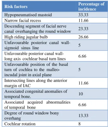
Table 3: Incidence of risk factors based on imaging finding.
Risk factors Percentage of
incidence
Hypopneumatised mastoid 33.33 Narrow facial recess 11.66 Descending segment of facial nerve
canal overhanging the round window 23.33 High riding jugular bulb 26.66 Unfavourable posterior canal wall-
sigmoid sinus line 5 Unfavourable posterior canal
wall-long axis cochlear basal turn lines 6.66 Unfavourable position of the basal
turn of cochlea to the malleo-incudal joint in axial plane
5 Intersecting lines along the anterior
margin of IAC 11.66
Associated congenital anomalies of
temporal bone. 10
Associated acquired abnormalities of temporal bone 6.66 Degree of round window bony
overhang 13.33
Table 4: Degree of surgical difficulty and its incidence
on patients based on PDS and surgical timing.
Score Surgical
time Grade
Number of patients
Imaging based prediction
0-3 60-90 1 37
No anticipated surgical difficulty 4-7 91-120 2 16
Anticipated surgical difficulty 8 and
above 121-200 3 7
Prolonged and difficult surgery Table 3 presents the frequency of risk factors in the patients as per 10 point scoring chart. Hypopneumatised mastoid was the common risk factor noticed in 33.33% patients followed by high riding jugular bulb (26.66%). All the 60 patients underwent cochlear implant surgery by the implant surgeon.
A total of 60 patients were evaluated for HRCT and MRI of the temporal bone in the study.
Table 2 presents the age and sex distribution of study participants. Out of 60, 27 were females and 33 were males. Majority of them were children of less than 5 years of age (n=41).
Table 5: Statistical correlation of imaging findings with surgical timings.
Imaging findings in scoring chart
Pearson correlation (r value)
P value
Degree of mastoid
pneumatisation 0.284 0.035 Facial recess anatomy 0.548 0.001 Descending segment of facial
nerve canal 0.341 0.024
Position of jugular bulb 0.160 0.032 Posterior wall of external
auditory canal or sigmoid sinus line
0.295 0.021 Posterior wall of external
auditory canal/long axis of the basal turn line
0.232 0.088 Relative position of basal
turn of cochlea to malleo- incudal joint in axial plane
0.299 0.016 Lines along anterior margin
of IAC 0.427 0.018
Associated congenital
anomalies of temporal bone. 0.403 0.022 Associated acquired
abnormalities of temporal bone
0.598 0.031 Degree of round window
bony overhang 0.242 0.024 Cochlear rotation 0.258 0.019
Total score 0.892 0.027
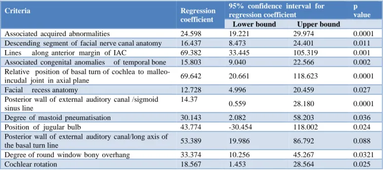
Table 6: Multiple linear regression showing predictors of surgical timings.
Criteria Regression
coefficient
95% confidence interval for regression coefficient
p value
Lower bound Upper bound
Associated acquired abnormalities 24.598 19.221 29.974 0.0001 Descending segment of facial nerve canal anatomy 16.437 8.473 24.401 0.011 Lines along anterior margin of IAC 69.382 33.445 105.319 0.001 Associated congenital anomalies of temporal bone 15.803 9.040 22.566 0.002 Relative position of basal turn of cochlea to malleo-
incudal joint in axial plane 69.642 20.661 118.623 0.0001
Facial recess anatomy 12.728 4.996 20.459 0.027
Posterior wall of external auditory canal /sigmoid sinus line
14.37
0.559 28.180 0.0001
Degree of mastoid pneumatisation 30.143 2.082 58.203 0.036 Position of jugular bulb 43.774 -30.454 118.002 0.024 Posterior wall of external auditory canal/long axis of
the basal turn line 53.389 19.986 86.792 0.088
Degree of round window bony overhang 33.374 10.256 45.267 0.0321
The time taken for the surgery ranges from 60-200 minutes. From Table 4 it was observed that patients with PDS score of 0-3 was graded as grade 1 patients (n=37) and had surgical timings within a range of 60-90 minutes. Those patients with PDS score of 4-7 was considered as grade 2 patients (n=16) and surgical timings ranged from 91-120 minutes and patients with PDS score of 8 and above graded as grade 3 patients with surgical timings ranges from 121-200 minutes (n=7).
In the present study, each imaging point was correlated with surgical time as given in Table 5.
Linear relationship was noted between scoring system and operative timings. Multiple linear regression analysis was done based on the above imaging criteria of PDS score and findings observed during HRCT and MRI (Table 6).
DISCUSSION
This study aims to evaluate the diagnostic efficacy of HRCT and MRI of temporal bone disorders based on 12-point scoring chart. The findings of the study showed that the both HRCT and MRI of the temporal bone had a high diagnostic value in patients with SNHL. The study included 60 patients with congenital sensorineural hearing loss belonging to age group of less than 10 years with most of them below 5 years of age. In elder age group patients, cause of sensory neural hearing loss was acquired mostly post infection and trauma.
In the present study, 12 point scoring chart was used to assess the challenges that arise during cochlear implant by using HRCT and MRI as diagnostic aids (Table 2). Similar to this Vaid et al previously proposed a grading system based on a 10-point scoring chart of HRCT and MRI imaging findings in patients being assessed preoperatively for cochlear implant.8
In this study ratio of male and female patients were almost equal. In our study male surpasses female by a small difference. This could be due to different demographic factors of place and patients attending this hospital.
In his study, out of the ten mentioned imaging points in the scoring chart determining the PDS, eight were found to correlate significantly with the surgical timings and, hence, had a direct impact on the degree of surgical difficulty. In this study two points were not correlating with the surgical timings. An insufficient cohort for this study was likely to be the cause for this observation. However in our study, twelve criteria were taken (Table 2) and studied out of which eleven criteria were found to be significant. The only factor found to be insignificant was- posterior wall of external auditory canal/long axis of the basal turn line which was similar to factor found insignificant in above mentioned study. However another
factor which was found insignificant in above mentioned study was position of jugular bulb which is found to be significantly correlating with surgical timings in our study.
Based on the grading system, in the present study, there were 37 patients (61.66 %) classified as Grade 1, 16 patients (26.67 %) classified as Grade 2 and 7 patients (11.67 %) classified as Grade 3. Out of the 37 patients in Grade 1, it was observed that 32 patients had uneventful surgery and five patients had prolonged surgery (sensitivity: 86.5 %; positive predictive value: 100%; specificity: 100%; negative predictive value: 86.5%). Out of these five patients, two patients had dense mastoid sclerosis, two had blood dyscrasias that were not identified on routine, preoperative, haematological investigations and one patient had congested middle ear mucosa.
Out of the 16 patients assigned as Grade 2,12 patients had minor surgical difficulties as predicted by specific imaging findings on the scoring chart, and four had uneventful surgery (sensitivity: 100%; positive predictive value: 75%; specificity: 90%; negative predictive value: 100%). Out of these three, two patients (despite having a hypopneumatized mastoid) had an uneventful surgery. One patient with labyrinthitis ossificans Balkany grade 1 had an easy insertion. One patient with narrow facial recess on imaging had easy access to the round window. All seven patients assigned as Grade 3 had prolonged and difficult surgery (sensitivity: 100%; positive predictive value: 100%; specificity: 100%; negative predictive value: 100%). The above observations were almost similar to the findings of Vaid et al.8
CONCLUSION
Cochlear implant is an accepted and popular treatment for patients with profound bilateral SNHL. The rate of success of the surgery would be good if the degree of associated difficulties were known preoperatively. With this, in mind, the authors of the study designed 12 point imaging based scoring chart and a grading system which alerts the surgeonfor likely problems and difficulties they may encounter during the surgery. After grading the patients based on imaging findings, the results of the current study concluded that, patients who have a potential difficulty score between 0 and 3 are more likely to have uneventful and uncomplicated surgery with the lowest intraoperative times. Scores between 4 and 7 alert the surgeon to moderate surgical difficulty and longer intraoperative times. Scores of eight and above indicate a high chance of a prolonged and difficult surgery.
Funding: No funding sources Conflict of interest: None declared
REFERENCES
1. McClay JE, Booth TN, Parry DA, Johnson R, Roland P. Evaluation of pediatric sensorineural hearing loss with magnetic resonance imaging, Arch. Otolaryngol Head Neck Surg. 2008;134(9):945-52.
2. Saki N, Bayat A, Hoeinabadi R, Nikakhlagh S, Karimi M, Dashti R. Universal newborn hearing screening in southwestern Iran. Int J Pediatr Otorhinolaryngol. 2017;97:89-92.
3. Parry DA, Booth T, Roland PS. Advantages of magnetic resonance imaging over computed tomography in preoperative evaluation of pediatric cochlear implant candidates. Otol Neurotol. 2005;26:976-82.
4. Joshi VM, Navlekar SK, Kishore GR, Reddy KJ, Kumar EC. CT and MR imaging of the inner ear and brain in children with congenital sensorineural hearing loss. Radiographics. 2012;32(3):683-98. 5. Witte RJ, Lane JI, Driscoll CL, Lundy LB,
Bernstein MA, Kotsenas AL, et al. Pediatric and adult cochlear implantation. Radiographics. 2003;23:1185-200.
6. Taha T, Wahba H, Ibrahim AS, Abd Elazim Y. Cochlear implant tailored imaging protocol: What clinicians need to know. Egyptian J Radiol Nucl Med. 2015;46:33-43.
7. Hanafi MG, Saki N, Shanehsaz F. Diagnostic value of CT and MRI of Temporal Bone in Cochlear Implantation Candidates. Int J Pediatr. 2019;7(7):9693-700.
8. Vaid S, Vaid N, Manikoth M, Zope A. Role of HRCT and MRI of the Temporal Bone in Predicting and Grading the Degree of Difficulty of Cochlear Implant Surgery. Indian J Otolaryngol Head Neck Surg. 2015;67(2):150-8.