Original citation:
Jaggers, Ross and Bon, Stefan A. F.. (2017) Temporal and spatial programming in soft
composite hydrogel objects. Journal of Materials Chemistry B, 5 (36). pp. 7491-7495.
Permanent WRAP URL:
http://wrap.warwick.ac.uk/92635
Copyright and reuse:
The Warwick Research Archive Portal (WRAP) makes this work of researchers of the
University of Warwick available open access under the following conditions. Copyright ©
and all moral rights to the version of the paper presented here belong to the individual
author(s) and/or other copyright owners. To the extent reasonable and practicable the
material made available in WRAP has been checked for eligibility before being made
available.
Copies of full items can be used for personal research or study, educational, or not-for-profit
purposes without prior permission or charge. Provided that the authors, title and full
bibliographic details are credited, a hyperlink and/or URL is given for the original metadata
page and the content is not changed in any way.
Publisher statement:
First published by Royal Society of Chemistry 2017
http://doi.org/10.1039/c7tb02011b
A note on versions:
The version presented here may differ from the published version or, version of record, if
you wish to cite this item you are advised to consult the publisher’s version. Please see the
‘permanent WRAP URL’ above for details on accessing the published version and note that
access may require a subscription.
Journal Name
COMMUNICATION
Received 00th January 20xx, Accepted 00th January 20xx
DOI: 10.1039/x0xx00000x
www.rsc.org/
Temporal and spatial programming in soft composite hydrogel
objects
Ross W. Jaggers and Stefan A. F. Bon*a
Soft composite hydrogel objects formed from the biopolymer sodium alginate, the enzyme urease, and oil droplets are formed by a simple gelation procedure to produce autonomous bodies with both time and spatial programming. These continuous objects of non-‐uniform dimensional composition selectively respond to an environmental stimulus of urea and disintegrate at pre-‐defined locations within the hydrogel structure after pre-‐ defined time intervals. The spatial and temporal responses of these hydrogels to an environmental stimulus are valuable tools in areas such as soft robotics.
Soft, flexible and deformable structures are commonplace in nature, where evolution has developed elegant and efficient designs to accommodate a wide range of functions.1 For many years, nature has inspired the creation of machines capable of ‘life-‐ like’ tasks,2 and these efforts have spawned the field of research known as soft robotics. This discipline seeks to create soft structures capable of adaptation, with a sensitivity and agility that can supersede mechanical, hard-‐bodied robots.3,4
Soft robots can have the ability to perform shape transformations,5,6 for example, muscles like actuation,7,8 or
undergo locomotion,9–11 and can interact with their
environments.12,13 They are typically composed of hydrogels,14,15
polymers16,17, including elastomers,18–20 and must be robust.21,22
However, the majority of soft robots are still composed of both soft and rigid counterparts, the latter being required for power and control.23,24
A key challenge in this field is the design of entirely soft robots, in which responsive autonomous behaviour and function is built in and programmed. An impressive soft robotic system is the octobot, capable of the catalytic decomposition of an on-‐board fuel to power arm movement.25 This example offers a soft analogue of the
previously existing hardware version of itself, and shows that soft systems can be programmed to act in an autonomous, robotic fashion. Furthermore, large (millimetre scale) soft objects have been designed to interact with their environments, such as the soft-‐ robotic ray that swims in response to a light source,26 the hydrogel
actuator that walks in an electric field,14 the tissue-‐engineered
jellyfish that propels in an electric field,27 and the sequentially self-‐
folding shapes formed from shape memory polymers.28
Critically, a soft object of this nature should be capable of non-‐ uniform, autonomous behaviour when provided with a fuel. Large soft objects need to be built in a way that their structures can vary, in order to introduce different functionalities into different parts of the object and allow for more complex behaviours, such as spatial or temporal programming.29
In this work, we show that soft hydrogel-‐based objects can be programmed to carry out non-‐uniform actions across their structures when exposed to a single fuel source, based on principles outlined in our previous work.30 We demonstrate the
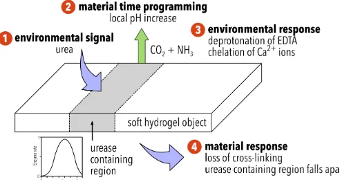
Figure 1 Schematic describing the behaviour of ionically cross-‐linked hydrogel objects: (1) the semi-‐permeable soft object converts the fuel source, urea, to ammonia, due to the inclusion of urease in selected parts of its structure (2) after a defined period of time, urea containing regions of the object generate a local pH increase. Time-‐programming is possible due to the bell-‐shaped activity curve of urease, depicted in the figure37 with an enzyme rate, v' = (1 + 2 x 10-‐9
/[H+] + [H+]/5 x 10-‐6) -‐1 (3) partially protonated ethylenediaminetetraacetic acid
[image:2.595.307.553.513.643.2]COMMUNICATION Journal Name
temporal and spatial responsive nature of the soft bodies programmed by non-‐uniform distribution of the enzyme urease to introduce triggerable active zones in the otherwise dormant jelly. These active zones process the trigger fuel, urea, to create local pH gradients that alter the object’s structure, as depicted in figure 1. In its simplest form (Generation 1) we show a temporal and spatial colour change, associated with a local change in pH. The concept is then extended to control of material disintegration (Generation 2).
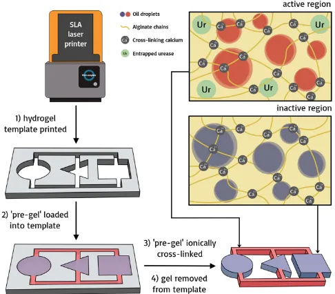
Hydrogel objects are made from viscous aqueous solutions of the biopolymer sodium alginate, sourced from brown algae. Gels are easily formed upon exposure to Ca2+ (aq).30,31 Sodium alginate is used widely in the pharmaceutical and food industries due to its lack of toxicity and high biocompatibility,32 and is a good candidate for building soft sustainable materials.33 We used a templating strategy to fabricate centimeter-‐sized structures, typically a few millimeters thick. Templates corresponding to the desired soft object shape were printed using a FormLab 2 SLA microstereolithographic printer. Aqueous sodium alginate formulations, termed ‘pre-‐gels’, were loaded manually into designated regions of the printed templates. These were immediately ionically cross-‐linked by spraying a liquid aerosol of calcium chloride solution onto them (see Figure 2 for schematic).
The ‘pre-‐gel’ formulations used contained different ingredients such as coloured oil droplets and/or pH indicator dye, and specified amounts of the enzyme urease. Urease is introduced to these soft objects to spatially control pH over time. The enzyme is trapped inside the gel and does not migrate or diffuse (see supporting information, figure 1).34,35 Urease raises the pH by converting urea into ammonia and carbon dioxide, which we introduce as the
trigger into the liquid environment in which the gels are placed.
The regional concentration of entrapped urease regulates the local increase in pH as a function of time. Time-‐programming is afforded due to the bell-‐shaped pH-‐activity curve of urease and its associated auto-‐catalytic behaviour. Low enzyme activity is
Figure 3 A continuous soft object is formed from both inactive (white) regions that contain no urease, and active regions (coloured) that contain urease at a concentration of 20 g L-‐1 and the pH indicator bromothymol blue. This object is placed in a bath of EDTA and urea at concentrations of 0.1 and 0.45 mol dm-‐3,
respectively, at a pH of 3.5 (hence the yellow regions). Once the active regions have processed the urea into ammonia, the pH locally rises (above the indicating pH of 7.6) to produce a blue colour. See supporting video for the full sequence. Scale bar = 1 cm. An RGB line scan is taken across the width of the most left leaf at the following time intervals: t = 0 n, t = 26 l, t = 38 p, t = 68 Ð, t = 136 ê, and t = 263 É. From these, the hue angle is calculated (used as a numerical representation of colour) and plotted against the distance of the line scan in graph A. This graph shows the change in colour across the width of the leaf. Plotting the hue angle over time at selected locations across these line scans (signified by the dashed lines in graph A and as follows: pixel distance = 4 n, = 10 l, = 18 p, = 30 ê, and = 40 Ð) produces graph B. This graph depicts the change in colour of the leaf over time.
Figure 2 Schematic depicting the synthesis of soft responsive objects. 1) The object template is designed and printed using CAD and an SLA laser printer 2) The ‘pre-‐gel’ is loaded into the template in the desired arrangement 3) the pre-‐gel is ionically cross-‐linked upon exposure to a 0.1 mol dm-‐3 aqueous
[image:3.595.307.550.65.278.2] [image:3.595.87.552.431.677.2]observed at low pH values; the generation of base elevates the pH into the higher activity window of the bell-‐shaped pH-‐activity curve, providing a positive feedback mechanism. As the initial increase in pH follows sigmoidal behaviour, the introduction of a dormancy period by variation of urease concentration is possible (see figure 1).
We will first discuss Generation 1 hydrogels where the temporal and spatial pH responses manifest themselves as colour changes.
When placed into acidic water of pH 3.5 in the presence of urea (0.45 mol dm-‐3), a urease (20 g L-‐1) and pH indicator dye containing hydrogel, in the form of leaves on an inactive jelly tree, evidently generate a pH increase in excess of 7.6, as determined by the pH colorimetric indicator bromothymol blue (see figure 3 and supporting video). We are able to quantify this change in colour by measuring the hue angle of the corresponding HSL histogram of a line scan from one of the leaves. The leaf starts with a hue angle of ca. 65°, corresponding to a yellow colour, and finishes with an angle of ca. 220°, that of the colour blue (N.B. the hue is the angle around the central vertical axis of an HSL cylindrical coordinate representation of a colour. The indicator bromothymol blue changes colour with pH, and as we are able to track this numerically, we are able to quantify its colour). As graph A of figure 3 shows, the colour change starts at the outside of the leaf and progresses inwards, as urea diffuses into the object. By comparing the hue angle over time at different points across this line scan, the change of colour over time is observed, as in graph B of figure 3. This curve is indicative of the pH increase over time generated by the activity of urease. As figure 3 shows, this response is not only time programmed, but also spatially programmed. By having a non-‐ uniform distribution of urease across the hydrogel structure, we are able to create a single, millimetre-‐sized soft object with anisotropic behaviour when exposed to a single stimulus.
In Generation 2 systems, we utilise this temporal and spatial programming in the selective and coordinated disintegration of a hydrogel object (see figure 1), paired in some cases with the release
of oil droplets. We achieve this by adding the sodium salt of ethylenediaminetetraacetic acid (EDTA) to the hydrogel environment as a chelating agent for calcium ions, acting as a dormant Ca2+-‐sink. Objects are placed into an aqueous environment containing both EDTA and urea set at a pH value of 3.50. EDTA binds Ca2+ ions best at high pH, when all 6 of its donor groups (4 carboxylic acid groups and 2 nitrogens) are ionized. At a pH of 7.5, only a fraction of the EDTA is in this ionized form and its affinity for Ca2+ ions is reduced. As the pH is lowered further, the affinity of this chelator continues to decreases.36 This was verified by enzyme loaded hydrogel objects into an EDTA solution of pH 3.50 without urea. Without the ability to raise its pH, it remained intact over a period of at least 14 days. These ionically cross-‐linked objects are stable for months in the absence of EDTA. Upon a pH increase inside and in close proximity of the object (as a result of the programmed enzymatic reaction), EDTA transitions to its Ca2+ chelating form and thus promotes cation exchange. This results in the eventual disintegration of the alginate-‐gel matrix and subsequent release of any entrapped oil droplets.
To recap this system (also summarised in figure 1), the object converts urea, the fuel, to ammonia, due to the inclusion of urease in its structure. After a defined time period, dependent on the concentration of urease, the object generates a local increase in pH. Partially protonated ethylenediaminetetraacetic acid (EDTA) is present in the low pH (3.50) environment. As the pH of the active sites on the hydrogel object increases locally, EDTA is deprotonated locally and chelation of calcium ions results in a loss of cross-‐linking and disintegration of the object, as well as the release of oil droplets if they are present.
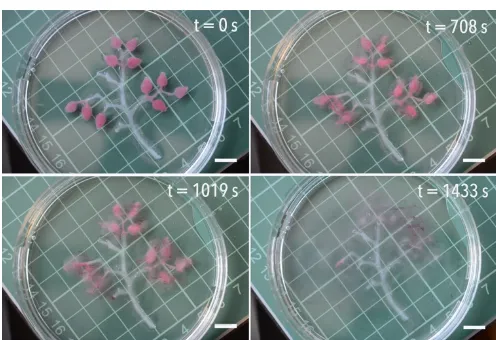
Figure 4 and the supporting video demonstrate time and spatial programming in a millimetre-‐sized soft object that make use of this autonomous, enzyme-‐powered system when exposed to a fuel source of urea. A 5 wt. % alginate skeleton has 1 wt. % alginate leaves attached to it that contain a coloured emulsified oil (Oil Red O in vegetable oil) and urease at a concentration of 20 g L-‐1. Introduction to an EDTA and urea environment causes the leaves to
Figure 5 A continuous soft object containing three distinct regions of different enzyme concentrations is immersed in an EDTA/urea environment (0.1 and 0.45 mol dm-‐3, respectively) at pH 3.5. Due to local pH increases, segment 1
containing emulsified blue oil droplets and 20 g L-‐1 urease, disintegrates first,
followed by segment 2 that contains emulsified red oil droplets and 10 g L-‐1
urease, and then segment 3 that contains emulsified yellow oil droplets and 5 g L-‐1 urease. See supporting video for the full sequence. Scale bar = 1 cm. Figure 4 A hydrogel tree branch is formed from an inactive branch structure
(clear, no urease) and active leaves (red emulsified oil droplets, 20 g L-‐1 urease).
When immersed in an EDTA/urea environment (0.1 and 0.45 mol dm-‐3,
[image:4.595.43.291.517.687.2] [image:4.595.310.556.521.688.2]COMMUNICATION Journal Name
disintegrate and ‘fall off’ the tree, thus decreasing the object’s size and changing its shape. Disintegration of the leaves results in the release of the emulsified oil, the droplets of which are no longer stabilised once liberated from the alginate matrix. All 11 leaves start to disintegrate at the same time, marked by the release of oil droplets at 220 seconds, and all disappear at approximately the same time (see supporting video). This design concept allows for the generation of a soft object that is programmed to change shape over time, thus allowing it to exist in more than one form during its lifetime. In this example, the enzyme concentration builds in a dormancy period prior to objects transformation, the length of which is determined by the concentration of urease at a fixed concentration of urea. As a result, a single destabilisation event is observed for all regions containing a single urease concentration. If, however, a single object contains three distinct regions of different urease concentrations, it will respond at three pre-‐defined time intervals in response to one urea stimulus.
Figure 5 and the supporting video show a composite gel object with regions that contain urease at a concentration of 20, 10 and 5 g L-‐1, and blue, red and yellow oil droplets, respectively. Following the introduction of the object to an EDTA and urea environment, the object sequentially disintegrates region by region (figure 5 and supporting video). At 160 seconds, the first segment, containing 20 g L-‐1 urease, starts to disintegrate. This is followed by the second segment, containing 10 g L-‐1 urease, at 300 seconds, and finally the third segment, containing 5 g L-‐1 urease, at 780 seconds. These three segments have fully disintegrated after 550, 1110, and 4140 seconds, respectively, releasing their oil droplets in the process. Reducing the urease concentration both increases the dormancy period before material decomposition and the length of the decomposition time for a given region in the object. This clearly demonstrates that one object can be programmed to exhibit three distinct responses with both spatial and temporal programming.
Conclusions
In conclusion, we demonstrate the design and fabrication of continuous soft hydrogel objects that are programmed to spatially and temporally respond to the environmental cue of a fuel source, urea. The enzyme urease provides the objects with in-‐built time-‐programming, allowing the objects to produce their own local pH fields after pre-‐defined time periods by means of varied enzyme concentrations. By controlling the distribution of the enzyme urease across the body of the object, regions of the object selectively disintegrate upon activation of the enzyme. This results in an object with autonomous control over its response to an environmental stimulus. Such a material is valuable in the design and fabrication of entirely soft robots, offering a way to selectively build responsive characteristics into entirely soft objects.
References
1 M. H. Dickinson, C. T. Farley, R. J. Full, M. A. R. Koehl, R. Kram and S. Lehman, Science, 2000, 288, 100–106. 2 A. W. Feinberg, Annu. Rev. Biomed. Eng., 2015, 17, 243–
265.
3 D. Rus and M. T. Tolley, Nature, 2015, 521, 467–475. 4 S. Kim, C. Laschi, B. Trimmer and B. Trimmer, Trends
Biotechnol., 2013, 31, 287–294.
5 A. D. Marchese, R. Tedrake and D. Rus, Int. J. Rob. Res., 2016, 35, 1000–1019.
6 B. Mazzolai, L. Margheri, M. Cianchetti, P. Dario and C. Laschi, Bioinspir. Biomim., 2012, 7, 25005.
7 M. Cianchetti, M. Calisti, L. Margheri, M. Kuba and C. Laschi, Bioinspir. Biomim., 2015, 10, 35003.
8 A. Wang, H. Yu and S. Cang, J. Franklin Inst., 2017, 354, 1759–1783.
9 M. T. Tolley, R. F. Shepherd, B. Mosadegh, K. C. Galloway, M. Wehner, M. Karpelson, R. J. Wood and G. M.
Whitesides, Soft Robot., 2014, 1, 213–223.
10 A. D. Marchese, C. D. Onal and D. Rus, Soft Robot., 2014, 1, 75–87.
11 C. D. Onal and D. Rus, Bioinspir. Biomim., 2013, 8, 26003. 12 D. Han, H. Gu, J. Kim and S. Yokota, Sensors Actuators A
Phys., 2017, 257, 47–57.
13 Y. J. H. Kin, E. Dong, M. Xu, C. Liu, G. Alici, Smart Mater.
Struct., 2016, 25, 85026.
14 D. Morales, E. Palleau, M. D. Dickey, O. D. Velev, S. J. Candau, R. A. Mashelkard, A. K. Leled, M. J. Tauberc, G. Aryab and S. Varghese, Soft Matter, 2014, 10, 1337–1348. 15 L. Ionov, Adv. Funct. Mater., 2013, 23, 4555–4570. 16 I. A. Anderson, T. A. Gisby, T. G. McKay, B. M. O’Brien and
E. P. Calius, J. Appl. Phys., 2012, 112, 41101. 17 A. Argiolas, B. C. Mac Murray, I. Van Meerbeek, J.
Whitehead, E. Sinibaldi, B. Mazzolai and R. F. Shepherd,
Soft Robot., 2016, 3, 101–108.
18 H.-‐T. Lin, G. G. Leisk and B. Trimmer, Bioinspir. Biomim., 2011, 6, 26007.
19 C. Larson, B. Peele, S. Li, S. Robinson, M. Totaro, L. Beccai, B. Mazzolai and R. Shepherd, Science, 2016, 351, 1071– 1074.
20 D. Hughes and N. Correll, Bioinspir. Biomim., 2015, 10, 55002.
21 C. Lee, M. Kim, Y. J. Kim, N. Hong, S. Ryu, H. J. Kim and S. Kim, Int. J. Control. Autom. Syst., 2017, 15, 3–15. 22 K. Suzumori, Rob. Auton. Syst., 1996, 18, 135–140. 23 K. B. Shimoga and A. A. Goldenberg, Proc. IEEE Int. Conf. on
Robotics and Automation, 1992, 1300–1305.
24 S. W. Kwok, S. A. Morin, B. Mosadegh, J.-‐H. So, R. F. Shepherd, R. V. Martinez, B. Smith, F. C. Simeone, A. A. Stokes and G. M. Whitesides, Adv. Funct. Mater., 2014, 24, 2180–2187.
Qi, Sci. Rep., 2015, 5, 13616.
29 X. He, J. Fan, J. Zou, K. L. Wooley, X. Li, G. Yu, J. Zhu, A. Li, K. Seetho, X. He, D. J. Pochan, K. L. Wooley, Z. Fang, S. O. Kelley, J. W. Gilman and A. Javey, Chem. Commun., 2016,
52, 8455–8458.
30 R. W. Jaggers and S. A. F. Bon, Mater. Horiz., 2017, 4, 402– 407.
31 A. Johnson and J. B. Speakman, J. Soc. Dye. Colour., 1946,
62, 97–100.
32 K. Y. Lee and D. J. Mooney, Prog. Polym. Sci., 2012, 37, 106–126.
33 M. Szekalska, A. Puciłowska, E. Szymańska, P. Ciosek, K. Winnicka, P. Ciosek and K. Winnicka, Int. J. Polym. Sci., 2016, 2016, 1–17.
34 A. R. DeGroot and R. J. Neufeld, Enzyme Microb. Technol., 2001, 29, 321–327.
35 N. Das, A. M. Kayastha and O. P. Malhotra, Biotechnol.
Appl. Biochem., 1998, 27, 25–9.
36 Y. V. Griko, Biophys. Chem., 1999, 79, 117–127. 37 G. Hu, J. A. Pojman, S. K. Scott, M. M. Wrobel and A. F.