Original Article
The study of the prognostic value of mixed lymphocyte
reaction blocking factor (MLR-Bf) for the pregnancy
outcome after lymphocyte immunization in
different kinds of patients with unexplained
recurrent spontaneous abortion
Ke Yang, Bing Zhang, Gui-Yu Lou, Hong-Dan Wang, Qiao-Fang Hou, Miao He, Yan Chu, Yu-Qin Chen, Qian-Cheng Li, Chao-Yang Zhang, Yin-Pei Song
Medical Genetic Institute of Henan Province, Henan Provincial People’s Hospital, People’s Hospital of Zhengzhou University, Zhengzhou 450003, PR China
Received September 12, 2016; Accepted September 28, 2016; Epub January 1, 2017; Published January 15, 2017
Abstract: The study was conducted to access the prognostic value of mixed lymphocyte reaction blocking factors (MLR-Bf) for the pregnancy outcome after lymphocyte immunization (LIT) in different kinds of patients with unex-plained recurrent spontaneous abortion (URSA). Following LIT, the patients were divided into two groups according to the time when the miscarriage occurs (group 1: before the demonstration of embryonic cardiac activity; group 2: after the demonstration of embryonic cardiac activity) and the times of URSA (patients with two URSA and three or more URSA). We respectively compared the success rate in immunized patients who showed MLR-Bf with control in
each group. For patients with two URSA, there was no significant difference between treatment group and control in each group; for patients with three or more URSA, the pregnancy outcome was significantly improved in treatment
group in group 1 (67.2% vs 53.1%, P=0.0004). Whereas no significant difference was detected in group 2 (60.3%
vs 56.6%, P=0.5684). The obtained data demonstrated that the presence of MLR-Bf is a good prognostic criterium for the pregnancy outcome in patients with three or more URSA who had never had the demonstration of embryonic cardiac activity after LIT.
Keywords: MLR-Bf, habitual abortion, immunotherapy
Introduction
Recurrent spontaneous abortion (RSA) is a common complication of pregnancy affecting about 1-3% couples [1], which is defined as two or more clinical miscarriages documented by ultrasonography or histopathologic examinati- on (not necessarily consecutive) by the Ameri- can Society for Reproductive Medicine (ASRM) [2]. Factors such as genetic impairment, struc-tural or functional abnormalities in genital organs, hormonal deficiency, infectious dis-ease, metabolic disorder, autoimmune abnor-malities, abnormal pre-thrombotic state are thought to be associated with the RSA [3]. However, in most women who suffered from RSA, no cause can be identified. The factors inducing miscarriage are still unclear. Alloim-
mune mechanisms have been proposed as the cause of some or all of these losses which prevent mothers from developing immunologi-cal responses essential for the survival of the embryo.
rate of successful pregnancy [9-13], while some researchers considered it had no benefit at all [14, 15]. The results from clinical trials have been problematic [9, 10, 14, 15]. In previ-ous studies, M LR-Bf is the most common used parameter for monitoring the treatment effec-tiveness of URSA in the world. While the prog-nostic value of it was controversial [14, 16, 17]. Before an effective treatment can be institut-ed, the reason of RSA must be determined. The causes for RSA of different gestational weeks are various. The first trimester miscarriage is hypothesized that this may due to chromosom-al abnormchromosom-alities, endocrinologicchromosom-al disorders, immunological abnormalities and abnormal pre-thrombotic state. The main reasons for mis-carriage occurring between 12-28 weeks incl- udes abnormal pre-thrombotic state, infec-tions, abnormal fetal appurtenances and so on. So in our study, to investigate the efficacy of LIT for URSA, we selected patients who had miscarriages in first trimester. The different time when the miscarriage occurs indicate that they may have different causes. In previous studies, we noticed that no one has evaluated the efficacy of LIT by dividing the patients into different groups according to the time when the miscarriage occurs, which may suggest the presence of specific cause. So in our study, we classified the patients into the following two groups according to the time when the miscar-riage occurs: group 1: before the demonstra-tion of embryonic cardiac activity; group 2: after the demonstration of embryonic cardiac activi-ty. At the same time, as is known that the suc-cess rate of untreated patients with only two URSA is very high, the patients involved in each group were divided into two subgroups for detailed analysis. The first group consisted of women with only two URSA, the second one included women with three or more URSA. We respectively compared the success rate in immunized patients who showed positive MLR-Bf with control in each group, and reassess the prognostic value of MLR-Bf.
Materials and methods
Patients
A total of 1993 patients were recruited at our center between July 2007 and July 2015. The selection criteria were: 1. Each participant cohabiting with a single partner had
experi-enced two or more confirmed first trimester (i.e.before 14 weeks of gestation) miscarriages that were not of chromosomally abnormal fetuses; 2. No MLR-Bf detected; 3. Age 40 years or younger at the time of recruitment; 4. Each participant’s partner was negative for hepatitis B surface antigen (HbsAg), human immunodeficiency virus (HIV), Hepatitis C Virus (HCV) and syphilis; 5. No identifiable causes for the previous miscarriages including chro- mosomal abnormalities, structural functional abnormalities in genital organs, endocrinologi-cal disorders. Our study center had approval from the institute ethical committee, and all patients gave their consent to participate in the study.
Design and procedures
According to the time when the previous mis-carriage occurs, the patients were divided into the following two groups: group 1: before the demonstration of embryonic cardiac activity; group 2: after the demonstration of embryonic cardiac activity. URSA patients had underwent two or more miscarriages, so some patients might have a complicated pregnancy history, for example one patient’s first miscarriage occurred after the demonstration of embryonic cardiac activity, second miscarriage occurred before the demonstration of embryonic cardiac activity; these kinds of patients were excluded from the investigation. The sufficient informa-tion of the effect of LIT was given to every par-ticipant. The patients who request LIT were considered to be the treatment group. Whereas the other patients who did not desire it were considered as controls. In this study, no other concomitant therapies for recurrent miscar-riage were used. Then we counted the patients who gave birth to normal healthy children.
Analysis of chromosome of aborted villi
Chromosomal analysis of aborted villi of the patients were conducted by G-banding karyo-typing and array comparative genomic hybrid-ization (array CGH).
G-banding karyotyping
37°C, in a 5% CO2 incubator. They were harve- st one week later. Metaphase chromosomes were prepared according to standard cytoge-netic protocols. Karyotypes were described according to the International System for Hu- man Cytogenetic Nomenclature 2013 (ISCN 2013).
Chromosomal copy number analysis by array CGH
Conception tissues were rinsed in normal saline solution for three times. Then 10 mg of the tissue was selected to obtain genomic DNA (Tiangen, China). Genomic DNA samples were fluorescently labelled and competitively hybrid-ized to CytoChip Focus Constitutional micro- arrays (Illumina, USA) with a normal male con-trol gDNA in an array CGH experiment format. A laser scanner InnoScan w710AL (Innopsys, France) was used to excite the hybridized fluo-rophores and read and store the resulting imag-es of the hybridization. Scanned imagimag-es were then analyzed and quantified by an algorithm with fixed settings in BlueFuse Multi Software (Illumina, USA) (available protocol at www.cyto-chip.com).
The detection of MLR-Bf
The detection of MLR-Bf was conducted by the method provided by Khonina NA [16].
Lymphocyte immunotherapy
Twenty mL fresh peripheral blood was drawn from the blood donor, then peripheral blood lymphocytes were obtained by density gradient centrifugation. After two washing steps (using normal saline), lymphocytes were suspended in 2 mL normal saline and was injected intramus-cular on the upper arm for 6 sites. The immuni-zation was carried out once every three weeks
erence in the age and the number of spontane-ous abortions between the treatment group and control in each group. Success rates were compared by chi-squared analysis. Values of
P<0.05 were considered to be statistically significant.
Results
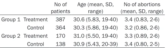
There were no significant differences of the age and number of spontaneous abortions between the treatment group and control in group 1 and group 2 (Table 1).
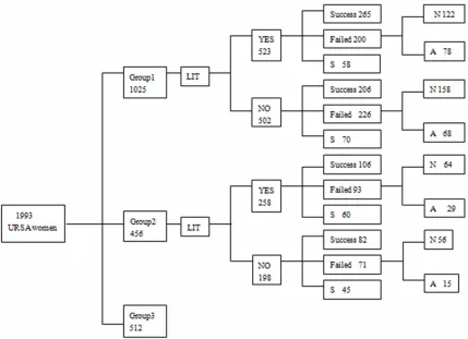
The patients recruited were shown in Figure 1. Among 1993 URSA patients, 1025 had preg-nancy history without demonstrated cardiac activity (group 1), 456 had pregnancy history with the demonstrated cardiac activity (group 2), 512 had a complicate pregnancy history (group 3). The details of the patients are in shown in Figure 1. In group 1, following LIT, 387 showed positive MLR-Bf (we had excluded the ones with abnormal karyotypes). In group 2, 170 showed positive MLR-Bf (we also had excluded the ones with abnormal karyotypes).
Comparison of success rate in treated patients with positive MLR-Bf and control in group 1 and 2
As shown in Table 2, the pregnancy outcome was significantly improved in the treatment patients with positive MLR-Bf than control in group 1 (68.5% vs 56.6%, P=0.0008). Unfor- tunately, no statistically significant differences were detected in group 2 (62.4% vs 59.4%,
P=0.5997).
Comparison of success rate of patients with two URSA in each group
[image:3.612.90.359.97.174.2]For patients with two URSA, patients with posi-tive MLR-Bf showed no significantly higher suc-cess rate as compared to control (Table 3). Table 1. Characteristics of Patients in the treatment group
and control in each group
No of
patients Age (mean, SD, range) (mean, SD, range)No of abortions Group 1 Treatment 387 30.6 (5.83, 19-40) 3.4 (0.83, 2-6)
Control 364 30.3 (5.86, 19-40) 3.2 (0.86, 2-6) Group 2 Treatment 170 31.0 (5.50, 19-40) 3.3 (0.89, 2-6) Control 138 30.9 (5.43, 20-39) 3.4 (0.80, 2-5)
There were no significant differences of the age and number of spontaneous
abortions in group 1 and 2.
for five times. MLR-Bf was mea-sured 2 or more weeks after the last immunization. Then they were advised to conceive within 12 months. In each groups, the failed ones whose fetus chromosome were abnormal or unsuccessful were excluded from the study.
Statistical analysis
Comparison of success rate of patients with three or more URSA in each group
As is shown in Table 4, in patients who had under-went three or more URSA, we can see the pregnan- cy outcome was significa- ntly improved in group 1- 2 (67.2% vs 53.1%, P= 0.0004), but not in group 2-2 (60.3% vs 56.6%, P= 0.5684).
Discussion
[image:4.612.93.522.72.383.2]In our study, to investiga- te the prognostic value of MLR-Bf for URSA, we select-Figure 1. The details of patients. The 1993 URSA patients were dividing into three groups. In group 1, 265 im-munized ones who showed positive MLR-Bf delivered healthy children, 122 aborted again. 206 unimim-munized pa-tients had improved pregnancy outcome, 158 did not. In group 2, 106 immunized ones with positive MLR-Bf had advanced pregnancy outcome, 64 did not. 82 unimmunized patients delivered healthy children, 56 did not. We had excluded the patients whose karyotype analyses were unsuccessful or abnormal; the immunized ones with negative MLR-Bf were also excluded from the study. S: Patients who were negative for MLR-Bf and did not get pregnant within 12 months. N: Patients who had normal chromosome karyotype. A: Patients who had abnormal or unsuccessful chromosome karyotype.
Table 2. Comparison of pregnancy outcome in group 1 and 2
Success rate P OR 95% CI Group 1 Treatment 68.5% 387 (265) 0.0008 0.6002 0.4454~0.8088
Control 56.6% 364 (206)
Group 2 Treatment 62.4% 170 (106) 0.5997 0.8841 0.5580~1.4007 Control 59.4% 138 (82)
The pregnancy outcomes were significantly improved in treatment group in group 1
(68.5% vs 56.6%, P=0.0008). Whereas no statistically significant differences were
detected in group 2 (62.4% vs 59.4%, P=0.5997).
Table 3. Comparison of pregnancy outcome in patients with two URSA in group 1 and 2
Success rate P OR 95% CI Group 1-1 Treatment 74.6% 67 (50) 0.4719 0.7650 0.3683~1.5886
Control 69.2% 78 (54)
Group 2-1 Treatment 69.2% 39 (27) 0.8082 0.8889 0.3432~2.3023 Control 66.7% 39 (26)
[image:4.612.92.382.504.571.2] [image:4.612.89.386.646.711.2]ed the URSA patients who had miscarriages in first trimester, we divided the patients into two different groups according to the time when the miscarriage occurs. Although we followed an identical protocol by adopting the same selection criteria, the same technical detail and the same immunotherapy procedure in the dif-ferent groups, the outcome was still difdif-ferent in each group. In group 1, the success rates were significantly higher among immunized patients than the ones who did not request for it in group 1 (68.5% vs 56.6%, P=0.0008). Whereas no significant differences were detected in group 2 (62.4% vs 59.4%, P=0.5997). After we divid-ed the patients into two sections according to the times of URSA in each group, we found that there was no significant difference in success rate between the treated and untreat-ed patients with only two URSA in each group. However for patients who had underwent three or more URSA, the pregnancy outcomes were significantly improved in treatment group in group 1-2 (67.2% vs 53.1%, P=0.0004), but not in group 2-2 (60.3% vs 56.6%, P=0.5684). Thus, the study can provide an useful clue to find an explanation for the previous conflicting results about LIT: they didn’t divide the URSA patients into different groups according to the different status of the embryo when the mis- carriage occurs [14-16]. Meanwhile, because of the definition of the URSA is different in different country, the patients involved in differ-ent study might be differdiffer-ent. ASRM defines URSA as two or more clinical miscarriages doc-umented by ultrasonography or histopathologic examination(not necessarily consecutive) [2], while Royal College of Obstetricians and Gyna- ecologists (RCOG) defines it as three or more consecutive pregnancy loss before 24 weeks of gestation [18]. There also may be other rea-sons for the discrepancy between their data and ours: 1. Lymphocyte cells they used were
reaction-blocking factor (MLR-bf), anti-HLA antibody, anti paternal lymphocyte antibodies (APLA) and antipaternal cytotoxic antibodies (APCA) for patient selection for immunother- apy, the treatment may be given to somebody who didn’t need it at all [14]. 3. The procedure of the immunotherapy they adopted were dif-ferent. Immunotherapy was performed only once or twice before pregnancy in most of the controversial studies, whereas in most suc-cessful studies, immunotherapy was perfor- med three to four times or more at the regular intervals of 3 to 6 weeks [21], 4. The technical details of the treatment were different, such as the immunizing cell dose, number of treatment, route of lymphocytes delivery.
[image:5.612.90.388.96.164.2]In former studies, there are many researchers used anti HLA antibody as the prognostic crite-ria for evaluating the efficacy of LIT. Previous studies showed that anti HLA antibodies are present in approximately 33% of normal suc-cessful pregnancies [22-24], however for pa- tients with primary RSA, not more than 10% of them showed positive anti HLA antibody [25-27]. The difference fostered a hypothesis that a failure to produce HLA-antibodies was part of the underlying cause of URSA [25]. However, there is a disputable view that anti HLA anti-bodies in pregnant women are considered to be the consequences instead of causes of successful pregnancies [28]. Nowadays redu- ced MLR-Bf production is always considered to be a possible cause of URSA [16], and MLR-Bf is the most commonly used indicator for evalu-ating the efficacy of LIT in the world, while the prognostic value of it was controversial. So in our study, a study was conducted to explore the prognostic value of MLR-Bf for evaluating the efficacy of LIT. As we know, LIT is believed to promote a favorable environment for the pater-nal alloantigen-bearing embryos by modulating the immunity of the patients [29], as it could Table 4. Comparison of pregnancy outcome in patients with three or
more URSA in group 1 and 2
Success rate P OR 95% CI Group 1-2 Treatment 67.2% 320 (215) 0.0004 0.5540 0.3986~0.7699
Control 53.1% 286 (152)
Group 2-2 Treatment 60.3% 131 (79) 0.5684 0.8572 0.5048~1.4557 Control 56.6% 99 (56)
The success rates were significantly increased in treatment group in group 1-2 (67.2%
vs 53.1%, P=0.0004), but not in group 2-2 (60.3% vs 56.6%, P=0.5684).
provide the protect factors APCA and MLR-Bf which may protect the fetus from the toxic effect of the mother’s immune system [5], reduce the NK cell activity [30] and perform the shift from Th1-type reactivity to Th2-type reac-tivity [31], but the precise mechanism of it is still under investigation. From my point of view, MLR-Bf was a kind of antibodies which emerged after LIT. The different numbers, cell doses and routes of the immunization brings different changes to the immune environment of the patients. In our study, following this immunizing cell dose, route and number, at the time the immunized ones developed MLR-Bf, the differ-ence of the immune environment brought by the immunization can significantly improved the pregnancy outcome of the ones with three or more URSA who had never had the demon-stration of embryonic cardiac activity, but not for patients with three or more URSA who had had the demonstration of embryonic cardiac activity and the ones with only two URSA. In this study, although we followed an identical protocol in the different groups, the treatment effect was still different. Using MLR-Bf as the indicator that the patients could be ready for pregnancy, the pregnancy outcome was signifi-cantly improved in patients who suffer the mis-carriage early in the pregnancy, but not in patients who had miscarriages later in preg-nancy. In my opinion, there are two reasons responsible for it: 1. The inadequate protecting factors and the poor immune condition may be not the main reason for the miscarriage later in gestation. There may be other reasons respon-sible for the miscarriage. 2. Maybe there are other indicators for evaluating the efficacy of LIT in patients who had ever had demonstra-tion of embryonic cardiac activity, which need further investigation.
In this study, the production of MLR-Bf is a good prognostic criterium for patients with three or more URSA who had never had the demonstra-tion of embryonic cardiac activity, but not for patients with three or more URSA who had had the demonstration of embryonic cardiac activi-ty and the ones with only two URSA.
Acknowledgements
We thank the patients and the colleagues par-ticipating in the study. The study was accom-plished in Medical Genetics Institute of Henan
Provincial People’s Hospital. This study was funded by the Medical Science Research Projects of Henan Province (No. 201303162); National Natural Science Foundation of China (NO. 81450018 and NO. 81501336).
Disclosure of conflict of interest None.
Address correspondence to: Dr. Shi-Xiu Liao, Medi- cal Genetic Institute of Henan Province, Henan Provincial People’s Hospital, People’s Hospital of Zhengzhou University, Zhengzhou 450003, PR China. Tel: 15003826037; Fax: 0371-65580927; E-mail: [email protected]
References
[1] Rai R, Regan L. Recurrent miscarriage. Lancet 2006; 368: 601-11.
[2] Practice Committee of American Society for
Reproductive Medicine. Definitions of infertility
and recurrent pregnancy loss: a committee opinion. Fertil Steril 2013; 99: 63.
[3] Allison JL, Schust DJ. Recurrent first trimester pregnancy loss: revised definitions and novel
causes. Curr Opin Endocrinol Diabetes Obes 2009; 16: 446-50.
[4] Coulam CB, Goodman C, Roussev RG, Thomanson EJ, Beaman KD. Systemic CD 56+ cells can predict predict pregnancy outcome. Am J Reprod Immunol 1995; 33: 40-46. [5] Ramhorst R, Agriello E, Zittermann S, Pando
M, Larriba J, Irigoyen M, Cortelezzi M, Auge L, Lombardi E, Etchepareborda JJ, Contreras Ortiz C, Fainboim L. Is the paternal mononucle-ar cell’s immunization a successful treatment for recurrent spontaneous abortion? Am J Reprod Immunol 2000; 44: 129-35.
[6] Clark DA, Chaouat G, Mogil R, Wegmann TG. Prevention of spontaneous abortion in DBA/2 mated CBA/J mice by GM-CSF involves CD8+ cell-dependent suppression of natural effector cell cytotoxicity against trophoblast target cells. Cell Immunol 1994; 154: 143-52. [7] Taylor CG, Faulk WP, McIntyre JA. Prevention of
recurrent spontaneous abortions by leukocyte transfusions. J R Soc Med 1985; 78: 623-27. [8] Beer AE, Quebbeman JF, Ayers JW, Haines RF.
Major histocompatibility complex antigens, maternal and paternal immune responses, and chronic habitual abortions in humans. Am J Obstet Gyneol 1981; 141: 987-97.
[10] The Recurrent Miscarriage Immunotherapy Trialists Group, Coulam CB, Clark DA, Collins J, et al. Worldwide collaborative observational study and meta-analysis on allogenic leuko-cyte immunotherapy for recurrent spontane-ous abortion. Am J Reprod Immunol 1994; 32: 55-72.
[11] Pandey MK, Thakur S, Agrawal S. Lymphocyte immunotherapy and its probable mechanism in the maintenance of pregnancy in women with recurrent spontaneous abortion. Arch Gynecol Obstet 2004; 269: 161-72.
[12] Cavalcante MB, Costa Fda S, Araujo Júnior E, Barini R. Risk factors associated with a new pregnancy loss and perinatal outcomes in cas-es of recurrent miscarriage treated with lym-phocyte. J Matern Fetal Neonatal Med 2015; 28: 1082-6.
[13] Clark DA. Immunological factors in pregnancy
wastage: fact or fiction. Am J Reprod Immunol
2008; 59: 277-300.
[14] Ober C, Karrison T, Odem RR, Barnes RB, Branch DW, Stephenson MD, Baron B, Walker MA, Scott JR, Schreiber JR. Mononuclear-cell immunization in prevention of recurrent mis-carriages: a randomized trial. Lancet 1999; 354: 365-9.
[15] Wong LF, Porter TF, Scott JR. Immunotherapy for recurrent miscarriage. Cochrane Database Syst Rev 2014; 21: CD000112.
[16] Khonina NA, Broitman EV, Shevela EY, Pasman
NM, Chernykh ER. Mixed lymphocyte reaction blocking factors (MLR-Bf) as potential
biomark-er for indication and efficacy of patbiomark-ernal lym -phocyte immunization in recurrent spontane-ous abortion. Arch Gynecol Obstet 2013; 288: 933-937.
[17] Peña RB, Cadavid AP, Botero JH, García GP, Gallego MI, Ossa JE. The production of MLR-blocking factors after lymphocyte immunother-apy for RSA does not predict the outcome of pregnancy. Am J Reprod Immunol 1998; 39: 120-124.
[18] Royal College of Obstetricians and Gynaeco- logists. The Investigation and Treatment of Couples with Recurrent First-trimester and Second-trimester Miscarriage. RCOG Green-top Guideline 2011: 4.
[19] Clark DA, Yu G, Levy GA, Gorczynski RM. Procoagulants in fetus rejection: the role of the OX-2 (CD200) tolerance signal. Semin Immunol 2001; 13: 255-263.
[20] Clark DA. Shall we properly re-examine the sta-tus of allogeneic lymphocyte therapy for recur-rent early pregnancy failure? Am J Reprod Immunol 2004; 51: 7-15.
[21] Cauchi MN, Lim D, Young DE, Kloss M, Pepperell RJ. Treatment of recurrent aborters by immunization with paternal cells--controlled trial. Am J Reprod Immunol 1991; 25: 16-7. [22] Ahrons S. HLA antibodies: influence on the hu
-man foetus. Tissue Antigen 1971; 1: 129-136. [23] BalaschJ, Ercilla G, Vanrell JA, Vives J,
González-Merlo J. Effects of HLA antibodies on pregnancy. Obstet Gynecol 1981; 57: 444-446.
[24] Regan L, Braude PR, Hill DP. A prospective study of the incidence, time of appearance
and significance of anti-paternal lymphocyto -toxic antibodies in human pregnancy. Hum Reprod 1991; 6: 294-298.
[25] Beard RW, Braude P, Mowbray JF, Underwood JL. Protective antibodies and spontaneous abortion. Lancet 1983; 2: 1090.
[26] Johnson PM, Barnes RM, Hart CA, Francis WJ. Determinants of immunological respon-siveness in recurrent spontaneous abortion. Transplantation 1984; 38: 280-284.
[27] Power DA, Catto GR, Mason RJ, MacLeod AM, Stewart GM, Stewart KN, Shewan WG. The fe-tus as an allograft: evidence for protective an-tibodies to HLA-linked paternal antigens. Lancet 1983; 2: 701-704.
[28] Yuan MM, Du MR, Wang MY, Duan ZL, Meng Y, Jin LP, Li MQ, Li DJ. Combination of CD4(+) CD25(+)CD127(-) regulatory T cells with
MLC-BE and MLC-BE-Ab2: an efficient evaluation of the
therapy of paternal lymphocyte induced immu-nization in unexplained recurrent spontaneous abortion patients. Int J Clin Exp Pathol 2015; 8: 4022-4032.
[29] Liang P, Mo M, Li GG, Yin B, Cai J, Wu T, He X, Zhang X, Zeng Y. Comprehensive analysis of peripheral blood lymphocytes in 76 women with recurrent miscarriage before and after lymphocyte immunotherapy. Am J Reprod Immunol 2012; 68: 164-74.
[30] Kwak JY, Gilman-Sachs A, Moretti M, Beaman KD, Beer AE. Natural killer cell cytotoxicity and paternal lymphocyte immunization in women with recurrent spontaneous abortions. Am J Reprod Immunol 1998; 40: 352-8.