Original Article
Celecoxib attenuates retinal angiogenesis in a mouse
model of oxygen-induced retinopathy
Ningning Liu, Lei Chen, Na Cai
Department of Ophthalmology, The First Affiliated Hospital of China Medical University, Shenyang, Liaoning Province, China
Received February 7, 2015; Accepted April 10, 2015; Epub May 1, 2015; Published May 15, 2015
Abstract: This study aimed to investigate the anti-angiogenic effects of Celecoxib on the expression of vascular
endothelial growth factor (VEGF) and hypoxia-inducible transcription factor 1α (HIF-1α) in a mouse model for
oxygen-induced retinopathy (OIR). The OIR mice were exposed to 75% oxygen from postnatal day 7 (P7) to P12, after which the mice were randomly assigned to two groups (Celecoxib and vehicle) and were brought to room air for additional
five days. Celecoxib or vehicle was administered from P12 to P17. Age-matched mice maintained in room air from birth to P17 were administered vehicle from P12 to P17 (RA group). Blood vessel profiles in the retina were used to count by histologic methods. Retina protein and mRNA of VEGF and HIF-1α were assessed by immunohisto -chemistry, western-blot and RT-PCR. Compared with the RA group, the OIR mice exhibited over-expression in VEGF
and HIF-1α mRNA and protein. In addition, they had a positive and spatial correlation. Celecoxib- treated OIR mice reduced the retinal neovascular tufts and the levels of VEGF and HIF-1α. These data suggest that Celecoxib inhibits retinal pathogenic angiogenesis through down-regulating HIF-1α expression which suppressing VEGF transcription.
Celecoxib could potentially serve as a portent pharmaceutical agent to inhibit retinal angiogenesis.
Keywords:Hypoxia-inducible factor-1α, vascular endothelial growth factor, retinal neovascularization, a selective
cyclooxygenase-2 inhibitor
Introduction
Pathological ocular angiogenesis, or ocular neovascularization (NV), is a central feature of retinopathy of prematurity, diabetic retinopa-thy, retinal vein occlusion and neovascular age-related macular degeneration, and is the lead-ing cause of irreversible blindness in developed countries [1]. It is clear that ischemia-induced hypoxia is a central etiological factor in retinal NV. In response to hypoxia, various pro-angio-genic growth factors are produced, among which the VEGF is the major one promoting reti-nal NV [2]. Hypoxia has been shown to increase VEGF expression at the level of gene transcrip-tion, mRNA stability, translatranscrip-tion, and protein secretion [3]. VEGF transcription is largely regu-lated by hypoxia-inducible transcription factor 1 (HIF-1), which consists of a hypoxically induc-ible subunit HIF-1α and constitutively expressed unbunit HIF-1β. In normoxia, the HIF-1α pro-teins are degraded causing no detectable
HIF-1α protein. During hypoxia, however, HIF-HIF-1α becomes stabilized and dimerizes with HIF-1β to form a complex which becomes transcrip-tionally active [4]. VEGF binds with VEGF recep-tors (VEGFR-1 and VEGFR-2) expressed on the endothelial cells, initiating signal transduction cascades and leading to angiogenic endothelia cell behaviors [5]. Increased levels of VEGF and HIF-1α were detected in the vitreous humor and in fibrovadcular tissues from eyes with PDR [6-9]. VEGF and HIF-1α were indicated to pro-vide targets for therapeutic intervention on reti-nal neovascularization [10].
Celecoxib inhibits ocular neovascularization
induced under inflammatory conditions and cytokines, tumor promoters and plays a key role in regulating angiogenesis through the induction of prostanoid synthesis [12]. Studies suggest that COX-2-induced prostanoids exhib-it angiogenic effects both up and downstream of growth factor production. Upstream, the prostanoids induce VEGF and bFGF [13]. Downstream, pro-angiogenic factors (e.g. hypoxia, VEGF) are believed to induce endothe-lial cell expression of COX-2 [14], and COX-2-derived prostanoids stimulated proliferation, migration and formation in human vein endo-thelial cells [15].
Nonsteroidal anti-inflammatory drugs (NSAIDs) such as Aspirin and Naproxen inhibit the forma-tion of both COX-1 and COX-2 and are common-ly used for the treatment of pain and arthritis. Studies demonstrated that administration of high dose Aspirin reduced the incidence of reti-nopathy in patients with diabetes, retinal vas-cular leakage in diabetic rats and retinal vascu-lar abnormalities in diabetic dogs [16-18], indi-cating benefits of COX inhibition in ameliorating diabetic retinopahy. However, there is evidence that NSAIDs produce serious gastrointestinal side effects due to the blockade of COX-1. COX-2 specific inhibitors were thus developed as anti-inflammatory and anti-angiogenic agents. Celecoxib, a highly selective COX-2 inhibitor, has been shown to effectively decrease tumor angiogenesis, reduce tumor growth in a variety of experimentally-induced tumors [19], and prevent pathological angio-genesis in the cornea and retina [20]. However, the exact mechanisms through which the COX-2 inhibition down-regulate pathological NV remain unclear. In the present study, we used the laboratory animal model of oxygen-induced retinopathy (OIR) that is a generally used meth-od to observe the retinal microvascular compli-cations in retinopathy of prematurity and dia-betic retinopathy to test our hypothesis that Celecoxib is a preventive agent against patho-logical retinal NV by inhibiting the HIF-1α and its downstream gene VEGF expression.
Materials and methods Mouse model of OIR
C57BL/6J mice from the Laboratory Animal Center of China Medical University (Shenyang,
China) were obtained. All animal experiments were conducted according to the ARVO Statement for the Use of Animals in Ophthalmic and Vision Research and were approved by the Institutional Animal Care and Use Committee of Medical University Ophthalmic Center. The methodology used for OIR model was consis-tent with that described by Smith et al [21]. In general, postnatal day 7 (P7) mice and their nursing mother were exposed to hyperoxia (75 ± 3% oxygen) for 5 days (P7 to P 11) and then removed into normoxic condition for an addi-tional 5 days (P12 to P17). The airtight incuba-tor temperature was maintained at 23 ± 2°C, and oxygen levels were monitoredusing a por-table oxygen analyzer (Model CY-12C, Electrochemical Analytical Instruments, Ltd, Meicheng, Zhejiang, China).
Celecoxib preparation and administration
As described previously [22], Celecoxib (Sigma Chemical Co., St. Louis, MO) were suspended in vehicle to a 0.5-ml suspension (0.5% methylcel-lulose, 0.1% polysorbate 80 in water). From P12 to P17, the OIR mice were randomly assigned to two groups: Celecoxib (n = 24) and Vehicle (OIR) groups (n = 24), where 3 mg/kg/ day celecoxib or equivalent volume of vehicle was administered through a gastric feeding tube. Age-matched mice maintained in room air from birth to P17 (RA group, n = 24) were treat-ed with equivalent volume of vehicle daily from P12 to P17. The mice in all three groups were euthanized at P17 and their eyeballs or retinas were prepared for morphological study and bio-chemical assays.
cell layer (GCL), and inner plexiform layer (IPL). A BVP was defined as an endothelial cell or a blood vessel with a lumen.
Retinal immunohistochemistry
Randomly chosen ten paraffin sections from each group were deparaffinized and rinsed with
[image:3.612.89.522.75.510.2]3% hydrogen peroxide for 10 min and then incu-bated for 30 min at room temperature with blocking serum. The sections were subse-quently incubated overnight at 4°C with prima-ry antibodies (rabbit polyclonal antibodies; Santa Cruz, CA): HIF1-α (1:200 in PBS), VEGF (1:200 in PBS), respectively. After washing with 0.1 M PBS (three 2-min washes), the sections Figure 1. Hematoxylin and eosin stained cross-section from all the groups. Retinal NV was resolved by counting the
number of extraretinal cell nuclei anterior to the ILM on P17. Neovascular tufts (BVPs) were proved by extending into the vitreous. A. No nuclei were detected in the RA group which extending to the vitreous, compare with OIR
group. B. In contrast, there are many of pathologic neovascularizar tufts beyonding the ILM into the vitreous (ar
-rows). C. The group treated with Celecoxib displays a significant reduction of the number of retinal neovascular cell nulei, compared with OIR group, but more than that of the RA group (arrows). Magnification, ×200. D. The average
numbers of BVPs in the inner retina of the Celecoxib group mice were compared with OIR group and RA group mice
using one-way ANOVA. Data are expressed as means ± SD (n = 8). ***P < 0.001 compared with RA group; **P <
Celecoxib inhibits ocular neovascularization
were incubated with secondary antibody goat anti-rabbit Ig G (1:200 in PBS, Santa Cruz, CA) for 20 min at room temperature and were visu-alized by 0.05% diaminobenzidine (DAB) solu-tion and stained with Mayer hematoxylin, and negative controls were taken by omitting the primary antibodies. Cells positive for HIF1-α and VEGF showed light yellow or dark brown coloration in the cytoplasm. The integrated areas of HIF1-α and VEGF staining were ana-lyzed by using automatic microscope and image analysis system (LUZEX-F, Japan) and identified the integral optical density (IOD) values as an indicator of HIF1-α and VEGFexpression. Western blot analysis
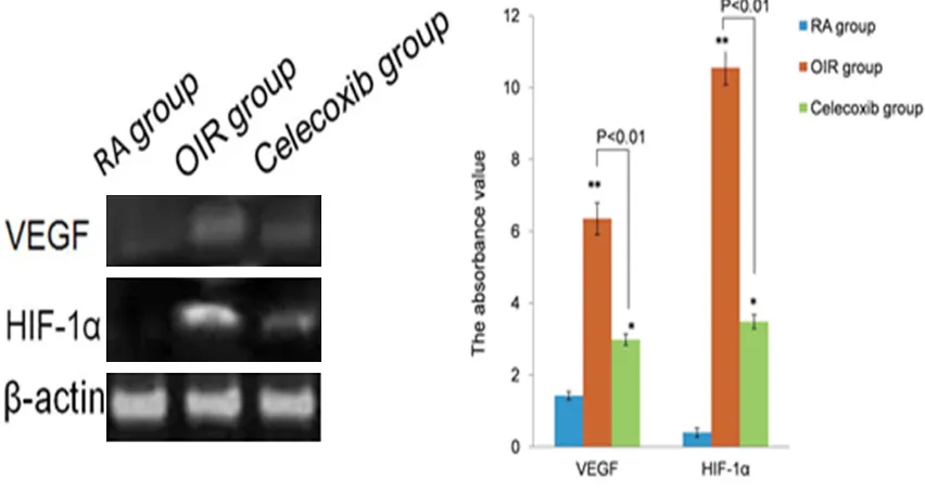
Retinas were isolated from eight mice per group and were lysed in 200 μL of lysis buffer with protease inhibitors (Sigma Chemical Co., St. Louis, MO). Protein concentration was deter-mined using the BCA protein assay kit (Pierce Biotechnology, Rockford. IL). 40 μg of total pro-tein extract from each sample were analyzed by SDS-PAGE and transferred onto a nitrocellulose membrane that was incubated with rabbit poly-clonal anti-HIF1-α antibody (1:400, Santa Cruz,
CA) or polyclonal anti-VEGF antibody (1:300, Santa Cruz, CA) and β-actin monoclonal anti-body (1:400 Santa Cruz, CA) overnight at 4°C followed by horseradish peroxidase-conjugated secondary antibody (goat anti-rabbit antibody, 1:1500, Santa Cruz, CA). The proteins were detected with enhanced chemiluminescence-detection (Santa Cruz, CA). β-actin served as the internal standard. Experiments were repeated three times.
Reverse transcription-polymerase chain reac-tion
Total retinal RNA was isolated from all the three groups (n = 8) using Tri-Zol reagent (Life Technologies, Glasgow, UK) according to the manufacturer’s protocol. RT-PCR assays were described previously [25]. Reverse transcrip-tion (RT) was performed with approximately 0.5 μg of total RNA, reverse transcriptase (SuperScript II; Life Technologies, Gaithersburg, MD), and 5.0 μm oligo-d (T) primer. Aliquots of cDNA were used for polymerase chain reaction (PCR) amplification with primers specific for VEGF, HIF1-α and β-actin. The primer sequenc-Figure 2. Immunohistochemical analysis for the protein expression of HIF-1a and VEGF in all the experimental groups. Low HIF-1a protein expressions were detected in GCL of the RA group retina (A, arrows). In contrast, there was strong HIF-1a protein expression in the inner nuclear layer (INL), the ganglion cell layer (GCL), as well as neo
-vascularization breaking through the inner retinal layer in OIR mice (B, arrows); In the Celecoxib- treated OIR group, the HIF-1α immunolabeling was reduced, only showed in the cytoplasm of some ganglion cells or small amount
extraretinal cell nuclei anterior to the ILM (C, arrows). The protein levels of VEGF were only weak present in the GCL
[image:4.612.93.521.72.292.2]es were VEGF (127 bp): sense 5’-CTG TGC AGG CTG CTG TAA CG-3’, antisense 5’-GAT CATT CGC TCT ATG TGG TGGC-3’; HIF1-α (460 bp): sence 5’-ACTTGATGTTCATCGTCCCTC-3’, antisense 5’-CGGCGGAAGG AAAGAGT-3’; and β-actin (450 bp): sence 5’-TCT ACA ATG AGC TGC GTG TGG-3’, antisense 5’-GGA ACC GCT CAT TGC CAA TG-3’. Standard PCR amplification was per-formed at 95°C for 2 min, 58°C for 1 min, and 72°C for 1 min for 30 cycles, 7 min at 72°C. The amplified products were visualized on 2% agarose gels. Calculation method: absorbance of target gene/β-actin absorbance value. Statistical analysis
The results were expressed as mean ± stan-dard deviation. Analysis of correlation between HIF1-α and VEGF expression was detected by linear correlation. One way ANOVA was used to analysis multiple variables and with student’s t tests. A P value < 0.05 was considered to be statistically significant.
Results
Effects of celecoxib on preretinal neovascular nuclei
The degree of preretinal NV, vascular cell nuclei on the vitreal side of the internal limiting
mem-brane (ILM) was assessed quantitatively through counting at P17. In RA group, no nuclei of new vascular endothelial cells were broken through the inner retina (0.21 ± 0.06) (Figure 1A).In contrast, all mice in the OIR groups had large pathologic neovascular tufts that were adherent to the ILM (53.14 ± 1.06, P < 0.001) (Figure 1B). The preretinal NV in the Celecoxib group was significantly reduced than that in the OIR group (10.65 ± 1.82, P < 0.01), but more than that of the RA group (P < 0.05) (Figure 1C). Immunohistochemistry for HIF1-α and VEGF protein expression
[image:5.612.89.525.73.303.2]RA group retinas expressed low levels of HIF1-α and VEGF in ganglion cells layer (GCL) (IOD = 1.22 ± 0.50, IOD = 2.36 ± 0.31, respectively) (Figure 2A, 2D). In OIR group retinas, there was higher expression for HIF1-α and VEGF in the INL and GCL and neovascularization breaking through the inner retina compare with RA group (IOD = 8.42 ± 0.13 and IOD = 10.37 ± 0.17 respectively. P < 0.05) (Figure 2B, 2E). However, Celecoxib-treated OIR retinas displayed a sig-nificantly reduced expression in HIF1-α (IOD = 3.78 ± 0.56) and VEGF (IOD = 3.4 2 ± 0.23) compared with the OIR group (P < 0.05). HIF1-α and VEGF protein were detected in the cyto-plasm of some ganglion cells and small amount Figure 3. Western blot analysis for HIF-1α and VEGF protein levels in the three experimental groups. Both of the protein expression levels were increased greatly in the OIR group compared with the RA group (**P < 0.01). After
the treated with Celecoxib in OIR group, the expression of these protein was significantly decreased compared with
Celecoxib inhibits ocular neovascularization
extraretinal cell nuclei anterior to the ILM (Figure 2C, 2F).
Celecoxib Inhibits HIF1-α and VEGF protein levels
In the RA group, HIF1-α protein was not detect-ed but the expression of VEGF was relatively strong. A significantly increased protein expres-sion in HIF1-α and VEGF was seen in OIR group when compared with RA group (P < 0.01). In the Celecoxib-treated retina, the protein expres-sion levels for HIF1-α and VEGF were markedly reduced compared to OIR (P < 0.01), but remained stronger than those in the RA group (P < 0.05) (Figure 3).
Effect of Celecoxib on HIF1-αand VEGF mRNA expression in the mouse OIR model
Overexpression of HIF1-α and VEGF was detect-ed in the OIR group, treatment of Celecoxib sig-nificantly inhibited the mRNA expression of HIF1-α and VEGF. The level of HIF1-α mRNA expression in OIR group was higher than that in the RA group (3.94-flod, P < 0.01) and higher than those in the ROP group treated with Celecoxib (1.42-flod, P < 0.01). The VEGF mRNA expression was increased in OIR group than those in RA group (7.78-flod, P < 0.01) and
high-er than those in the ROP group treated with Celecoxib (1.51-flod, P < 0.01) (Figure 4). Correlation analysis
The correlation between the levels of expres-sion in HIF1-α, VEGF protein and their mRNA in the OIR group were statistically significant. (P < 0.001, r = 0.725 and P < 0.001, r = 0.537, respectively).
Discussion
OIR is a well-established animal model to inves-tigate hypoxia-induced ocular NV. Using this method, the present study provided further evi-dence that hypoxia does up-regulate the HIF1-α and VEGF expressions. More importantly, our study for the first time demonstrated that the potent anti-angiogenic activity of celecoxib was associated with decreased expression of HIF1-α and VEGF on both protein and mRNA levels in retinal NV as assessed using western blot and RT-PCR. The suppression of retinal NV by celecoxib is, at least partially, the result of reduced VEGF and HIF1-α expression.
Today, various therapeutic strategies such as laser photocoagulation, photodynamic therapy, anti-VEGF therapy shows regression of the reti-Figure 4. HIF-1α and VEGF mRNA expression in all the groups were detected by RT-PCR. There was marked elevated of HIF-1α and VEGF mRNA in ROP mice compared with those of the RA group (**P < 0.0.01). There was a signifi
-cantly suppressed of HIF-1α and VEGF mRNA in the retinas of ROP mice treated with Celecoxib, compared with
[image:6.612.95.521.75.302.2]nal NV, severe undesirable side effects have been also reported such as the damage of reti-na and its photoreceptors, potential neuroreti-nal and glial toxicity, that do not cure the disease completely [26]. Thus, it is necessary to thor-ough understand mechanisms of NV in order to develop effective therapeutic targets. Clinical and experimental evidence suggests that reti-nal NV is often the result of ischemia-induced hypoxia. Several angiogenic growth factors and cytokines that stimulate angiogenesis partici-pate in the pathogenesis of retinal neovascu-larization [27].
VEGF, an essential angiogenic factor, plays a crucial role in tumor angiogenesis and progres-sion. Expression of the VEGF is up-regulated is up-regulated in the retina and vitreous of patients or the model animals with ischemic retinopathies. COX-2 expression has been found in various ocular tissues: cornea, iris, cil-liary body, neuroretina and the retinal pigment epithelium, and its function is achieved through its prostanoid products which subsequently induce production of VEGF and other grow fac-tors [28-30]. Cancer literature has shown that COX-2 and its prostanoid metabolites, in par-ticular PGE2, induce the expression of VEGF and bFGF thus promote angiogenesis during tumor growth [31]. In eyes, it has been found that hypoxia induces COX-2, prostanoid produc-tion, and VEGF synthesis in Műller cells, and that VEGF production is at least partially COX-2 dependent [32]. Using wild-type mouse Műller cells and COX-2 null mouse Műller cells, one study found that in response to hypoxia, the production in COX-2 and its prostanoid was increased in wild-type cells. VEGF levels were also found to be increased with a temporal sequence that lagged behind COX-2 induction and activity, suggesting that there is COX-2 dependent aspects of VEGF production, where-as COX-2 null mouse Műller cells treated with hypoxia produced significant less VEGF. It was also revealed that prostanoid PEG2, signaling through the EP2 and/or EP4 receptor and PKA, mediates the VEGF response of Műller cells [33]. As COX-2 and its prostanoids induce VEGF production, theoretically COX-2 inhibitors reduce VEGF production; therefore inhibit hypoxia-induced ocular NV. Several reports showed that Celecoxib, a highly selective COX-2 inhibitor, was a potent antiangiogenic agent in vitro and in vivo [34]. Oral Celecoxib inhibited angiogenesis by 79% in a rat model of
bFGF-induced corneal angiogenesis [35]. Celecoxib inhibited diabetes-induced retinal VEGF expres-sion and vascular leakage in vivo [36]. Our study showed that the protein and mRNA levels of VEGF were increased in the ROP group and were significantly decreased in the ROP group treated with Celecoxib. This finding is consis-tent with studies demonstrating that treatment with Celecoxib decrease expression of VEGF in several other tissues.
As mentioned, HIF-1α is a principal regulator responsible for the transcription of VEGF and other angiogenic factors. Our study showed that under hypoxic condition, the expression of HIF-1α was increased along with over-expres-sion in VEGF. Consistent with the research [38], it has been shown that tumor angiogenesis occurs partly by elevating the expression of VEGF, along with the up-regulation of HIF1-α. Inhibition of COX-2 by Celecoxib caused a reduced expression in HIF-1α and VEGF on both mRNA and protein levels. Retinal angio-genesis reduction was also showed. The find-ings suggest that, under hypoxic condition, COX-2 may play a vital part in promoting VEGF transcription by up-regulating HIF-1α expres-sion. The anti-angiogenic effect of Celecoxib is achieved, at least partially, by down-regulating HIF-1α, thus reducing VEGF production. To our knowledge, this is the first study which demon-strated Celecoxib inhibited VEGF expression at transcriptional level through down-regulating HIF-1α expression. The application of our find-ings is that Celecoxib may be a promising anti-angiogenic pharmacological agent for inhibition of retinal NV.
In our study, Celecoxib was effective in reduc-ing retinal neovascularization through the inhi-bition of VEGF and HIF-1α expression. However, suppression of retinal neovascularization was incomplete in ROP mice treated with Celecoxib, indicating that angiogenesis is a complex pro-cess, and there are other factors involved in these complex angiogenic cascades. A number of molecules that play role in hypoxia-induced angiogenesis remain to be further examined. The molecular mechanisms through which Celecoxib suppresses HIF-1α expression also need further investigation.
Celecoxib inhibits ocular neovascularization
sion which subsequently suppressing VEGF production. Although the molecular mecha-nisms of how COX-2 inhibition suppresses HIF-1α remains nuclear, our study provided evi-dence that Celecoxib can be a potentially prom-ising potent anti-angiogenic agent for retinal NV.
Acknowledgements
This research was supported by Doctoral Scientific Research Foundation of China (No. 20091115).
Disclosure of conflict of interest
None.
Address correspondence to: Dr. Na Cai, Department of Ophthalmology, The First Affiliated Hospital of
China Medical University, 155 Nanjing North Street,
Heping Area, Shenyang 110001, Liaoning Province,
China. Tel: 86-24-83282928; E-mail: cnln1688@- 126.com
References
[1] Rencová E. Angiopathy and the eye. Vnitr Lek 2010; 56: 333-9.
[2] Lee HS, Jun JH, Jung EH, Koo BA, Kim YS.
Epigalloccatechin-3-gallate inhibits ocular
neo-vascularization and vascular permeability in
human retinal pigment epithelial and human retinal microvascular endothelial cells via sup-pression of MMP-9 and VEGF activation. Molecules 2014; 19: 12150-72.
[3] Chen MH, Ren QX, Yang WF, Chen XL, Lu C, Sun J. Influences of HIF-lα on Bax/Bcl-2 and VEGF
expressions in rats with spinal cord injury. Int J Clin Exp Pathol 2013; 6: 2312-22.
[4] Scholz CC, Taylor CT. Targeting the HIF pathway in inflammation and immunity. Curr Opin
Pharmacol 2013; 13: 646-53.
[5] Napione L, Pavan S, Veglio A, Picco A, Boffetta G, Celani A, Seano G, Primo L, Gamba A,
Bussolino F. Unraveling the influence of endo -thelial cell density on VEGF-A signaling. Blood 2012; 119: 5599-607.
[6] Han XX, Guo CM, Li Y, Hui YN. Effects of beva
-cizumab on the neovascular membrane of pro -liferative diabetic retinopathy: reduction of en-dothelial cells and expressions of VEGF and
HIF-1α. Mol Vis 2012; 18: 1-9.
[7] Loukovaara S, Koivunen P, Inglés M, Escobar J, Vento M, Andersson S. Elevated protein
car-bonyl andHIF-1α levels in eyes with prolifera -tive diabetic retinopathy. Acta Ophthalmol 2014; 92: 323-7.
[8] Al-Salam S, Balalaa N, Faour I, Akhter S,
Alashari M. HIF-1α, VEGF and WT-1 are pro -tagonists in bilateral primary angiosarcoma of breast: a case report and review of literature. Int J Clin Exp Pathol 2012; 5: 247-53.
[9] Lim JI, Spee C, Hinton DR. A comparison of hy
-poxia-inducible factor-α in surgically excised
neovascular membranes ofpatients with dia-betes compared with idiopathic epiretinal membranes in nondiabetic patients. Retina 2010; 30: 1472-8.
[10] Jiang J, Xia XB, Xu HZ, Xiong Y, Song WT, Xiong SQ, Li Y. Inhibition of retinal neovascularization
by gene transfer of small interfering RNA
tar-geting HIF-1alpha and VEGF. J Cell Physiol
2009; 218: 66-74.
[11] Bretz CA, Savage S, Capozzi M, Penn JS. The
role of the NFAT signaling pathway in retinal
neovascularization. Invest Ophthalmol Vis Sci
2013; 54: 7020-7.
[12] Smith WL, DeWitt DL, Garavito RM.
Cyclooxygenases: structural, cellular, and mo-lecular biology. Annu Rev Biochem 2000; 69: 145-82.
[13] Dagouassat M, Gagliolo JM, Chrusciel S, Bourin MC, Duprez C, Caramelle P, Boyer L, Hue S, Stern JB, Validire P, Longrois D, Norel X, Dubois-Randé JL, Le Gouvello S, Adnot S, Boczkowski J. The cyclooxygenase-2-prosta -glandin E2 pathway maintains senescence of
chronic obstructivepulmonary disease fibro -blasts. Am J Respir Crit Care Med 2013; 187: 703-14.
[14] Zhang C, Gao GR, Lv CG, Zhang BL, Zhang ZL, Zhang XF. Protease-activated receptor-2 induc-es exprinduc-ession of vascular endothelial growth factor andcyclooxygenase-2 via the mitogen-activated protein kinase pathway in gastric cancer cells. Oncol Rep 2012; 28: 1917-23. [15] Namkoong S, Lee SJ, Kim CK, Kim YM, Chung
HT, Lee H, Han JA, Ha KS, Kwon YG, Kim YM.
Prostaglandin E2 stimulates angiogenesis by activating the nitric oxide/cGMP pathway inhu-man umbilical vein endothelial cells. Exp Mol Med 2005; 37: 588-600.
[16] Idris I, Warren G, Donnelly R. Association be
-tween thiazolidinedione treatment and risk of
macular edema among patientswith type 2 di-abetes. Arch Intern Med 2012; 172: 1005-11. [17] Qaum T, Xu Q, Joussen AM, Clemens MW, Qin
W, Miyamoto K, Hassessian H, Wiegand SJ, Rudge J, Yancopoulos GD, Adamis AP.
VEGF-initiated blood-retinal barrier breakdown in early diabetes. Invest Ophthalmol Vis Sci 2001; 42: 2408-13.
[18] Kern TS, Engerman RL. Pharmacological inhi-bition of diabetic retinopathy: aminoguanidine
and aspirin. Diabetes 2001; 50: 1636-42.
chemotherapy on serum levels of vascular en-dothelial growth factor and cyclooxygenase-2 in patients with gastric cancer. Biomed Rep 2014; 2: 183-187.
[20] Pakneshan P, Birsner AE, Adini I, Becker CM,
D’Amato RJ. Differential suppression of vascu -lar permeability and corneal angiogenesis by
nonsteroidal anti-inflammatory drugs. Invest
Ophthalmol Vis Sci 2008; 49: 3909-13. [21] Smith LE, Wesolowski E, McLellan A, Kostyk
SK, D’Amato R, Sullivan R, D’Amore
PA.Oxygen-induced retinopathy in the mouse. Invest Ophthalmol Vis Sci 1994; 35: 101-11.
[22] Ozaki NK, Beharry KD, Nishihara KC, Akmal Y, Ang JG, Sheikh R, Modanlou HD. Regulation of
retinal vascular endothelial growth factor and receptors in rabbits exposed to hyperoxia. Invest Ophthalmol Vis Sci 2002; 43: 1546-57. [23] Wilkinson-Berka JL, Alousis NS, Kelly DJ,
Gilbert RE. COX-2 inhibition and retinal angio-genesis in a mouse model of retinopathy of prematurity. Invest Ophthalmol Vis Sci 2003; 44: 974-979.
[24] Moravski CJ, Kelly DJ, Cooper ME, Gilbert RE,
Bertram JF, Shahinfar S, Skinner SL,
Wilkinson-Berka JL. Retinal neovascularization is pre -vented by blockade of the renin-angiotensin
system. Hypertension 2000; 36: 1099-104.
[25] Liu CH, Chang SH, Narko K, Trifan OC, Wu MT, Smith E, Haudenschild C, Lane TF, Hla T. Overexpression of cyclooxygenase-2 is suffi -cient to induce tumorigenesis in transgenic mice. J Biol Chem 2001; 276: 18563-9. [26] Henaine-Berra A, Garcia-Aguirre G,
Quiroz-Mercado H, Martinez-Castellanos MA. Retinal fluorescein angiographic changes following in -travitreal anti-VEGF therapy. J AAPOS 2014; 18: 120-3.
[27] Nakajima T, Anayama T, Koike T, Shingyoji M,
Castle L, Kimura H, Yoshino I, Yasufuku K.
Endobronchial ultrasound doppler image fea-tures correlate with mRNA expression of
HIF1-α and VEGF-C in patients with
non-small-cell lung cancer. J Thorac Oncol 2012; 7: 1661-7.
[28] Damm J, Rau T, Maihöfner C, Pahl A, Brune K. Constitutive expression and localization of
COX-1 and COX-2 in rabbit iris and ciliary body. Exp Eye Res 2001; 72: 611-21.
[29] Maihöfner C, Schlötzer-Schrehardt U, Gühring H, Zeilhofer HU, Naumann GO, Pahl A, Mardin
C, Tamm ER, Brune K. Expression of cyclooxy-genase-1 and -2 in normal and glaucomatous human eyes. Invest Ophthalmol Vis Sci 2001; 42: 2616-24.
[30] Ju WK, Neufeld AH. Cellular localization of cy -clooxygenase-1 and cyclooxygenase-2 in the normal mouse, rat, andhuman retina. J Comp Neurol 2002; 452: 392-9.
[31] Condamine T, Ramachandran I, Youn JI, Gabrilovich DI. Regulation of tumor metastasis
by myeloid-derived suppressor cells. Annu Rev Med 2015; 66: 97-110.
[32] Yanni SE, McCollum GW, Penn JS. Genetic de -letion of COX-2 diminishes VEGF production in
mouse retinal Müller cells. Exp Eye Res 2010;
91: 34-41.
[33] Vosooghi M, Amini M. The discovery and devel-opment of cyclooxygenase-2 inhibitors as
po-tential anticancer therapies. Expert Opin Drug Discov 2014; 9: 255-67.
[34] Pakneshan P, Birsner AE, Adini I, Becker CM,
D’Amato RJ. Differential suppression of vascu -lar permeability and corneal angiogenesis by
nonsteroidal anti-inflammatory drugs. Invest
Ophthalmol Vis Sci 2008; 49: 3909-13. [35] Ayalasomayajula SP, Kompella UB. Celecoxib,
a selective cyclooxygenase-2 inhibitor, inhibits retinal vascular endothelial growthfactor ex-pression and vascular leakage in a
strepto-zotocin-induced diabetic rat model. Eur J
Pharmacol 2003; 458: 283-9.
[36] Luo HQ, Xu M, Zhong WT, Cui ZY, Liu FM, Zhou KY, Li XY. EGCG decreases the expression of HIF-1α and VEGF and cell growth in MCF-7