Original Article
Expression of phosphorylated mTOR and its clinical
significances in small cell lung cancer
Ji Hyun Lee1, Kyung Woo Kang1, Hyoun Wook Lee2
1Division of Pulmonary and Critical Care Medicine, Department of Medicine, Samsung Changwon Hospital,
Sungkyunkwan University School of Medicine, Changwon, South Korea; 2Department of Pathology, Samsung
Changwon Hospital, Sungkyunkwan University School of Medicine, Changwon, South Korea
Received December 17, 2014; Accepted February 20, 2015; Epub March 1, 2015; Published March 15, 2015
Abstract: The mammalian target of rapamycin (mTOR) is a serine/threonine kinase regulating the cell cycle and protein synthesis, and is an attractive molecule for novel molecular targeting therapy in various cancers, including non-small cell lung cancer (NSCLC). In contrast with NSCLC, mTOR expression has not been fully investigated in SCLC. In this study, we evaluated the correlations between mTOR expression and clinical characteristics in SCLC. Immunohistochemical staining for phosphorylated mTOR (p-mTOR) was performed and histoscores were calculated on 115 SCLC tissue specimens. Based on the distribution of the data, a histoscore of 60 was used as a cutoff to dichotomize SCLCs into low versus high expression groups. Extended-stage SCLCs showed significantly lower p-mTOR expression than those of a limited-stage (P = 0.008). Lymph node metastasis was more frequently detected in the low than high expression group (P = 0.074). The high p-mTOR expression group had a weak tendency toward prolonged overall survival, but the difference was not statistically significant (P = 0.170). We found that there is a sig-nificant difference in p-mTOR expression between different clinical stages in SCLC. This result indicates that p-mTOR might play a more pivotal role in the biologic behavior of early SCLCs than advanced ones and the effectiveness of mTOR inhibitors might vary according to the extent of disease.
Keywords: Small cell lung cancer, mammalian target of rapamycin, immunohistochemistry, prognosis
Introduction
Small cell lung cancer (SCLC) accounts for approximately 15% to 20% of all lung cancers [1]. SCLC is one of the most aggressive can-cers. Only 15% of patients with SCLC have a local disease at the time of diagnosis, whereas more than 55% have metastasized. The five year survival rate for the metastasized stage of the disease is 1-2% [2]. More recently, molecu-lar target agents, including epidermal growth factor receptor (EGFR) tyrosine kinase inhibi-tors have improved the survival rate for non-SCLC (Nnon-SCLC), especially adenocarcinoma. However, any effective target therapy for SCLC has not been developed yet.
The mammalian target of rapamycin (mTOR) is a serine/threonine kinase located in mammali-an cell lysosomes, nuclei, mitochondria, plas-ma membranes, and cytoplasm. Phos- phorylated mTOR (p-mTOR) regulates the cell
a phase II study of everolimus as a treatment for relapsed SCLC showed limited antitumor activity [11]. Moreover, the correlation between p-mTOR expression and the prognosis of patients with SCLC has not been described yet. Thus, results regarding p-mTOR expression in SCLC are controversial and limited.
In this study, therefore, we aimed to investigate the expression of p-mTOR in SCLCs and ana-lyzed its association with a variety of clinical characteristics and patient survival.
Materials and methods
Patients and tissue samples
We retrieved from the pathology archives of 183 SCLC patients with tumor specimens obtained by surgical resection, CT-guided trans-thoracic lung biopsy, or bronchoscopic biopsy at Samsung Changwon Hospital from January 2002 to December 2009. We excluded patients who had a previous diagnosis of any other can-cer; who had received chemotherapy or radio-therapy prior to enrollment in this study; and those whose accurate medical records were not available. In addition, we excluded speci-mens obtained from recurrent or metastatic tumors and those that were inadequate for immunohistochemical staining. Ultimately, 115 SCLC patients were included in this study. Clinicopathological data such as age, sex, smoking status, performance status, tumor
[image:2.612.92.391.71.283.2]graded alcohol series. For antigen retrieval, sections were heated in an autoclave for 13 minutes in 10 mM citrate buffer (pH 6.0). After blocking the endogenous peroxidase activity with 3% hydrogen peroxide for 10 minutes, slides were incubated with the primary anti-body was for 30 minutes at room temperature. The primary antibody was a rabbit monoclonal antibody (clone 49F9, 1:50, Cell Signaling Technology, Danvers, MA, USA) for p-mTOR. A DAKO EnVision Kit (Dako, Carpinteria, CA, USA) was used as the secondary antibody, which was applied during a 15 minute room tempera-ture incubation period. After washing the slides in phosphate buffered saline for 10 minutes, 3, 3’-diaminobenzidine was used as a chromogen, and Mayer’s hematoxylin counterstain was applied. Colon carcinoma was used as the posi-tive control. The negaposi-tive control was stained with a buffer instead of the primary antibody. Immunostained slides were interpreted by a single observer (HWL) who was blinded to the clinicopathological data. Histoscores were cal-culated by multiplying the intensity score (0 = negative; 1 = weak; 2 = moderate; 3 = strong) and proportion score (percentage of positive tumor cells; range = 0-100), and ranged from 0 to 300 [5, 7]. When histoscores were classified into two groups for statistical analyses, we used a cutoff of 60 on the basis of the distribu-tion of the data (Figure 1). Cases with a his-toscore of 60 or lower were classified as low
Figure 1. Distribution of p-mTOR histoscores.
size, lymph node metastasis, initial stage, and survival data were obtained from medical records. The dura-tion of overall survival (OS) was defined as the time interval between the date of diagnosis and the date of last follow-up or death. The study was approved by the Institutional Review Board of our medical institution.
Immunohistochemisty
expression, while cases with a histoscore high-er than 60 whigh-ere classified as high expression.
Statistical analysis
Data are presented as median and range for continuous variables, and as numbers and per-centages for categorical variables. The relation-ships between the categorical variables were analyzed by the chi-square test or Fisher’s exact test. The impacts of parameters on OS were analyzed by the Kaplan-Meier method, and differences were compared by the log-rank test. In all statistical analyses, a P-value of <0.05 was considered statistically significant. All data were analyzed using IBM SPSS Statistics Ver. 19 (SPSS, Chicago, IL, USA). Results
Clinical characteristics of SCLC patients
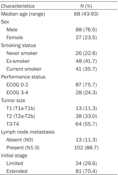
The 115 patients with SCLC were composed of 88 males and 27 females. At the time of diag-nosis, the median age of these patients was 68 years (range 49 to 93 years). Eighty-eight patients (77.4%) were current or past smokers, while 26 (22.6%) were never smokers. The
per-formance statuses of 87 patients (75.7%) were ECOG 0-2 and 28 (24.3%) were ECOG 3-4. Seventy-five tumors (65.2%) were extended dis-ease and 40 (34.8%) were limited disdis-ease. According to the 7th edition of the TNM staging system, 64 tumors (55.7%) were T3-T4 stage, while 51 (44.3%) were T1-T2 stage. Nodal metastasis was detected in 101 (87.8%) cases. These clinical characteristics are summarized in Table 1.
Correlation between p-mTOR expression and clinical characteristics
Of the 115 tumors, 101 (87.8%) were positive for p-mTOR with cytoplasmic immunostaining (Figure 2). Histoscores ranged from 0 to 240, with a median of 70. As previously stated, a his-toscore of 60 or lower was considered low p-mTOR expression, and a histoscore higher than 60 was considered high expression. We analyzed the correlation between p-mTOR expression and clinical characteristics (Table 2).
The level of p-mTOR expression inversely cor-related with the tumor stage, and p-mTOR expression was significantly higher in limited-stage than in extended-limited-stage SCLCs (P = 0.008). In addition, p-mTOR had higher expres-sion in SCLCs without nodal metastasis com-pared with those with nodal metastasis, although the difference was not statistically significant (P = 0.074). No significant associa-tion was observed between p-mTOR expression and other clinical characteristics.
Correlation between p-mTOR expression and patient survival
The median follow-up period was 69.5 days (range 4 to 3304 days). During follow-up, 113 (98.3%) of the 115 patients died from disease progression. The median duration of diagnosis to death was 5.8 months. The 2-year cumula-tive survival rate was 7.9%. SCLC patients with low p-mTOR expression had worse 2-year cumu-lative survival rates compared to those with high p-mTOR expression, although the differ-ence was not statistically significant (3.6% vs. 12.1%; P = 0.170) (Figure 3).
Discussion
[image:3.612.91.289.94.387.2]SCLC is the second most common primary lung cancer, accounting for approximately 15-20% Table 1. Demographic characteristics of 115
patients with small cell lung cancer
Characteristics N (%)
Median age (range) 68 (43-93) Sex
Male 88 (76.5)
Female 27 (23.5)
Smoking status
Never smoker 26 (22.6)
Ex-smoker 48 (41.7)
Current smoker 41 (35.7) Performance status
ECOG 0-2 87 (75.7)
ECOG 3-4 28 (24.3)
Tumor size
T1 (T1a-T1b) 13 (11.3) T2 (T2a-T2b) 38 (33.0)
T3-T4 64 (55.7)
Lymph node metastasis
Absent (N0) 13 (11.3)
Present (N1-3) 102 (88.7) Initial stage
Limited 34 (29.6)
of all lung cancers [1]. Because of its aggres-sive growth and extenaggres-sive metastasis, the main therapy for SCLC has been chemoradiation [1, 12]. SCLC shows a better response to initial chemotherapy, as compared to NSCLC. However, SCLC has a higher recurrence rate and worse prognosis than NSCLC. In recent years, molecular targeting agents, especially for EGFR and ALK-EML4, have been developed for the treatment of adenocarcinoma, but not SCLC [13]. New therapeutic strategies that can improve the survival of SCLC patients have not been developed yet.
mTOR is involved in various cellular processes including cell growth, metabolism, proliferation, and survival by regulating protein synthesis, and is activated by numerous molecules such as growth factors, hormones, cytokines, and other signaling molecules. mTOR is one of the key molecules of the PI3K/AKT/mTOR pathway that contributes to the development of various human cancers [3, 4]. For this reason, mTOR has been regarded as an attractive potential target of anticancer agents.
[image:4.612.92.525.72.361.2]Recent studies have shown the influences of the PI3K/AKT/mTOR pathway on SCLC patho-genesis. Tsurutani et al. [14] reported that acti-vation of PI3K/AKT/mTOR pathway plays a criti-cal role in cellular survival and induces resis-tance to imatinib, and Krystal et al. [15] report-ed that PI3K/AKT signaling induces a resis-tance to etoposide-mediated cell death. In both studies, mTOR inhibitors showed inhibition of growth and survival of human SCLC cells. However, two phase II clinical trials have shown conflicting results. The study using temsirolim-us (CCL-779), a novel mTOR inhibitor, did not show any significant increase in progression-free survivals in patients with extensive-stage SCLC [16]. Similarly, the other phase II study with everolimus (RAD001) failed to show a sig-nificantly improved survival rate in previously treated and relapsed SCLCs [11]. These results suggest that all SCLCs do not respond well to mTOR inhibitors, and that there might be a spe-cific SCLC group for which mTOR inhibitors would be more effective. We speculated that p-mTOR expression in tumor tissues could pro-vide a clue to identify the specific SCLC group that is more sensitive to mTOR inhibitors.
However, the association between p-mTOR expression and the clinical features of SCLC still remains unclear.
We found that p-mTOR expression is signifi-cantly higher in limited-stage than extended-stage SCLCs, and that lymph node metastasis was more frequently detected in the low p-mTOR expression group, as compared to the high expression group, although the difference was not statistically significant. In our study, only two patients were alive after the study period. They both had a limited-stage tumor with high p-mTOR expression. SCLC patients with high p-mTOR expression showed better survival rates than those with low p-mTOR expression, but it was not statistically signifi-cant. Our results are in accordance with previ-ous studies on neuroendocrine tumors of the lung [10, 17]. They demonstrated that high-grade neuroendocrine carcinomas, such as
large cell neuroendocrine carcinoma (LCNEC) or SCLC, have lower levels of p-mTOR expres-sion compared with low-grade neuroendocrine tumors such as typical or atypical carcinoid, and that p-mTOR expression inversely corre-lates with tumor size in high-grade neuroendo-crine carcinomas.
Interestingly, the above results in SCLC contra-dict previous data regarding other types of can-cers. In various cancers, it is well-established that activated AKT/mTOR pathway promotes growth, proliferation, and survival of tumor cells, leading to carcionogenesis and chemore-sistance [18-20], and that high p-mTOR expres-sion is associated with unfavorable prognostic factors [5-8]. A possible hypothesis to explain our unexpected results is that activated mTOR might have a crucial role for initial SCLC cell transformation, but thereafter downstream molecules of mTOR signaling process or anoth-er signaling pathways, including fatty acid syn-thase (Fas) and Ras/Raf/MEK/ERK pathway, might dominate in their contribution to tumor proliferation and progression, and negatively regulate mTOR in advanced SCLCs [21, 22]. Further molecular studies are needed to iden-tify cross-talk between intracellular pathways that could alter the oncogenic potential of mTOR in SCLC.
On the basis of our results, mTOR inhibitors might be more effective for limited-stage SCLCs with high p-mTOR expression, but do not have a definite advantage in treatment for extended-stage SCLCs with low p-mTOR expression. This may be why previous phase II clinical trials with mTOR inhibitors failed to show improved sur-vival rates in advanced or relapsed SCLC patients. In SCLC, the therapeutic value of mTOR inhibitors seems to be limited because novel target therapies are far more necessary for advanced or chemoresistant SCLCs than for early ones.
[image:5.612.91.288.109.452.2]Our study has some limitations. First, this is a retrospective study with a small number of enrolled cases. Second, we could not evaluate the correlation between mTOR and related can-didate molecules. Finally, our study does not provide any information about the response to mTOR inhibitors according to the expression level of p-mTOR in SCLCs. Despite the limita-tions, however, our study showed that p-mTOR is expressed higher in a specific SCLC group Table 2. Correlation between p-mTOR
ex-pression and clinical characteristics in 115 patients with small cell lung cancer
Characteristics p-mTOR expression Low (%) High (%) P-value
Total 57 (100) 58 (100)
Age (years)
≤65 19 (33.3) 13 (22.4) 0.193 >65 38 (66.7) 45 (77.6)
Sex
Male 46 (80.7) 42 (72.4) 0.380 Female 11 (19.3) 16 (27.6)
Smoking status
Never smoker 9 (15.8) 17 (29.3) 0.161 Ex-smoker 24 (42.1) 24 (41.4)
Current smoker 24 (42.1) 17 (29.3) Performance status
ECOG 0-2 46 (80.7) 41 (70.7) 0.278 ECOG 3-4 11 (19.3) 17 (29.3)
Tumor size
T1 (T1a-T1b) 6 (10.5) 7 (12.1) 0.393 T2 (T2a-T2b) 17 (29.8) 21 (36.2)
T3-T4 34 (59.7) 30 (51.7) Lymph node metastasis
Absent (N0) 3 (5.3) 10 (17.2) 0.074 Present (N1-3) 54 (94.7) 48 (82.8) Initial stage
with a limited stage and without lymph node metastasis. This result indicates that p-mTOR might play a more pivotal role in the biologic behavior of early SCLCs than advanced ones and the effectiveness of mTOR inhibitors might vary according to the extent of disease. New tri-als incorporating the response to mTOR inhibi-tors need to consider the preceding evaluation of the expression level of p-mTOR in SCLC. Acknowledgements
This study was supported by a grant from the Dong-A ST.
Disclosure of conflict of interest None.
Address correspondence to: Dr. Hyoun Wook Lee, Department of Pathology, Samsung Changwon Hospital, School of Medicine, Sungkyunkwan University 50, Hapsung-dong, Masan Hoewon-gu, Changwon 630-723, South Korea. Tel: +82-55-290-6497; Fax: +82-55-290-6418; E-mail: sudowo@ naver.com
References
[1] Amini A, Byers LA, Welsh JW, Komaki RU. Progress in the management of limited-stage small cell lung cancer. Cancer 2014; 120: 790-798.
[2] Argiris A, Murren JR. Staging and clinical prog-nostic factors for small-cell lung cancer. Cancer J 2001; 7: 437-447.
[3] Marinov M, Fischer B, Arcaro A. Targeting mTOR signaling in lung cancer. Crit Rev Oncol Hematol 2007; 63: 172-182.
[4] Sun SY, Fu H, Khuri FR. Targeting mTOR signal-ing for lung cancer therapy. J Thorac Oncol 2006; 1: 109-111.
[5] Dhillon T, Mauri FA, Bellezza G, Cagini L, Barbareschi M, North BV, Seckl MJ. Overexpression of the mammalian target of ra-pamycin: a novel biomarker for poor survival in resected early stage non-small cell lung can-cer. J Thorac Oncol 2010; 5: 314-319.
[6] An JY, Kim KM, Choi MG, Noh JH, Sohn TS, Bae JM, Kim S. Prognostic role of p-mTOR expres-sion in cancer tissues and metastatic lymph nodes in pT2b gastric cancer. Int J Cancer 2010; 126: 2904-2913.
[7] Zhang YJ, Dai Q, Sun DF, Xiong H, Tian XQ, Gao FH, Xu MH, Chen GQ, Han ZG, Fang JY. mTOR signaling pathway is a target for the treatment of colorectal cancer. Ann Surg Oncol 2009; 16: 2617-2628.
[8] Sun CH, Chang YH, Pan CC. Activation of the PI3K/Akt/mTOR pathway correlates with tu-mour progression and reduced survival in pa-tients with urothelial carcinoma of the urinary bladder. Histopathology 2011; 58: 1054-1063.
[9] Albanell J, Dalmases A, Rovira A, Rojo F. mTOR signalling in human cancer. Clin Transl Oncol 2007; 9: 484-493.
[10] Righi L, Volante M, Rapa I, Tavaglione V, Inzani F, Pelosi G, Papotti M. Mammalian target of ra-pamycin signaling activation patterns in neuro-endocrine tumors of the lung. Endocr Relat Cancer 2010; 17: 977-987.
[11] Tarhini A, Kotsakis A, Gooding W, Shuai Y, Petro D, Friedland D, Belani CP, Dacic S, Argiris A. Phase II study of everolimus (RAD001) in previ-ously treated small cell lung cancer. Clin Cancer Res 2010; 16: 5900-5907.
[12] Califano R, Abidin AZ, Peck R, Faivre-Finn C, Lorigan P. Management of small cell lung can-cer: recent developments for optimal care. Drugs 2012; 72: 471-490.
[13] Ulivi P, Zoli W, Capelli L, Chiadini E, Calistri D, Amadori D. Target therapy in NSCLC patients: Relevant clinical agents and tumour molecular characterisation. Mol Clin Oncol 2013; 1: 575-581.
[14] Tsurutani J, West KA, Sayyah J, Gills JJ, Dennis PA. Inhibition of the phosphatidylinositol 3-ki-nase/Akt/mammalian target of rapamycin pathway but not the MEK/ERK pathway atten-uates laminin-mediated small cell lung cancer cellular survival and resistance to imatinib me-sylate or chemotherapy. Cancer Res 2005; 65: 8423-8432.
[15] Krystal GW, Sulanke G, Litz J. Inhibition of phosphatidylinositol 3-kinase-Akt signaling
[image:6.612.88.290.67.251.2]blocks growth, promotes apoptosis, and en-hances sensitivity of small cell lung cancer cells to chemotherapy. Mol Cancer Ther 2002; 1: 913-922.
[16] Pandya KJ, Dahlberg S, Hidalgo M, Cohen RB, Lee MW, Schiller JH, Johnson DH, Eastern Cooperative Oncology Group (E1500). A ran-domized, phase II trial of two dose levels of temsirolimus (CCI-779) in patients with exten-sive-stage small-cell lung cancer who have re-sponding or stable disease after induction chemotherapy: a trial of the Eastern Cooperative Oncology Group (E1500). J Thorac Oncol 2007; 2: 1036-1041.
[17] Alì G, Boldrini L, Capodanno A, Pelliccioni S, Servadio A, Crisman G, Picchi A, Davini F, Mussi A, Fontanini G. Expression of p-AKT and p-mTOR in a large series of bronchopulmonary neuroendocrine tumors. Exp Ther Med 2011; 2: 787-792.
[18] Kim D, Dan HC, Park S, Yang L, Liu Q, Kaneko S, Ning J, He L, Yang H, Sun M, Nicosia SV, Cheng JQ. AKT/PKB signaling mechanisms in cancer and chemoresistance. Front Biosci 2005; 10: 975-987.
[19] Oki E, Baba H, Tokunaga E, Nakamura T, Ueda N, Futatsugi M, Mashino K, Yamamoto M, Ikebe M, Kakeji Y, Maehara Y. Akt phosphoryla-tion associates with LOH of PTEN and leads to chemoresistance for gastric cancer. Int J Cancer 2005; 117: 376-380.
[20] Liu SQ, Yu JP, Yu HG, Lv P, Chen HL. Activation of Akt and ERK signalling pathways induced by etoposide confer chemoresistance in gastric cancer cells. Dig Liver Dis 2006; 38: 310-318. [21] Shimizu M, Kondo M, Ito Y, Kume H, Suzuki R,
Yamaki K. Soluble Fas and Fas ligand provide new information on metastasis and response to chemotherapy in SCLC patients. Cancer Detect Prev 2005; 29: 175-180.