Int J Clin Exp Pathol 2015;8(5):5379-5386 www.ijcep.com /ISSN:1936-2625/IJCEP0006372
Original Article
A missense mutation in TMEM67 causes Meckel-Gruber
syndrome type 3 (MKS3): a family from China
Manli Zhang1*, Jing Cheng2*, Aijun Liu3, Longxia Wang4, Lihua Xiong1, Meixia Chen1, Yi Sun5, Jianzhong Li6, Yu Lu2, Huijun Yuan2, Yali Li1, Yanping Lu1
1Department of Obstetrics and Gynecology, Chinese PLA General Hospital, Beijing 100853, China; 2Institute of Otolaryngology, Chinese PLA General Hospital, Beijing 100853, China; 3Department of Pathology, Chinese PLA General Hospital, Beijing 100853, China; 4Department of Ultrasound, Chinese PLA General Hospital, Beijing 100853, China; 5Department of Otolaryngology, Wuhan General Hospital, Wuhan 430070, China; 6Fuzhou Gen-eral Hospital of Nanjing Command PLA, Fuzhou 350025, China. *Equal contributors.
Received January 26, 2015; Accepted March 23, 2015; Epub May 1, 2015; Published May 15, 2015
Abstract: Meckel-Gruber syndrome (MKS) is a lethal autosomal recessive condition characterized by renal cysts and variably associated features, including developmental anomalies of the central nervous system (typically encepha-locele), hepatic ductal dysplasia and cysts, and polydactyly. Genetic heterogeneity has been demonstrated at eleven loci, MKS1-11. Here, we present the clinical and molecular characteristics of a Chinese MKS3 family with occipital encephalocele and kidney enlargement. DNA sequencing of affected fetuses revealed a homozygous c.1645C>T substitution in exon 16 of TMEM67, leading to a p.R549C substitution in meckelin. The R549 residue is highly
conserved across human, rat, mouse, zebrafish, chicken, wolf and platypus genomes. Hha I restriction analysis demonstrated that the c.1645C>T mutation was absent in 200 unrelated control chromosomes of Chinese back-ground, supporting the hypothesis that it represents causative mutation, not rare polymorphism. Our data provide additional molecular and clinical information for establishing a better genotype-phenotype understanding of MKS.
Keywords: MKS3, TMEM67, meckelin, mutation
Introduction
Meckel-Gruber syndrome (MKS; MIM 24900) is a lethal autosomal recessive disorder charac-terized by various severe malformations. The minimal diagnostic criteria are cystic dysplasia
of the kidneys, with fibrotic changes in the liver
and occipital encephalocele or some other mal-formations of the central nervous system. Polydactyly is also frequently reported in MKS patients. Patients with classic MKS phenotype usually die in the perinatal period. MKS is genetically heterogenous [1, 2], with eleven causative genes: MKS1 (OMIM 249000), MKS1 (OMIM 609883), 17q23; MKS2 (OMIM 603194), TMEM216 (OMIM 613277), 11q13; MK- S3 (OMIM 607361), TMEM67 (OMIM 609884) 8q21.13-q22.1; MKS4 (OMIM 611134), CEP290 (OMIM 610142) 12q21.3; MKS5 (OMIM 611561), RPGRIP1L (OMIM 610937) 16q-12.2; MKS6 (OMIM 612284), CC2D2A (OMIM 612013) 4p15; and MKS7 (OMIM
267010), NPHP3 (OMIM 608002), 3q22; MKS8 (OMIM 613885), TCTN2 (OMIM 613846); MKS9 (OMIM 614209), B9D1 (OMIM 614144); MKS10 (OMIM 614175), B9D2 (OMIM 611951); and MKS11 (OMIM 615397), TMEM231 (OMIM 614949). All genes involved in MKS are associ-ated with ciliary functions [4-7].
To date, a total of 25 TMEM67 pathogenic mu- tations have been reported in TMEM67 spec-trum disease (http://omim.org/entry/609884)
and seven of them identified in MKS3 families.
In China, patients with MKS phenotypes have
been reported, but no mutation was identified.
In this report, we present detailed clinical,
genetic and pathological findings of four fetus -es in one family who had clinical characteristics of MKS3 and exhibited pathogenic TMEM67
mutations.
Materials and methods
Ethics statement
Written informed consents were obtained from
to their participation in the study, and all research procedures were approved by the Research Ethics Committee of the Chinese PLA General Hospital. We also have the consent to use the tissues of the aborted normal fetus at 17 gestational week (gw) because the pregnant woman was diagnosed as primary pulmonary
hypertension during her first trimester.
Patients
The Chinese family MKS-H01 was from Beijing, China. The proband was a 14gw fetus (III-3) with occipital encephalocele and kidney en- largement detected by ultrasound. This was the third pregnancy of this couple. The other three pregnancies of this couple carried similar
mal-Figure 1. Clinical features of two affected fetuses in Chinese MKS3 family. (A) Ultrasound scans of III-3 at 14 gw.
Occipital encephalocele was observed and confirmed by autopsy for III-3 (B). Parietal absence, distended abdomen
[image:2.612.90.525.71.428.2]MKS3 from China
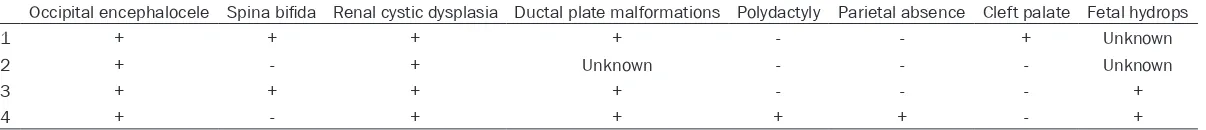
Table 1. Clinical features observed in four MKS fetuses from the Chinese family
Occipital encephalocele Spina bifida Renal cystic dysplasia Ductal plate malformations Polydactyly Parietal absence Cleft palate Fetal hydrops
III-1 + + + + - - + Unknown
III-2 + - + Unknown - - - Unknown
III-3 + + + + - - - +
III-4 + - + + + + - +
mosome analysis for the first and second fetus -es were performed in another hospital and both were normal. Therapeutic termination of pregnancy was performed for the four affected fetuses after ultrasound diagnosis of MKS dur-ing the second trimester of pregnancy, followed
by autopsies of the first, the third and the fourth
affected fetus.
Isolation of genomic DNA and RNA
Peripheral venous blood samples (3 ml) were drawn from family participants and 200 healthy controls for genomic DNA extraction using the Genomic DNA isolation kit (Qiagen).
After abortion, fetal tissues of brain, liver and kidney from III-3 and III-4 were stored in liquid nitrogen. DNA was isolated using Gentra Puregene Mouse Tail kit (Qiagen).
The formalin-fixed, paraffin-embedded tissues
of brain, liver and spleen from the III-1 fetus were applied to isolate genomic DNA using the QIAamp DNA FFPE Tissue Kit (Qiagen), following the protocol supplied by the manufacturer.
cDNA preparation
Total RNA was isolated from aborted proband’s fetal kidney using RNeasy Mini Kit (Qiagen) and
cDNA was obtained by RT-PCR amplification
using SuperScript™ III Reverse Transcriptase (Invitrogen) following the manufacturer’s inst- ructions.
DNA sequencing
After gel purification, each of the amplified PCR
fragments was directly sequenced in both
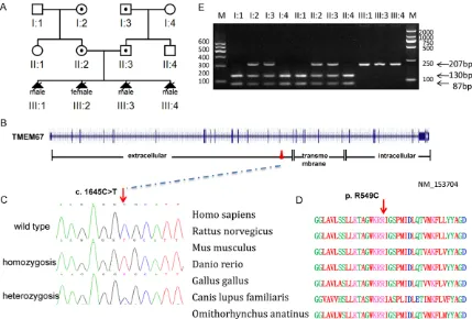
for-Figure 2. Mutation analysis of the Chinese MKS3 family. A. Pedigree of the Chinese MKS3 family. B. Genomic structure of TMEM67 (NM_153704). The 28 exons of TMEM67, encompassing 62 kb of genomic sequence, are shown as vertical bars, with their approximate sizes and positions indicated. Positions of the start codon (ATG) at nucleotide +1 and the stop codon (TAA) are indicated. Correspondence between TMEM67 exons and predicted meckelin domains is depicted at the bottom. C. Sequence chromatogram of affected individual (homozygous), car-rier (heterozygous) and control (wild type). D. Conservation analysis showing that Arg549 in TMEM67 is conserved across human (Homo sapiens), rat (Rattus norvegicus), mouse (Mus musculus), zebrafish (Danio rerio), chicken (Gallus gallus), wolf (Canis lupus familiaris) and platypus, (Omithorhynchus anatinus). E. Endonuclease digestion
[image:4.612.92.521.72.362.2]MKS3 from China
ward and reverse directions using BigDye Terminator chemistry and ABI Prism Sequencer 3100 (Applied Biosystems).
Endonuclease digestion
In the fast assay of the c.1645C>T mutation in fetal tissues and in 200 normal controls, Hha I
restriction analysis of PCR fragment covering the mutation site was applied. We designed the primers (Forward: 5’gtttttgaacaccgatgacaga 3, reverse: 5’agaaggatccagaatggtcaaa 3) to amp- lify a 217bp PCR product. Hha I digestion distin-guishes the homozygote (217 bp) from the
wild-type (130, 87 bp) and heterozygote (217, 130, 87 bp) on agarose gel electrophoresis.
Immunohistochemistry
[image:5.612.91.518.67.459.2]Immunohistochemistry was conducted with the fetal tissues (liver and kidney) obtained from III-4, as well as a 17 gw fetus as a normal control. Standard procedures were used for staining tis-sue slides with the antibodies: primary antibod-ies (Rabbit polyclonal to meckelin ab76786 (Abcam) at 100 dilutions in PBS, matched to appropriate pre-immune negative control sera). The secondary antibody (general two-stage me-
Figure 3. Meckelin expression detected by immunostaining. A. In the liver of the MKS3 fetus (III-4), meckelin was expressed in epithelial cells of enlarged intrahepatic bile ducts around the portal area in the MKS3 fetus. B. In the control, the same location was seen but without bile ducts dilated. Hepatocytes showed slight staining in both af-fected fetuses and in the control. C. In the kidney of the MKS fetus (III-4), showed intense staining was seen in cysts epithelium but not in the remnant glomerulus. D. In the control, meckelin was expressed in renal tubules but not in
thod, PV9000 Zhongshan, China) was applied at a dilution of 1:100.
Results
Clinical features
The proband fetus (III-3) was found to have occipital encephalocele and kidney enlarge-ment by ultrasound (Figure 1) at 14 gw and was aborted one week later. The III-4 fetus was diagnosed at 13 gw by ultrasound and aborted at 14 gw. The couple had two similarly mal-formed fetuses (III-I, III-2) before. Four fetuses had similar clinical characteristics, including occipital encephalocele and renal cystic dys-plasia, as well as some diverse phenotypes.
Table 1 shows the detailed clinical features of the four affected fetuses of this Chinese MKS family. In fetus III-4, we detected parietal absence and polydactyly (Figure 1), which did
not show in the first three fetuses. The absence
of polydactyly is typical phenotype of MKS3.
Mutation identification
Based on the clinical features of this family, we
firstly chose the MKS3 candidate gene,
TMEM67, for mutation screening. We per-formed direct sequencing of full-length cDNA extracted from fetal brain and renal tissues of proband of Chinese family MKS-H01 (III-3). A homozygous C to T transition at position 1645
in exon 16 was identified, resulting in p.R549C
substitution in meckelin (Figure 2). Sequencing
analysis of the formalin-fixed, paraffin-embed -ded tissue from III-1 and the fresh tissue from
III-4 confirmed this finding (we did not have tis -sue from III-2). The Arg residue at 549 in meck-elin is conserved across human, rat, mouse,
zebrafish, chicken, wolf and platypus. Both of
the parents were found to be for the carriers of this substitution. Hha I restriction analysis demonstrated that the c.1645C>T substition was absent in 200 unrelated control chromo-somes with Chinese background, supporting the hypothesis that it represents a causative mutation, not a rare polymorphism.
Immunohistochemitry of meckelin
We further visualized the localization of
meck-elin in paraffin-embedded fetal tissues (liver
and kidney from III-4 and the normal control from a 17 gw fetus. In the normal control,
mod-erate to high levels of meckelin were localized at the renal tubule epithelia, but not in the glomeruli. In the MKS3 fetus, intense staining was shown in the cysts epithelium of the kidney but not in the remnant glomeruli. In the liver, meckelin was expressed in the epithelial cell layer of enlarged intrahepatic bile ducts around the portal area in MKS3 fetus. In the control, the same locations were seen, but without bile ducts dilated (Figure 3).
Discussion
In the present study, we report the identifica -tion of a TMEM67 mutation in a Chinese MKS3 family with four affected individuals. All affect-ed fetuses displayaffect-ed renal cystic dysplasia and occipital encephalocele. Ductal plate malfor-mations with proliferation of bile duct were showed in the last three affected fetuses. Comparison of the clinical features of MKS3-linked cases with MKS1-MKS3-linked cases suggest-ed that polydactyly and possibly encephalocele are less common in MKS3-linked families [9,
10]. The absence of polydactyly in the first
three affected fetuses suggested that TMEM67
was the good candidate gene for mutation screening in this family. Sequence analysis and
Hha I restriction analysis demonstrated that a homozygous c.1645C>T mutation in TMEM67
was cosegregated with the MKS phenotype in this family and the parents were heterozygous carriers. Note that the fourth affected fetus, which carried the same TMEM67 mutation, had additional polydactyly which showed phenotyp-ic heterogeneity in the Chinese MKS3 family.
MKS3 from China
16 of the TMEM67 gene in Chinese family, which encodes the extracellular region of
meck-elin. This is the first report of TMEM67 muta-tion in Chinese populamuta-tion. The same substitu-tion was reported by Katharina Hopp as a highly likely mutation [7]. It is possible that, within the range of exons 8 to 16, all encoding extracellu-lar fragments are located just before the corre-sponding transmembrane fragments. Except for lethal MKS, primary cilia-related disease, including Joubert syndrome, Bardet-Biedl syn-drome and COACH synsyn-drome, were accompa-nied with some structural abnormality at the fetal stage, such as ‘molar tooth sign’ (MTS)-cerebellar vermis hypo-dysplasia, thickening and horizontalization of superior cerebellar peduncles and deepening of the interpeduncu-lar fossaon MRI, Dandy-Walker malformation and polydactyly [12, 13]. Because many genes are involved in these disorders, traditional
methods of mutation screening are difficult to
identify the causative genes. With the develop-ment of a new generation of sequencing strate-gies, we can test all ciliapathy-related genes
simultaneously following ultrasound identifica -tion of features associated with ciliopathy by prenatal diagnosis [14].
We also visualized the localization of meckelin in the MKS fetus and normal controls. As previ-ously reported there was strong staining in the epithelial cell layer of the renal tubule but not in the glomerulus [6]. Our results showed intense staining of renal cysts in the MKS3 fetus. Dawe
et al. reported that meckelin immunostaining was absent in the renal tissue of the MKS3 fetus who carried a homozygous c.1127A>C (p.Q376P) mutation in TMEM67 [6]. In our study, meckelin staining was present in both kidney tissues of affected fetus with homozygo- us c.1645C>T (p.R549C) mutation in TMEM67
and normal control.
In summary, we have identified a homozygous
TMEM67 mutation in a Chinese family exhibit-ing clinical characteristics of MKS3. Statistically, this couple would be predicted to have a 25% chance of producing an affected embryo. However, four previous natural pregnancies of this couple all turned to be affected MKS3 fetuses detected by ultrasound, and the couple repeatedly opted to terminate the pregnancies
by artificial abortions. The identification of the
causative mutation of TMEM67 in this family
provided a ground for PGD procedure for this family. Further efforts will be focused on devel-oping a PGD protocol to the couples at risk of conceiving a pregnancy affected with MKS3 and other known monogenic diseases.
Acknowledgements
This work was supported by the Key Project of National Natural Science Foundation of China to Huijun Yuan (No. 81030017), National Science Fund for Distinguished Young Scholars to Huijun Yuan (No. 81125008). Science Fund from Chinese PLA General Hospital to Yanping Lu (No. 2012FC-CXYY-1001). We sincerely thank the family members for their participa-tion and support in this study.
Disclosure of conflict of interest
None.
Address correspondence to: Huijun Yuan, Institute of Otolaryngology, Chinese PLA General Hospital, Beijing 100853, China. Tel: +86 10 6693 8147; Fax: 86 10 6815 6974; E-mail: yuanhj301@163.com; Yali Li or Yanping Lu, Department of Obstetrics and Gynecology, Chinese PLA General Hospital, Beijing 100853, China. E-mail: li_Yali@hotmail.com (YLL); yanpinglu569@163.com (YPL)
References
[1] Alexiev BA, Lin X, Sun CC, Brenner DS. Meckel-Gruber syndrome: pathologic manifestations, minimal diagnostic criteria, and differential di-agnosis. Arch Pathol Lab Med 2006; 130: 1236-1238.
[2] Barker AR, Thomas R, Dawe HR. Meckel-Gru-ber syndrome and the role of primary cilia in kidney, skeleton, and central nervous system development. Organogenesis 2014; 10: 96-107.
[3] Kyttala M, Tallila J, Salonen R, Kopra O, Kohlschmidt N, Paavola-Sakki P, Peltonen L, Kestilä M. MKS1, encoding a component of
the flagellar apparatus basal body proteome,
is mutated in Meckel syndrome, Nat Genet 2006; 38: 155-157.
The transmembrane protein meckelin (MKS3) is mutated in Meckel-Gruber syndrome and the wpk rat. Nat Genet 2006; 38: 191-196. [5] Dawe HR, Smith UM, Cullinane AR, Gerrelli D,
Cox P, Badano JL, Blair-Reid S, Sriram N, Kat-sanis N, Attie-Bitach T, Afford SC, Copp AJ, Kelly DA, Gull K, Johnson CA. The Meckel-Gru-ber Syndrome proteins MKS1 and meckelin interact and are required for primary cilium formation. Hum Mol Genet 2007; 16: 173-186.
[6] Garcia-Gonzalo FR, Corbit KC, Sirerol-Piquer MS, Ramaswami G, Otto EA, Noriega TR, Seol AD, Robinson JF, Bennett CL, Josifova DJ, Gar-cía-Verdugo JM, Katsanis N, Hildebrandt F, Re-iter JF. A transition zone complex regulates mammalian ciliogenesis and ciliary membrane composition. Nat Genet 2011; 43: 776-784. [7] Hopp K, Heyer CM, Hommerding CJ, Henke SA,
Sundsbak JL, Patel S, Patel P, Consugar MB, Czarnecki PG, Gliem TJ, Torres VE, Rossetti S, Harris PC. B9D1 is revealed as a novel Meckel syndrome (MKS) gene by targeted exon-en-riched next-generation sequencing and dele-tion analysis. Hum Mol Genet 2011; 20: 2524-34.
[8] Baala L, Romano S, Khaddour R, Saunier S, Smith UM, Audollent S, Ozilou C, Faivre L, Lau-rent N, Foliguet B, Munnich A, Lyonnet S, Salo-mon R, Encha-Razavi F, Gubler MC, Boddaert N, de Lonlay P, Johnson CA, Vekemans M, Anti-gnac C, Attie-Bitach T. The Meckel-Gruber drome gene, MKS3, is mutated in Joubert syn-drome. Am J Hum Genet 2007; 80: 186-194. [9] Consugar MB, Kubly VJ, Lager DJ, Hommerding
CJ, Wong WC, Bakker E, Gattone VH 2nd, Tor-res VE, Breuning MH, Harris PC. Molecular di-agnostics of Meckel-Gruber syndrome high-lights phenotypic differences between MKS1 and MKS3. Hum Genet 2007; 121: 591-599. [10] Khaddour R, Smith U, Baala L, Martinovic J,
Clavering D, Shaffiq R, Ozilou C, Cullinane A,
Kyttälä M, Shalev S, Audollent S, d’Humières C, Kadhom N, Esculpavit C, Viot G, Boone C, Oien C, Encha-Razavi F, Batman PA, Bennett CP, Woods CG, Roume J, Lyonnet S, Génin E, Le Merrer M, Munnich A, Gubler MC, Cox P, Mac-donald F, Vekemans M, Johnson CA, Attié-Bit-ach T; SOFFOET (Société Française de Foeto-pathologie). Spectrum of MKS1 and MKS3 mutations in Meckel syndrome: a genotype-phenotype correlation. Hum Mutat 2007; 28: 523-524.
[11] Iannicelli M, Brancati F, Mougou-Zerelli S,
Maz-zotta A, Thomas S, Elkhartoufi N, Travaglini L,
Gomes C, Ardissino GL, Bertini E, Boltshauser E, Castorina P, D’Arrigo S, Fischetto R, Leroy B, Loget P, Bonnière M, Starck L, Tantau J, Genti-lin B, Majore S, Swistun D, Flori E, Lalatta F, Pantaleoni C, Penzien J, Grammatico P; Inter-national JSRD Study Group, Dallapiccola B, Gleeson JG, Attie-Bitach T, Valente EM. Novel TMEM67 mutations and genotype-phenotype correlates in meckelin-related ciliopathies. Hum Mutat 2010; 31: E1319-E1331.
[12] Maria BL, Hoang KB, Tusa RJ, Mancuso AA, Hamed LM, Quisling RG, Hove MT, Fennell EB, Booth-Jones M, Ringdahl DM, Yachnis AT, Creel G, Frerking B. “Joubert syndrome” revisited: key ocular motor signs with magnetic reso-nance imaging correlation. J Child Neurol 1997; 12: 423-430.
[13] Saleem SN, Zaki MS, Soliman NA, Momtaz M. Prenatal magnetic resonance imaging diagno-sis of molar tooth sign at 17 to 18 weeks of gestation in two fetuses at risk for Joubert syn-drome and related cerebellar disorders. Neu-ropediatrics 2011; 42: 35-38.