Original Article
miR-30a inhibits breast cancer progression
through the Wnt/β-catenin pathway
Yu Miao1*, Lei Wang2*, Xin Zhang1, Rong-Ge Xing1, Wei-Wei Zhou1, Chun-Rong Liu1, Xiao-Ling Zhang1, Liang Tian1
1Department of Pathology, Central Hospital of Cangzhou, Cangzhou, China; 2Cangzhou Medical College, Yingbin South Avenue and 307 National Highway Intersection in Cangzhou Canal District, China. *Equal contributors. Received October 25, 2018; Accepted November 23, 2018; Epub January 1, 2019; Published January 15, 2019
Abstract: Background: miR-30a is a microRNA associated with the progression of malignant tumors such as gastric cancer, colon cancer, prostate cancer, and lung cancer, and can regulate the proliferation and migration of breast
cancer (BC) cells in vitro. However, its expression, function, clinical significance and relationship with the
Wnt/β-catenin pathway in human BC were still unclear. Methods: Immunohistochemistry, Western blotting and real-time
quantitative PCR (RT-qPCR) were used to measure the expressions of miR-30a and β-catenin in 114 pairs of human
BC tumor tissues and adjacent normal tissues which were collected from March 2014 to October 2015. The effect
of miR-30a on the expression of β-catenin was studied in the MCF-7 cells in vitro. Results: The expression levels of miR-30a in human BC tumor tissues were significantly lower than they were in the adjacent normal tissues (P < 0.001), and significantly higher in β-catenin protein (P < 0.001), but there was no significant different in β-catenin mRNA (P = 0.3816). The immunohistochemistry results showed that β-catenin protein was only expressed on the cell membrane in paracancerous normal tissues, but β-catenin protein was expressed on the cell membrane and cy
-toplasm in BC tumor cells. In addition, there was a significantly negative correlation (r = -0.816, P < 0.001) between the expression miR-30a and β-catenin protein in BC tissues. The age of onset, PR expression, ER expression, and HER-2 expression of the BC patients were not related to miR-30a or β-catenin protein expression (P > 0.05). Tumor diameter, histological grade, lymph node metastasis, TNM stage, and the prognosis of BC patients (P < 0.05) were significantly related to miR-30a or β-catenin protein expression. In MCF-7 cells, miR-30a regulated the accumula
-tion of β-catenin protein by inhibiting the expression of BCL9 in BC cells. Conclusion: miR-30a was lowly expressed in breast cancer tissues and highly in β-catenin protein, and miR-30a might block the Wnt/β-catenin pathway by inhibiting the accumulation of β-catenin, and then inhibiting breast cancer progression.
Keywords: miR-30a, breast cancer, progression, β-catenin
Introduction
Since 2010, the morbidity and mortality of can-cer in the Chinese have been rising. According to the latest data released by the National Cancer Center, the new cancer growth rate in China is 3%, accounting for 1/4 of new cancer cases worldwide [1]. According to the cancer
statistics of China in 2015 [2], the five most
common cancers among women were breast cancer (BC), lung and bronchial cancer, stom-ach cancer, colorectal cancer and esophageal cancer, and of these 15% were breast cancer. microRNA is a small non-coding RNA that plays an important role in regulating the transcription of genes and can regulate cell proliferation, dif-ferentiation, apoptosis, metabolism, and
β-Catenin is a key molecule in the Wnt signaling
pathway, and many human malignancies have been found to be associated with abnormally
activated Wnt/β-catenin pathways [19, 20]. In
addition, miR-30a was found to affect the
acti-vation of the Wnt/β-catenin pathway by regulat -ing BCL9 expression in myeloma cells [21] or PRDM1 in glioma cells [22]. However, the
rela-tionship between miR-30a and the
Wnt/β-catenin pathway in breast cancer has not been
confirmed. In this paper, we measured the expression of miR-30a and β-catenin in BC tis -sues and studied their regulatory relationship
in MCF-7 cells.
Materials and methods
Tissue and statement
114 breast cancer patients (all female) were selected as subjects for this study, which was
conducted from January 2014 to December 2015 in Changzhou Central Hospital. The patients’ tumor tissue and adjacent normal tis-sues were removed surgically and collected. The tissues were divided into three parts, one
for making paraffin sections, and the other two
were stored in liquid nitrogen. The ages of the 114 breast cancer patients ranged from 42 to 71 years, and 63 was the median age. Other clinical data is shown in Table 1. In addition, all subjects (or their guardians) included in this study consented to the research protocol and signed an informed consent form. The ethics committee of Changzhou Central Hospital approved this research protocol.
Real-time quantitative PCR
[image:2.612.91.521.84.454.2]Trizol was used to extract the total RNA of the tissues or cells. The extracted RNA was reverse transcribed into cDNA using a PrimeScript™RT
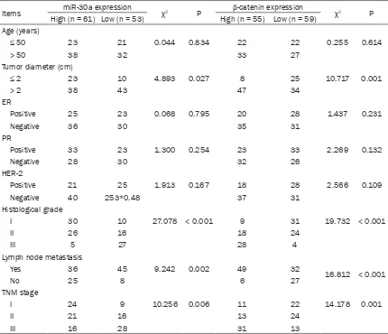
Table 1. Relationship between miR-30a or β-catenin and the clinical pathology of BC patients
Items miR-30a expression χ2 P β-catenin expression χ2 P
High (n = 61) Low (n = 53) High (n = 55) Low (n = 59) Age (years)
≤ 50 23 21 0.044 0.834 22 22 0.255 0.614
> 50 38 32 33 27
Tumor diameter (cm)
≤ 2 23 10 4.893 0.027 8 25 10.717 0.001
> 2 38 43 47 34
ER
Positive 25 23 0.068 0.795 20 28 1.437 0.231
Negative 36 30 35 31
PR
Positive 33 23 1.300 0.254 23 33 2.269 0.132
Negative 28 30 32 26
HER-2
Positive 21 25 1.913 0.167 18 28 2.566 0.109
Negative 40 253*0.48 37 31
Histological grade
I 30 10 27.078 < 0.001 9 31 19.732 < 0.001
II 26 16 18 24
III 5 27 28 4
Lymph node metastasis
Yes 36 45 9.242 0.002 49 32
16.812 < 0.001
No 25 8 6 27
TNM stage
I 24 9 10.256 0.006 11 22 14.178 0.001
II 21 16 13 24
Master Mix reverse transcription kit (RR036B, Takara, Beijing, China). The PCR parameters were set at: 37°C/60 minutes, 85°C/5
sec-onds. A 20 μl real-time fluorescence quantita -tive PCR (RT-qPCR) system was prepared according to the SYBR Green qPCR Master Mix kit instructions (638320, TakaRa, Beijing,
China) and amplified using ABI 7500 fluores -cence quantitative PCR instrument (Applied Biosystems, Maryland, USA). The PCR para- meters were set at: 95°C/30 s, [90°C/5 s, 65°C/30 s]-40 cycles. The PCR primers were:
miR-30a-F,
[image:3.612.94.509.73.507.2]5’-ACACTCCAGCTGGGTGTAAACATC-CTCGAC-3’, miR-30a-R, 5’-TGGTGTCGTGGAGT-
Figure 1. Expression of miR-30a and β-catenin. (A, B) RT-qPCR was used to detect the expression of miR-30a (A) and β-catenin mRNA (B) in BC tissues and paracancerous normal tissues; (C) Expression of β-catenin protein was
counted in BC tissues and paracancerous normal tissues; (D) T = neuroblastoma tissues, N = paracancerous
nor-mal tissues, nx = number x of patient with BC; (E) The expression miR-30a and β-catenin protein in the BC tissues. (Fa) β-catenin protein was only expressed on the cell membranes in paracancerous normal tissues; (Fb) Expression of β-catenin protein in BC tissues; (Fc) β-catenin protein was only expressed on the cell membranes in BC tissues (-); (Fd) β-catenin protein was expressed on the cell membranes and weak positive expression on the cytoplasms and cytoplasms in BC tissues (+); (Fe) β-catenin protein was expressed on the cell membranes and positive expres
-sion on the cytoplasms and the cytoplasms in BC tissues (++); (Ff) β-catenin protein was expressed on the cell
CG-3’; F, 5’-CTCGCTTCGGCAGCACA-3’, U6-R, AACGCTTCACGAATTTGCGT-3’; BCL9-F,
5’-GGCCATACCCCTAAAGCACTC-3’, BCL9-R, 5’-CG-
GAAATACTTCGCTCCCTTTT-3’; β-catenin-F, A-AAGCGGCTGTTAGTCACTCG-3’, β-catenin-R, 5’-CGAGTCATTGCATACTGTCCAT-3’; GAPDH-F, 5’-C-TGGGCTACACTGAGCACC-3’, GAPDH-F,
5’-AAG-TGGTCGTTGAGGGCAATG-3’.
Western blot
Tissue or cell lysates were separated by
SDS-page and then transferred to a PVDF mem -brane. Primary antibody: anti-BCL9 (1:500,
ab37305, Abcam, Cambridge, UK), or
anti-β-catenin (ab32572, 1:5000, Abcam, Cambridge, UK), or anti-GAPDH (ab9484, 1:3000, Abcam, Cambridge, UK). Second antibody: goat anti-rabbit (ab150077, 1:1000, Abcam, Cambridge, UK), or goat anti-rat (ab150117, 1:1000, Abcam, Cambridge, UK). The primary antibody
was incubated overnight at 4°C and the second antibody was incubated for 1 hour at room temperature.
Immunohistochemistry
Immunohistochemistry was used to measure
the β-catenin protein expression of the BC tis -sues or the adjacent normal tis-sues using a VECTASTAIN® Elite® ABC Kit (Vector Labora-
tories, Boston, MA, USA). A β-catenin antibody
(ab32572, 1:5000, Abcam, Cambridge, UK) was used as a primary antibody (PBS instead of primary antibody as a negative control) and incubated overnight at 4°C. Goat Anti-Rabbit IgG H & L (HRP) (1:1000, Abcam, Cambridge, UK) was incubated as a secondary antibody at
[image:4.612.91.520.73.399.2]37°C for 2 h. Finally, 5 fields per slice were pho -tographed and evaluated: no color was nega-tive (-), a faint yellow was weak posinega-tive (+), yel-low was positive (++), claybank or tan was
Figure 2. Expressions of 30a and β-catenin protein in different breast cancer tissues. (A-C) Comparison of
miR-30a expression in different histological grades (A), TNM stage (B) and lymph node metastasis (C) in breast cancer
strongly positive (+++). Two pathology deputy chief physicians who were blinded to the patients’ diagnoses evaluated the stained tissue.
Cell and cell transfection
MCF-7 cells (HTB-22, ATCC, VA, USA), from the
human breast adenocarcinoma cell line were cultured with DMEM medium (12491-15, Th-
ermoFisher, CA, USA), to which was added 10% fetal bovine serum (10100-147, ThermoFisher,
CA, USA) and 1% penicillin-streptomycin (156-
40055, ThermoFisher, CA, USA). miR-30a-NC,
the miR-30a-mimic, and the miR-30a-inhibitor were designed and synthesized by Shenggong Bioengineering Co., Ltd. (Shanghai, China), and were directly transferred into the cells using the Lipofectamine™ 2000 transfection reagent
(11668019, Invitrogen, CA, USA). For the wild
type or the mutation mRNA 3’-UTR of BCL9,
they were first connected to pisCHECK2
(Pro-mega, WI, USA) and then were transfected into the cells as miRNA.
Statistical analysis
The data were analyzed by SPSS 20.0 for sta-tistical analysis and were stated as the mean ± standard deviation or the frequency. Student’s
t-test or a chi-square test was used to compare the differences between two groups. Com- parisons between multiple groups were per-formed using the single factor ANVOA method, and the Duncan method was used for post hoc testing. The correlations between two groups was analyzed by Pearson. The Cox regression
model was used to test the univariate and mul-tivariate analyses for the survival times of the BC patients. The survival curves of the BC
patients based on the miR-30a or β-catenin
protein expressions were drawn by the Kaplan-Meier method. A log-rank test was used to
com-pare the differences of the survival curves. P < 0.05 indicated a significant difference.
Results
miR-30a and β-catenin expression in BC tis -sues
The expressions of miR-30a and β-catenin
were measured in 114 pairs of breast cancer (BC) tissues and paracancerous normal tis-sues, and we found that the expression of
miR-30a in the BC tissues was significantly lower
than it was in the paracancerous normal
tis-sues (P < 0.001) (Figure 1A), and there was a
significantly negative correlation (r = -0.816, P < 0.001) between the expression miR-30a and β-catenin protein in the BC tissues (Figure 1E).
Although there was no significant difference between the expression of β-catenin in the BC
tissues and the paracancerous normal tissues (P = 0.3861) (Figure 1B), the expression of
β-catenin protein in the BC tissues was signifi -cantly higher than it was in the paracancerous
normal tissues (P < 0.001) (Figure 1C, 1D). In addition, immunohistochemistry was used to
localize the expression of β-catenin in the BC
tissues and paracancerous normal tissues,
and we found that β-catenin protein was only
expressed on the cell membranes in the
para-Table 2. Univariate and multivariate Cox regression analysis for the prognosis of BC patients Clinical parameters Univariate analysis Multivariate analysis
OR 95% CI P OR 95% CI P
Age 1.245 0.417-3.658 0.624 0.586
Tumor diameter 2.029 0.809-4.627 0.083 0.823
ER positive 3.256 1.241-5.324 0.069 0.162
PR positive 3.164 0.986-4.238 0.062 0.159
HER-2 positive 3.342 0.429-04.824 0.041 0.084
Histological grade 3.514 0.912-11.254 0.028 2.739 1.127-12.414 0.031 Lymph node metastasis 3.435 1.020-5.184 < 0.001 2.685 1.432-9.867 0.024
TNM stage 4.022 1.615-11.231 0.003 2.612 1.004-10.628 0.047
MiR-30a expression 6.545 2.318-18.210 < 0.001 6.028 3.042-15.917 < 0.001
β-catenin expression 5.488 3.218-13.657 < 0.001 5.124 2.987-12.368 0.002
[image:5.612.92.522.85.244.2]cancerous normal tissues, but β-catenin pro -tein was expressed on the cell membranes and cytoplasms in the BC tissues. This indicates
that β-catenin was deposited from the cell
membranes into the cytoplasms and nuclei.
The relationship between miR-30a or β-catenin and the clinical pathology of BC patients
According to the expression levels of miR-30a
or the β-catenin protein in breast cancer tis -sues, tissues from 114 breast cancer patients were divided into two groups, and the miR-30a low expression group was the expression level
of miR-30a < the median value of 114 BC patients, and the same as β-catenin protein.
We analyzed the relationship between miR-30a
or β-catenin protein and the clinical pathology
of BC patients. The results showed that the age of onset, PR expression, ER expression, and HER-2 expression of the BC patients were not
related to miR-30a or β-catenin protein expres
-sion (P > 0.05). Tumor diameter, histological
grade, lymph node metastasis, and TNM stage
of BC patients (P < 0.05) were significantly related to miR-30a or β-catenin protein expres -sion (Table 1). With the increase of histological grade or TNM stage in BC patients, the expres-sion of miR-30a in the BC tissues was gradually decreased (Figure 2A, 2B), but the expression
of β-catenin protein in the BC tissues was grad -ually increased (Figure 2D, 2E). In addition, the expression of miR-30a in the BC tissues with
lymph node metastasis was significantly lower
than that in BC tissues without lymph node
metastasis (P < 0.001) (Figure 2C), but the
expression trend of β-catenin protein was just
the opposite (Figure 2F).
The effect of miR-30a or β-catenin expression on the prognosis of BC patients
114 BC patients were followed up at least once every four months or the patients came to
hos-pital for a review. The factors influencing the
survival times of BC patients were analyzed using the COX regression model. The results showed that (Table 2) the miR-30a expression level (OR = 6.028, 95% CI = 3.042-15.917) and
the β-catenin protein expression level (OR =
5.124, 95% CI = 2.987-12.368) were
indepen-dent risk factors influencing the prognosis of
BC patients. Postoperative survival in BC patients with low expression of miR-30a was
significantly lower than it was for patients with
high expression of miR-30a (P = 0.0309) (Figure 3A), and postoperative survival in BC
patients with high expression of β-catenin pro
-tein was significantly lower than it was in patients with low expression of β-catenin pro -tein (P = 0.0174) (Figure 3B).
miR-30a knockdown promoted the
accumula-tion of β-catenin protein in BC cells
In other cancer studies, such as those looking at multiple myeloma and prostate cancer,
miR-30a has been found to regulate the
[image:6.612.90.520.73.262.2]Wnt/β-catenin signaling pathway by targeting BCL9 protein expression. We searched for binding
sites for miR-30a and BCL9 from the starBase
database. To confirm that miR-30a could regu -late BCL9 expression by binding to the BCL9 3’-UTR end, we used the luciferase gene report-er system. The results showed that
transfec-tion of miR-30a-mimics significantly decreased WT type 3’-UTR luciferase activity (P < 0.001), and the miR-30a-inhibitor significantly increas-ed in MCF-7 cells. But it didn’t work in MUT. Further, as shown in Figure 4C, 4D, the miR-30a-inhibitor can increase the expression BCL9 mRNA and protein, but it only increases
the expression of β-catenin protein and does not work with β-catenin mRNA. This means that
miR-30a knockdown promotes the
accumula-tion of β-catenin protein in BC cells. Discussion
miR-30a is located at the 6q13 position on human chromosomes. Although it does not encode any protein, it plays an important role in many human diseases [23, 24], especially in cancer [25]. Previous research has shown that
miR-30a not only negatively regulates the TGF-β1-induced epithelial-mesenchymal transition and peritoneal fibrosis by targeting Snai1 [26],
but also functions as a tumor suppressor and novel therapeutic tool in many malignant tumors, such as gastric cancer [14], colon can-cer [15], prostate cancan-cer [16], and lung cancan-cer [17]. In this paper, we found that the expression of miR-30a in 114 samples of BC tissues was
significantly lower than it was in adjacent para -cancerous normal tissues, and was related to the tumor diameter, histological grade, lymph node metastasis, TNM stage, and the progno-sis of BC patients. It means that miR-30a plays an important role in the occurrence and devel-opment of breast cancer.
TNM staging, lymph node metastasis and histo-logical grading of the cancer are macroscopic representations of tumorigenesis and develop-ment, and their intrinsic nature is inseparable from the biological characteristics of tumor cell growth, proliferation, apoptosis and migra- tion, and even more in-depth molecular
mecha-Figure 4. miR-30a regulated β-catenin protein accumulation by targeting BCL9. (A) WT-BCL9 3’UTR luciferase re -porter vector, and a MUT-BCL9 3’UTR luciferase re-porter vector with mutations on miR-30a binding sites of the
[image:7.612.93.522.72.331.2]nisms may be closely related to multiple fac-tors such as oncogene activation, tumor sup-pressor gene mutation inactivation, and abnormal changes in multiple signaling path-ways [27, 28]. In this study, the expression of miR-30a in 114 samples of BC tissues de- creased with the increase of TNM staging and tissue grade and lymph node metastasis. In addition, many previous studies had reported that miR-30a inhibited the proliferation, migra-tion, and other biological functions of breast cancer cells in vitro by targeting Eya2 [18] and Notch1 [29], or via the p53/miR-30a/ZEB2 axis [30] and the miR-30a-5p/UBE3C axis [31]. Therefore, whether it is in breast cancer or in other malignant tumors, miR-30a exhibits the function of a tumor suppressor gene.
Because the development of tumors is the result of long-term interaction of multiple onco-genes and tumor suppressor onco-genes through multiple signaling pathways, a tumor suppres-sor gene or oncogene may participate in the growth, proliferation and migration of tumor cells through multiple signaling pathways [32,
33]. The Wnt/β-catenin pathway is highly con -served during biological evolution, and partici-pates in the regulation of cell growth, develop-ment and differentiation, and is regulated closely at the level of transcription and
post-transcriptional modification [34], and its abnor
-mal activation has been confirmed with the
occurrence of various tumors including breast cancer [35, 36]. More and more studies have
shown that the Wnt/β-catenin pathway plays
an important role in the development and pro-gression of breast cancer [37], invasion and metastasis [38, 39], and chemotherapy drug resistance [35], and many miRNAs play a key role in it, such as miR-34a [40] and miR-100 [41].
Although the relationship between miR-30a
and the Wnt/β-catenin pathway in breast can
-cer has not been confirmed, miR-30a was found to affect the activation of the
Wnt/β-catenin pathway by regulating BCL9 expression in myeloma cells [21] or PRDM1 in glioma cells [22]. In this study, we also found that there was
a significantly negative correlation between the expression miR-30a and β-catenin protein in
BC tissues, and miR-30a could regulate the
accumulation of β-catenin protein in BC cells in vitro. β-catenin has been found to play a key
role in the regulation of the biological
charac-teristics of stem cells at various stages of breast development, and the activation or
aggregation of the β-catenin signaling pathway
leads to abnormal cell growth, differentiation, metabolism and biology, and eventually to
breast cancer [42, 43]. β-Catenin is a key mol
-ecule in the Wnt/β-Catenin signaling pathway
and plays a dual role in the signal transduction
of the Wnt/β-Catenin signaling pathway: it can
be used as a marker of pathway activation and can also bind to E-cadherin on the membrane to regulate cell-to-cell interaction and adhe-sion, affecting the aggressiveness of cancer
cells [44]. In normal cells, β-catenin is localized
in the cell membrane and is used to bind to E-cadherin on the membrane to form a complex and the content in the cytoplasm remains low or even nil, and a small amount of unbound
β-catenin in cytoplasm is degraded by the APC/ GSK-3β/Axin complex. However, when the Wnt
pathway is abnormally activated, the
degrada-tion process of β-catenin is inhibited, allowing it
to accumulate in the cytoplasm and enter into
the nucleus to form a complex with TCF/LEF,
which promotes the transcriptional activity of the target gene downstream of the pathway, resulting in an increase in transcriptional activ-ity and abnormal biological characteristics in cells.
All in all, miR-30a was lowly expressed in breast
cancer tissues and highly expressed in
β-ca-tenin protein, and miR-30a might block the
Wnt/β-catenin pathway by inhibiting the accu
-mulation of β-catenin, and then inhibiting
breast cancer progression. Acknowledgements
This research was supported by the Cangzhou Central Hospital Project fund (162302069). Disclosure of conflict of interest
None.
References
[1] Ferlay J, Soerjomataram I, Dikshit R, Eser S, Mathers C, Rebelo M, Parkin DM, Forman D, Bray F. Cancer incidence and mortality world -wide: sources, methods and major patterns in GLOBOCAN 2012. Int J Cancer 2015; 136: E359-86.
[2] Chen W, Zheng R, Baade PD, Zhang S, Zeng H,
Bray F, Jemal A, Yu XQ, He J. Cancer statistics
in China, 2015. CA Cancer J Clin 2016; 66: 115-32.
[3] Sun W, Julie Li YS, Huang HD, Shyy JY, Chien S. microRNA: a master regulator of cellular pro-cesses for bioengineering systems. Annu Rev Biomed Eng 2010; 12: 1-27.
[4] Liston A, Linterman M, Lu LF. MicroRNA in the
adaptive immune system, in sickness and in health. J Clin Immunol 2010; 30: 339-46. [5] Gregory RI and Shiekhattar R. MicroRNA
bio-genesis and cancer. Cancer Res 2011; 65: 3509-3512.
[6] Wu W, Sun M, Zou GM, Chen J. MicroRNA and cancer: current status and prospective. Int J Cancer 2007; 120: 953-60.
[7] Kurozumi A, Goto Y, Matsushita R, Fukumoto I,
Kato M, Nishikawa R, Sakamoto S, Enokida H, Nakagawa M, Ichikawa T, Seki N. Tumor-sup-pressive microRNA-223 inhibits cancer cell mi-gration and invasion by targeting ITGA3/ITGB1 signaling in prostate cancer. Cancer Sci 2016; 107: 84-94.
[8] Goto Y, Nishikawa R, Kojima S, Chiyomaru T,
Enokida H, Inoguchi S, Kinoshita T, Fuse M,
Sakamoto S, Nakagawa M, Naya Y, Ichikawa T, Seki N. Tumour-suppressive microRNA-224 in-hibits cancer cell migration and invasion via targeting oncogenic TPD52 in prostate cancer.
FEBS Lett 2014; 588: 1973-82.
[9] Yang TS, Yang XH, Chen X, Wang XD, Hua J, Zhou DL, Zhou B, Song ZS. MicroRNA-106b in
cancer-associated fibroblasts from gastric can -cer promotes cell migration and invasion by
targeting PTEN. FEBS Lett 2014; 588: 2162-9.
[10] Qin J, Luo M. MicroRNA-221 promotes
colo-rectal cancer cell invasion and metastasis by
targeting RECK. FEBS Lett 2014; 588: 99-104.
[11] Shen H, Li L, Yang S, Wang D, Zhong S, Zhao J, Tang J. MicroRNA-29a contributes to drug-re-sistance of breast cancer cells to adriamycin
through PTEN/AKT/GSK3β signaling pathway.
Gene 2016; 593: 84-90.
[12] Wiggins JF, Ruffino L, Kelnar K, Omotola M, Pa -trawala L, Brown D, Bader AG. Development of a lung cancer therapeutic based on the tumor suppressor microRNA-34. Cancer Res 2010; 70: 5923-30.
[13] Naidu S, Magee P, Garofalo M. MiRNA-based therapeutic intervention of cancer. J Hematol Oncol 2015; 8: 68.
[14] Min J, et al. Abstract 3115: miR-30a-5p func-tions as a tumor suppressor gene in gastric cancer. Cancer Research 2015; 75 Suppl: 3115-3115.
[15] Liu M, Huang F, Zhang D, Ju J, Wu XB, Wang Y,
Wang Y, Wu Y, Nie M, Li Z, Ma C, Chen X, Zhou JY, Tan R, Yang BL, Zen K, Zhang CY, Chen YG,
Zhao Q. Heterochromatin protein HP1γ pro -motes colorectal cancer progression and is regulated by miR-30a. Cancer Res 2015; 75: 4593-604.
[16] Xu CG, Yang MF, Fan JX, Wang W. MiR-30a and
miR-205 are downregulated in hypoxia and modulate radiosensitivity of prostate cancer cells by inhibiting autophagy via TP53INP1. Eur Rev Med Pharmacol Sci 2016; 20: 1501-8. [17] Guan Y, Rao Z, Chen C. miR-30a suppresses
lung cancer progression by targeting SIRT1. Oncotarget 2018; 9: 4924-4934.
[18] Fu J, Xu X, Kang L, Zhou L, Wang S, Lu J, Cheng L, Fan Z, Yuan B, Tian P, Zheng X, Yu C, Ye Q, Lv
Z. miR-30a suppresses breast cancer cell pro-liferation and migration by targeting Eya2. Bio-chem Biophys Res Commun 2014; 445: 314-319.
[19] King ML, Lindberg ME, Stodden GR, Okuda H, Ebers SD, Johnson A, Montag A, Lengyel E,
Ma-cLean Ii JA, Hayashi K. WNT7A/β-catenin sig
-naling induces FGF1 and influences sensitivity
to niclosamide in ovarian cancer. Oncogene 2015; 34: 3452-3462.
[20] Guo M, Zhang X, Wang G, Sun J, Jiang Z, Kha-darian K, Yu S, Zhao Y, Xie C, Zhang K, Zhu M, Shen H, Lin Z, Jiang C, Shen J, Zheng Y. miR-603 promotes glioma cell growth via Wnt/β-catenin pathway by inhibiting WIF1 and CTNN -BIP1. Cancer Lett 2015; 360: 76-86.
[21] Zhao JJ, Lin J, Zhu D, Wang X, Brooks D, Chen M, Chu ZB, Takada K, Ciccarelli B, Admin S, Tao J, Tai YT, Treon S, Pinkus G, Kuo WP, Hideshima T, Bouxsein M, Munshi N, Anderson K, Carras-co R. miR-30-5p functions as a tumor suppres-sor and novel therapeutic tool by targeting the
oncogenic Wnt/β-catenin/BCL9 pathway. Can -cer Res 2014; 74: 1801-13.
[22] Wang X, Wang K, Han L, Zhang A, Shi Z, Zhang K, Zhang H, Yang S, Pu P, Shen C, Yu C, Kang C. PRDM1 is directly targeted by miR-30a-5p and
modulates the Wnt/β-catenin pathway in a
Dkk1-dependent manner during glioma grow- th. Cancer Lett 2013; 331: 211-9.
[23] Riess M, Fuchs NV, Idica A, Hamdorf M, Flory E,
Pedersen IM, König R. Interferons induce ex-pression of SAMHD1 in monocytes through down-regulation of miR-181a and miR-30a. J Biol Chem 2017; 292: 264-277.
[24] Hu E, Ding L, Miao H, Liu F, Liu D, Dou H, Hou
Y. MiR-30a attenuates immunosuppressive
functions of IL-1β-elicited mesenchymal stem cells via targeting TAB3. FEBS Lett 2015; 589:
[25] Zou Z, Ni M, Zhang J, Chen Y, Ma H, Qian S, Tang L, Tang J, Yao H, Zhao C, Lu X, Sun H, Qian J, Mao X, Lu X, Liu Q, Zen J, Wu H, Bao Z, Lin S,
Sheng H, Li Y, Liang Y, Chen Z, Zong D. miR-30a can inhibit DNA replication by targeting RPA1 thus slowing cancer cell proliferation. Biochem J 2016; 473: 2131-9.
[26] Zhou Q, Yang M, Lan H, Yu X. miR-30a nega
-tively regulates TGF-β1-induced epithelial-mesenchymal transition and peritoneal fibro -sis by targeting snai1. Am J Pathol 2013; 183: 808-19.
[27] Aleskandarany MA, Sonbul SN, Mukherjee A, Rakha EA. Molecular mechanisms underlying lymphovascular invasion in invasive breast cancer. Pathobiology 2015; 82: 113-123. [28] Bairi KE, Farah K, Hajar J. Lymphangiogenesis
in breast cancer: molecular mechanisms and therapeutic perspectives. International Jour-nal of Gynecological Cancer 2015; 25 Suppl 19: 705.
[29] Zhang HD, Jiang LH, Sun DW, Li J, Tang JH. miR-30a inhibits the biological function of breast cancer cells by targeting Notch1. Int J Mol Med 2017; 40: 1235-1242.
[30] di Gennaro A, Damiano V, Brisotto G, Armellin M, Perin T, Zucchetto A, Guardascione M, Spaink HP, Doglioni C, Snaar-Jagalska BE, San-tarosa M, Maestro R. A p53/miR-30a/ZEB2 axis controls triple negative breast cancer ag-gressiveness. Cell Death Differ 2018; 25: 2165-2180.
[31] Xiong J, Wei B, Ye Q, Liu W. MiR-30a-5p/UBE3C
axis regulates breast cancer cell proliferation and migration. Biochem Biophys Res Commun 2016; [Epub ahead of print].
[32] Vogelstein B, Papadopoulos N, Velculescu VE, Zhou S, Diaz LA Jr, Kinzler KW. Cancer genome landscapes. Science 2013; 339: 1546-1558. [33] Evan G. Cancer--a matter of life and cell death.
Int J Cancer 1997; 71: 709-11.
[34] Huang H and He X. Wnt/β-catenin signaling:
new (and old) players and new insights. Curr Opin Cell Biol 2008; 20: 119-125.
[35] Howe LR, Brown AM. Wnt signaling and breast cancer. Cancer Biol Ther 2004; 3: 36-41. [36] Wang K, Li N, Yeung CH, Li JY, Wang HY, Cooper
TG. Oncogenic Wnt/β-catenin signalling path -ways in the cancer-resistant epididymis have implications for cancer research. Mol Hum Re-prod 2013; 19: 57-71.
[37] Yang L, Tang H, Kong Y, Xie X, Chen J, Song C,
Liu X, Ye F, Li N, Wang N, Xie X. LGR5 promotes
breast cancer progression and maintains
stem-like cells through activation of
wnt/β-catenin signaling. Stem Cells 2015; 33: 2913-2924.
[38] Song L, Liu D, He J, Wang X, Dai Z, Zhao Y, Kang H, Wang B. SOX1 inhibits breast cancer cell growth and invasion through suppressing
the Wnt/β-catenin signaling pathway. Apmis
2016; 124: 547-555.
[39] Li Y, Jin K, van Pelt GW, van Dam H, Yu X,
Mesker WE, Ten Dijke P, Zhou F, Zhang L.
c-Myb enhances breast cancer invasion and
me-tastasis through the Wnt/β-catenin/Axin2
pathway. Cancer Res 2016; 76: 3364.
[40] Si W, Li Y, Shao H, Hu R, Wang W, Zhang K,
Yang Q. MiR-34a inhibits breast cancer prolif -eration and progression by targeting wnt1 in
wnt/β-catenin signaling pathway. Am J Med Sci
2016; 352: 191-199.
[41] Jiang Q, He M, Guan S, Ma M, Wu H, Yu Z, Jiang L, Wang Y, Zong X, Jin F, Wei M. MicroRNA-100
suppresses the migration and invasion of
breast cancer cells by targeting FZD-8 and in
-hibiting Wnt/β-catenin signaling pathway. Tu -mour Biol 2016; 37: 5001-5011.
[42] Morrow KA, Das S, Meng E, Menezes ME, Bai-ley SK, Metge BJ, Buchsbaum DJ, Samant RS, Shevde LA. Loss of tumor suppressor Merlin
results in aberrant activation of Wnt/β-catenin
signaling in cancer. Oncotarget 2016; 7: 17991-18005.
[43] Yadunandam AK, Yoon JS, Jeong YT, Kim WY, Lee SY, Kim GD. Differential effects of
tetrahy-dropyridinol derivatives on β-catenin signaling
and invasion in human hepatocellular and breast carcinoma cells. Int J Mol Med 2015; 36: 577-87.
[44] Sugioka K, Mizumoto K, Sawa H. Wnt regulates spindle asymmetry to generate asymmetric