Original Article
Effect of long-chain non-coding RNA H19 targeting
Mir-17 on invasion and migration of colon cancer
Zhi-Ning Liu, Lian-Bang Zhou, Heng Jiang, Sheng-Yun Wan, Yong Wang, Gang Yu
Department of General Surgery, The Second Hospital of Anhui Medical University, Hefei, Anhui Province, China Received March 10, 2017; Accepted May 9, 2017; Epub June 1, 2017; Published June 15, 2017
Abstract: Objective: To investigate the expression of H19 in colon cancer and the role and mechanism of H19 in invasion and migration of colon cancer cells. Methods: The expression of H19 in colon cancer and adjacent tissues and different colon cancer cells was detected by qPCR. Transwell invasion assay was used to detect the invasion ability of colon cancer cells after silencing H19. The migration ability of colon cancer cells after silencing H19 was detected by scratch test the expression of miR-17 in colorectal cancer and paracancerous tissues was detected by qPCR. Transwell invasion assay was used to detect the expression of miR-17 in silenced H19 cells. The expression of miR-17 was detected by double-luciferase reporter gene. The effect of miR-17 on the proliferation of colon can-cer cells after silencing H19 was detected by plate cloning assay. The effect of miR-17 on the proliferation of colon cancer cells was observed by scratch test. The effect of miR-17 on the tumor size and volume of colon cancer was detected after silencing H19. Western blotting was used to detect the expression of Notch pathway protein after si-lencing H19. Results: The expression of H19 in colorectal cancer cells was significantly higher than that in adjacent tissues, and the expression of H19 in colon cancer cells SW116 was the highest. Silencing H19 could inhibit the invasion and migration of colon cancer cells. H19 could be associated with 3RU of miR-17 specifically. The expres-sion of miR-17 was significantly increased in colon cancer tissues compared with adjacent tissues. Overexpresexpres-sion of miR-17 could promote the proliferation, invasion and migration of colon cancer cells after silencing H19; the tu-mor volume and weight of H19-siRNA + miR-17-mimic group were significantly increased compared with H19-siRNA group. The expression of miR-17 and Notch pathway protein was up-regulated after silencing H19. Conclusion: H19 plays an important role in the development and progression of colon cancer. It can target the regulation of miR-17 through Notch signaling pathway to regulate the proliferation, invasion and migration of colon cancer cells.
Keywords: H19, colon cancer, miR-17, transwell
Introduction
Colon cancer is one of the most common ma- lignant tumors of the digestive tract; the inci-dence of colon cancer gradually increased with the change in people’s eating habits, high fat and high protein intake increased, especially in developing countries. The incidence and mor-tality of colorectal cancer in China accounted for the third and fifth [1, 2] at present. Many researchers have shown that colon cancer is a multi-stage, multi-factor, multi-gene and signal pathway involved in the process [3]. Early colon cancer generally did not show obvious symp-toms, patients with obvious symptoms are often in the middle and late period. The prog- nosis of patients with colon cancer has impro- ved significantly with the rapid development of surgery based comprehensive treatment in
recent years. Researches have shown that 30- 40% of patients have had local infiltration and peripheral metastases at the time of treatment [5]. The invasion and metastasis of colon can-cer are the main causes of death, but the mechanism of tumor invasion and metastasis is not clear, so the mechanism of invasion and metastasis of colon cancer and related molecu-lar target research is the focus of colon cancer research.
can-cer, bladder cancan-cer, etc. H19 was found to have cytostatic activity in these malignancies, but researches have suggested that H19 also has potentiating activity [7-9], which can inhibit tumor proliferation, invasion and migration, so the biological function and function of H19 in colon cancer and its mechanism need to be fur-ther investigated. MicroRNA is a kind of evolu-tionarily highly conserved endogenous small fragment of non-coding single-stranded RNA. The expression of target gene is changed by binding to the target gene mRNA 3’UTR, which is an important molecule for post-transcription-al regulation of gene expression. MiRNAs have a very important role in various cell life activi-ties such as self-replication, proliferation, dif-ferentiation, apoptosis, etc. [11]. Many resear- chers have shown that miRNA abnormal ex- pression is closely related to human tumors, miR-17 in colorectal cancer in high expression, but miR-17 in colon cancer and its specific me- chanism is not clear.
This research was to investigate the expression of H19 in colon cancer, and to further explore the interaction between H19 and miR-17 and its correlation, and to clarify the role of H19 in the migration and invasion of colon cancer. Materials and methods
Samples collection
125 cases of colon cancer patients with tumor tissue and paracancerous tissue admitted to our hospital from March 2015 to May 2016 were collected. All patients had no chemother-apy or radiotherchemother-apy before surgery; pathologi-cal sections were confirmed by two patholo-gists. There were 27 cases of grade A, 51 cases of grade B, 31 cases of grade C, 16 cases of grade D, 25 cases of poorly differentiated, 72 cases of poorly differentiated and 28 cases of well differentiated according to the Dukes stage of colon cancer. 31 cases had lymph node metastasis, 94 cases had no lymph node metastasis. The tumor tissue was divided into three pieces in vitro, one piece was quickly put into RNA preservation solution, one was rinsed with cold phosphate buffer treated with coke diethylene glycol (DEPC) to remove blood and quickly put into liquid nitrogen, the other piece was put into 4% paraformaldehyde fixed. The tumor tissue was quickly put into RNA preser-vation solution.
Cell lines
Human colon cancer cells SW116, HT-29, HCT116, SW480 were purchased from the Wuhan Cell Collection, and cultured in DMEM medium containing 10% fetal bovine serum in 37°C, 5% CO2 incubator to cultivate and pas-sage. Fetal bovine serum, RPMI 1640 medium were purchased from Hyclone Corporation (Hyclone, Logan, UT). Transwell chamber was purchased from Millipore (Millipore, Billerica, MA); Matrigel was purchased from Bio-Rad (Bio-Rad, Madrid, Spain). Lipofectamine 2000, miR-17-inhibitor was purchased from (Gnen- pharma Co., Shanghai, China). Trierol was pur-chased from Ambion (Ambion Inc., Austin, TX, USA), reverse transcription kit (FSQ-101) was purchased from Japan TOYOBO Corporation (TOYOBO, FSQ-101, Japan), PCR kit was pur-chased from Sigma (KapaBiosystems Inc., Bos- ton, US). The luciferase activity assay kit was purchased from Promega (Promega Biotech Co., Beijing, China). The luciferase reporter vec-tor was synthesized by Promega Corporation (Promega Biotech Co., Beijing, China).
Quantitative real-time polymerase chain reac-tion
PCR reaction according to the PCR kit instruc-tions. Obtain the data to calculate the mRNA expression by RQ = 2-ΔΔCT.
Cell transfection
The cells were adjusted to logarithmic growth state and were seeded on 24-well plates at 36 h before transfection; the cell density reached 50% to 80% at day 2. The virus was set on ice and then diluted with the best MOI solution. 5 ng/mL of polybrene was then mixed gently into the cells. The miR-17-inhibitor and negative control cells were transfected according to the Lipofectamine 2000 Transfection Kit. The com-plete medium was replaced after incubation in the incubator for 12 h.
Transwell invasion assays
The Transwell chamber used in this experiment was purchased from BD, with a cell aperture of 8 μm. Matrigel was spoiled overnight at 4°C and the Matrigel gum was diluted in a 1:8 ratio in a serum-free pre-cooled 1640 medium. Matrigel was diluted with 100 μL and added to the Transwell chamber. The cells were pla- ced in 24-well plates and incubated at 37°C for 4 h so that it was gel-like, take the logarith-mic group of cells, with serum-free medium adjusted to 1 × 10⁶/ml cell suspension, take 0.2 ml into the upper chamber, the next room by adding 0.6 ml containing 10% fetal cattle. The cells were washed with 0.1% crystal violet for 2 minutes, and the cells were stained with 0.1% crystal violet for 2 minutes. The cells were washed with 0.1% crystal violet for 2 minutes, and the cells were washed with 0.1% crystal violet for 2 minutes. Buffer washed twice, ob- served under an inverted microscope and pho-tographed, each experiment repeated 3 times.
Wound-healing assays
The logarithmic phase cells were seeded in 6 well plates, cell density of 5 × 10⁴/ml, when cell wall into monolayer cells after disinfection with a 10 uL gun head uniform draw 4 lines, with PBS away draw the cells into culture medium containing 1.5% fetal bovine serum culture, set three complex the hole, placed in 5% CO2 in- cubation. Photographed under a microscope according to the experimental design of sam-pling time point. The initial distance measuring scratch was (0 time); Scratches were measured
and photographed after 24, 48, 72 h, distance migration rates were calculated. The test was repeated 3 times.
Luciferase activity assays
The luciferase reporter vector was co-transfect-ed with H19-siRNA to colon cancer cells. The transfected pRL-TK was used as standard in- ternal control. The cells were harvested after transfection for 36 h. The luciferase activity of colon cancer cells was detected by Promega’s luciferase activity assay kit. Calculate relative luciferase activity = firefly luciferase activity value/bloody luciferase activity value.
Western blotting assays
Cells were extracted from each group using the whole protein extraction kit and quantified. SDS-PAGE electrophoresis was performed on 50 μg of total protein per well. Then, the protein was transferred onto PVDF membrane and blocked with TBST containing 5% skimmed milk powder for 2 hours. Added the antibody to the overnight anti- °C incubation 1 h after wash- ing the membrane, washed film, ECL lumines-cence, Bio-Rad gel imaging system to collect images. Quantity one software was used for gray value analysis, and each experiment was repeated 3 times.
Plate cloning assays
Colon cancer xenografts
The colon cancer SW116 cells were
[image:4.612.93.371.73.362.2]transfect-est in different colon cancer cells (Figure 1B). The expression of miR-17 mRNA in colon can-cer tissues was significantly higher than that
Figure 1. A. The mRNA expression of H19 was detected by qPCR in colon cancer tissue and normal tissue. B. The mRNA expression of H19 were detected by qPCR in different colon cancer cells. C. The mRNA expression of miR-17 were detected by qPCR in colon cancer tissue and normal tissue. Error bars represent standard error. *P<0.05.
hibitor to 80-90%, respective-ly. The cells were made into a cell suspension with a cell concentration of 2 × 10⁸/ml. Nude mice are selected from 4 to 6 weeks old. Take 0.1 ml cell suspension injection in each nude mice left forelimb axillary subcutaneous, a total of 10, each group of 5. The survival rate, body weight and survival status of the mice were monitored within 4 weeks after injection. The nude mice were sacrificed and the tumor was taken out after 28 days. The size and weight of the tumor were measured.
Statistical analysis
SPSS 21.0 software was used for statistical analysis. The data were expressed as mean ± standard deviation. The measurement data were analyzed by one-way ANOVA. The variance was treated with Karuskal-Wallis method. The difference was considered statistically significant at P< 0.05.
Results
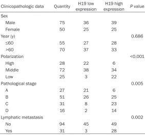
Expression of H19 mRNA and miR-17 mRNA in colon can-cer tissue and adjacent tis-sues and expression of H19 mRNA in colon cancer cells
The results of QPCR showed that (Figure 1A) the expres-sion of H19 mRNA in colon cancer tissues was signifi-cantly higher than that in adjacent tissues (P<0.05), and the difference was sta- tistically significant (P<0.05). The expression level of H19 in SW116 cells was the high-Table 1. Relationship between H19 expression and
clinicopatho-logical features of colon cancer
Clinicopathologic data Quantity expression H19 low expressionH19 high P value Sex
Male 75 36 39
Female 50 25 25
Year (y) 0.686
≤60 55 27 28
>60 70 37 33
Polarization <0.001
High 28 22 6
Middle 72 38 34
Low 25 3 22
Pathological stage 0.005
A 27 21 6
B 51 26 25
C 31 8 23
D 16 2 14
Lymphatic metastasis 0.002
No 94 45 49
[image:4.612.91.374.409.672.2]was statistically significant. These results sug-gested that H19 and miR-17 are highly expre- ssed in colon cancer. H19 and miR-17 played an important role in colorectal cancer. We se- lected colon cancer SW116 cells as further experimental cell lines.
Relationship between H19 expression and clinicopathological data of colon cancer
Statistical analysis showed that the express- ion level of H19 increased with the increase of pathological stage of colon cancer (Table 1). The expression level of H19 increased gradual-ly with the decrease of differentiation degree. The expression of H19 was significantly higher in colorectal carcinoma with the increase of
lymph node metastasis; H19 expression level has nothing to do with age and gender. The results showed that H19 was associated with pathological stage of colon cancer and lymph node metastasis, but not with sex, age and so on.
Effect of silencing H19 on invasion and migra-tion of human colon cancer cell line SW116
[image:5.612.92.517.83.519.2]Transwell results showed that: (Figure 2A) H19-siRNA group colon cancer cells SW116 Matrigel matrix by the number of cells was significantly increased compared with the NC group, the dif-ference was statistically significant (P<0.01), which indicated that H19-siRNA can inhibit the invasion of human colon cancer SW116 cells.
The results of scratches showed that the mobil-ity of H19-siRNA group was significantly lower than that of NC group at 24 h, 48 h and 72 h (P<0.01), indicating that H19-siRNA can inhibit the migration of human colon cancer SW116 cells.
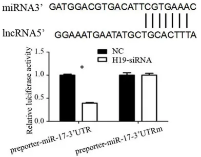
Relationship between H19 and miR-17 was detected by luciferase reporter gene
The use of bioinformatics predictors suggests that H19 may interact directly with miR-17 in order to clarify the situation of miRNAs associ-ated with H19. To demonstrate whether H19 binds to miR-17 3’UTR, we treated H19-siRNA with miR-17 co-transfected into colon cancer SW116 cells. Luciferase reporter gene results showed (Figure 3) that H19 significantly inhib-ited luciferase activity in miR-17. The results show that H19 could specifically bind to the 3’UTR of miR-17.
Effects of miR-17 on the invasion and migra-tion of human colon cancer SW116 cells after silencing H19
To clarify the effect of miR-17 on the invasion and migration of colon cancer cells, we used miR-17-mimic to overexpress miR-17 expres-sion, and to investigate the role of miR-17 in the invasion and migration of colon cancer cells. The Transwell results (Figure 4A) showed that the number of cells in the H19-siRNA + miR-17-inhibitor group was significantly increased by Matrigel matrix gel (P<0.01), indicating that overexpression of miR-17 can promote the invasion of human colon cancer SW116 cells after silencing H19.
The results of scratches (Figure 4B) showed that the mobility of H19-siRNA + miR-17-inhibi-tor group was significantly higher than that of H19-siRNA group at 24 h, 48 h and 72 h, the difference was statistically significant (P<0.01), indicating that overexpression of miR-17 can promote the migration of human colon cancer SW116 cells after silencing H19.
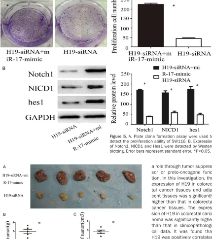
Effect of miR-17 on the proliferation of colon cancer cells after silencing H19 detected by plate cloning
The results of plate cloning showed (Figure 5A) that the number and size of H19-siRNA + miR-17-mimic cell clones were significantly increa- sed compared with H19-siRNA group, so the effect of H19 on colony cancer cell clone for- mation was dependent on the expression of miR-17.
Expression of Notch1, NICD1 and Hes1 pro-moted by silencing H19 and overexpression of miR-17
Researches have shown that Notch signaling pathways played a very important role in tumor invasion and migration. When Notch was acti-vated, it was enzymatically digested to form water-soluble NICD, which bind to the DNA binding protein CSL on the nucleus to form a transcription factor, thereby stimulating the transcription of the target gene Hes1 [12]. Western blotting showed that the expression of Notch1, NICD1 and Hes1 protein in H19-siRNA + miR-17-mimic group was significantly higher than that in H19-siRNA group, and the differ-ence was statistically significant (P<0.05), indi-cating that silencing H19, overexpression of miR-17 can promote the expression of Notch1, NICD1 and Hes1 and indicating that H19 and miR-17 can function through the Notch signal-ing pathway.
Impact of miR-17 on tumor growth detected in nude mice subcutaneous tumor after silencing H19
[image:6.612.92.288.73.226.2]Subcutaneous tumor 28 days after the death of the neck of the nude mice, the autopsy showed that the left armpit tumor growth, the tumor was gray white, solid, round or oval, the surface of nodular protrusions, profiles of fish samples, the rate of 100%.
Nude mice tumor growth (Figure 6A): the tumor size of H19-siRNA + miR-17-mimic group was significantly higher than that of H19-siRNA group. The difference was statistically signifi-cant (P<0.05). Comparison of tumor weight and volume (Figure 6B, 6C): the tumor volume and weight of H19-siRNA + miR-17-mimic group were significantly higher than those of H19-siRNA group. The difference was statistically significant (P<0.05).
Discussion
LncRNA has been considered to be no function, the researchers found more and more non-cod-ing RNA with the development of experimental
technology, especially the development of se- quencing technology. The length of more than 200 bt plays a very important role in the tran-scriptional regulation process. LncRNA as an important non-coding RNA, many molecular biological functions can be revealed. Resear- ches have shown that lncRNAs can be closely related to epigenetic modifications, transcrip-tional regulation, protein translation, and pro-tein degradation [13, 14].
[image:7.612.91.526.70.532.2]LncRNA H19 is transcribed by the H19 gene and is a lncRNA that is maternal in human tis-sue [15]. H10 has been found to be closely related to the process of emergence, develop-ment, invasion and migration of tumors. How-
ever, the specific mechanism of action in tu-
[image:8.612.89.519.87.572.2]mor is not clear. In different tumors, it may play ety of tumor cells to enhance the invasion and migration capacity. Leighton et al. [16] studies
[image:8.612.95.377.411.633.2]Figure 5. A. Plate clone formation assay were used to detect the proliferation ability of SW116. B. Expression of Notch1, NICD1 and Hes1 were detected by Western blotting. Error bars represent standard error. *P<0.05.
Figure 6. Effect of miR-17 on tumorigenesis after silencing H19 in vivo. A. Comparison of the tumor size of nude mice. B, C. Compare the volume and weight of the tumor in nude mice.
vari-have shown that H19 expression level and lung cancer staging, and H19 and lung cancer inva-sion depth and metastasis range was related. In this investigation, we found that the invasion and migration ability of colon cancer cells were weakened by silencing H19. The results of pre-vious experiments indicated that H19 played an important role in the development and pro-gression of colon cancer.
The research of LncRNA including bioinformat-ics needs to be combined with experimental validation and biochip screening combined with experimental validation at present. In this investigation, bioinformatics analysis confirm- ed that H19 and miR-17 have a direct interac-tion, further through the double luciferase reporter gene confirmed that H19 can be com-bined with miR-17 3’UTR. The expression level of miR-17 in colorectal cancer tissues and adja-cent tissues was detected and we found that miR-17 expression in colon cancer was signifi-cantly higher compared with the adjacent tis-sues. The miR-17~92 cluster is the first with the role of oncogene miRNA [17], consistent with the results of this investigation.
To further investigate the role of H19 and miR-17 in the biological function of colon cancer and the related mechanism, we used silencing H19, overexpressing miR-17 expression to fur-ther investigate the miR-17 in colon cancer cell invasion and migration process effect. The results showed that the expression of miR-17 overexpressing H19 could promote the prolif-eration, invasion and migration of colon cancer cells. Similar results were observed in vivo experiments using nude mice subcutaneously. Notch signaling pathway is a highly conserved signaling pathway in the process of biological evolution. Notch signaling pathway is closely related to biological behavior such as tumor proliferation, apoptosis, invasion and migration in the tumor. Yun et al. [18] found that MCF-7/ ADR had stronger ability to invade and migrate than MCF-7 compared to MCF-7 and MCF-7/ ADR while Notch-1 expression level is higher, indicating that the invasion and migration of breast cancer cells are associated with Notch-1. Jie et al. [19] found that Notch-1 expression was significantly increased with the progress of liver cancer staging by analyzing the different stages of liver cancer tissue and the high ex- pression of Notch-1 is closely related to lymph
node metastasis and distant metastasis. This investigation found that overexpression of miR-17 after silencing H19 revealed that overex-pression of miR-17 could promote the expres-sion of Notch1, NICD1 and Hes1, indicating that H19 and miR-17 may function through the Notch signaling pathway.
The expression of H19 and miR-17 in colon and adjacent tissues was detected by qPCR in this investigation. The relationship between H19 and miR-17 was further observed, and the effects of H19 and miR-17 on the proliferation, invasion and migration of colon cancer cells were further investigated. Researches have shown that H19 and miR-17 are up-regulated in colon cancer and H19 has a direct interaction with miR-17. H19 can target the invasion and migration of colon cancer by miR-17. H19 can regulate the expression of Notch1, NICD1 and Hes1, indicating indirectly that Notch signaling pathway plays a role in H19 regulation of miR-17 affect the biological function of colon can-cer cells. It suggests that H19 and miR-17 may be involved in the proliferation, invasion and migration of colon cancer cells, which may be a marker for predicting the progress of colon cancer, prognosis and monitoring of therapeu-tic effects.
Acknowledgements
Anhui Provincial Health Department Clinical Medicine Application Technology Project (20- 08B070).
Disclosure of conflict of interest
None.
Address correspondence to: Lian-Bang Zhou, De- partment of General Surgery, The Second Hospital of Anhui Medical University, 678 Fu Rong Road, Hefei, Anhui Province, China. Tel: +86-1515606- 0699; E-mail: [email protected]
References
[1] Schreuders EH, Ruco A, Rabeneck L, Schoen RE, Sung JJ, Young GP and Kuipers EJ. Colorec-tal cancer screening: a global overview of exist-ing programmes. Gut 2015; 64: 1637.
[3] Ricci-Vitiani L, Lombardi DG, Pilozzi E, Biffoni M, Todaro M, Peschle C and De MR. Identifica-tion and expansion of human colon-cancer-ini-tiating cells. Nature 2007; 445: 111-115. [4] Kogita A, Yoshioka Y, Sakai K, Togashi Y,
So-gabe S, Nakai T, Okuno K and Nishio K. Inter- and intra-tumor profiling of multi-regional co-lon cancer and metastasis. Biochem Biophys Res Commun 2015; 458: 52-56.
[5] Shen Y, Huang S and Xianrong HU. Clinical ef-fect of laparoscopic hepatectomy in treating liver metastasis of colon cancer: a systematic review and meta-analysis. Journal of Clinical Hepatology 2014; 30.
[6] Prensner JR and Chinnaiyan AM. The emer-gence of lncRNAs in cancer biology. Cancer Discov 2011; 1: 391.
[7] Li H, Yu B, Li J, Su L, Yan M, Zhu Z and Liu B. Overexpression of lncRNA H19 enhances car-cinogenesis and metastasis of gastric cancer. Oncotarget 2014; 5: 2318.
[8] Luo M, Li Z, Wang W, Zeng Y, Liu Z and Qiu J. Long non-coding RNA H19 increases bladder cancer metastasis by associating with EZH2 and inhibiting E-cadherin expression. Cancer Lett 2013; 333: 213-221.
[9] Byun HM, Wong HL, Birnstein EA, Wolff EM, Li-ang G and YLi-ang AS. Examination of IGF2 and H19 loss of imprinting in bladder cancer. Can-cer Res 2007; 67: 10753-10758.
[10] Reddy KB. MicroRNA (miRNA) in cancer. Can-cer Cell Int 2015; 15: 1-6.
[11] Gaidatzis D, Van NE, Hausser J and Zavolan M. Inference of miRNA targets using evolutionary conservation and pathway analysis. BMC Bio-informatics 2007; 8: 69.
[12] Zhang K, Zhang YQ, Ai WB, Hu QT, Zhang QJ, Wan LY, Wang XL, Liu CB and Wu JF. Hes1, an important gene for activation of hepatic stel-late cells, is regustel-lated by Notch1 and TGF-β/ BMP signaling. World J Gastroenterol 2015; 21: 878.
[13] Haerty W and Ponting CP. Mutations within ln-cRNAs are effectively selected against in fruit-fly but not in human. Genome Biol 2013; 14: R49.
[14] Li P, Yuan X, Jiang B, Tang Z and Li GC. Ln-cRNAs: key players and novel insights into cer-vical cancer. Tumor Biol 2016; 37: 2779-2788. [15] Kallen A, Zhou XB, Xu J, Qiao C, Ma J, Yan L, Lu L, Liu C, Yi JS and Zhang H. The imprinted H19 LncRNA antagonizes Let-7 MicroRNAs. Mol Cell 2013; 52: 101.
[16] Leighton PA, Saam JR, Ingram RS, Stewart CL and Tilghman SM. An enhancer deletion af-fects both H19 and Igf2 expression. Genes Dev 1995; 9: 2079-2089.
[17] Mendell JT. miRiad roles for the miR-17-92 cluster in development and disease. Cell 2008; 133: 217-222.
[18] Yun J, Espinoza I, Pannuti A, Romero D, Marti-nez L, Caskey M, Stanculescu A, Bocchetta M, Rizzo P and Band V. p53 modulates notch sig-naling in MCF-7 breast cancer cells by associ-ating with the notch transcriptional complex via MAML1. J Cell Physiol 2015; 230: 3115-3127.