Original Article
The expression of ERβ2, Bcl-xl and Bax in non-small cell
lung cancer and associated with prognosis
Junfeng Liu1*, Zhaoming Nie1*, Yiyan Lei2, Shibin Yang3, Zhaoguo Liu2
Departments of 1Pathology, 2General Thoracic Surgery, 3Gastrointestinal Surgery, The First Affiliated Hospital of
Sun Yat-sen University, Guangzhou, People’s Republic of China. *Equal contributors.
Received July 4, 2017; Accepted August 24, 2017; Epub September 1, 2017; Published September 15, 2017
Abstract: Lung cancer is now the leading cause of related death in the world, non-small lung cancer (NSCLC) in
pre-dominant type of lung cancer. In this study, we mainly discuss expression, distribution, and prognostic significance of ERβ2, Bcl-xl and Bax in NSCLC. The expression of ERβ2, Bcl-xl and Bax were detected by immunohistochemistry
(IHC), and then the staining was evaluated and correlated with clinic and pathologic characteristics, overall survival
(OS). ERβ2, Bcl-xl and Bax were localized in NSCLC, and they were over-expressed all in NSCLC (P<0.05) compared with BPL tissues. IHC results showed that ERβ2, Bcl-xl and Bax were not correlated with gender, age, smoking index, histological type, regional lymph node metastasis, whereas it was correlated with TNM staging of patients. In a Kaplan Meier analysis, the higher expression of ERβ2, Bcl-xl and Bax was correlated with good OS. ERβ2, Bcl-xl and Bax may be prognostic factors in NSCLC and useful to clinic trials.
Keywords: Estrogen receptors beta, non-small cell lung cancer, Bcl-xl, Bax
Introduction
Lung cancer is now the leading cause of can-cer-related mortality worldwide [1]. Non-small-cell lung cancer (NSCLC) is the most common type of the lung cancer. It accounts for about 85% of the lung cancer cases throughout the world and the five-year overall survival rate is almost 15% [2]. Through its therapy has seen much improvement, the prognosis of NSCLC patients remains poor. Even in early stages with no nodal or other metastatic involvement, there is little advancement in regards to distant recurrence and subsequent mortality [3]. In recent years, ERβ was found as an important maker in the disease progression of some can-cers, including some estrogen targeted organ cancers, such as prostate cancer and breast cancer, even some no estrogen targeted organ cancers, such as colon cancer and lung cancer [4-9]. Some studies suggested that ERβ may be a good prognosis maker for NSCLC, which is related to inhabit its progression [10, 11]. ERβ is one class of estrogen receptors that has five protein isoforms, including ERβ1, ERβ2, ERβ3, ERβ4 and ERβ5. The expression level, distribu-tion and funcdistribu-tion of them in organs are
differ-ent [8, 9, 12-14]. In the breast cancers, ERβ2 could predict better prognosis [15]. In our previ-ous studies, we found ERβ2 maybe had impor-tant function in NSCLC. But there are still some controversies on mechanisms of ERβ2 in tumors, which is necessary to be explored further.
non-squamous NSCLC [21]. Since anti-apoptotic proteins have always been described as being redundant, we underline the necessity of a bet-ter understanding of their relevance and com-mitment in lung cancers.
In our study, our study is mainly to detect the expression of ERβ2 and Bcl-xL, Bax via immu-nohistochemical staining, and observe the dis-tribution in NSCLC. We would analyze the rela-tionship between ERβ2, Bcl-xL and the pa- thology would be analyzed, the survival. The results obtained from this study maybe provide useful clinical data into the treatment in NSCLC.
Materials and methods
Human tissues
148 samples including 108 NSCLC (adenocar-cinoma (AC), squamous car(adenocar-cinoma (SC) and magnocellular carcinoma (MC)) and 40 benign pulmonary lung tissues (BPL) were taken upon surgical resection in the first affiliated hospital of Sun Yat-sen university from 2009 to 2011. All the samples were made into paraffin-em- bedded tissue specimens after normal dehy-dration and dehydehy-dration processing. All patients
selected had no liver, kidney or endocrine dis-eases, and had not received radiotherapy, che-motherapy or estrogen therapy before surgical resection. The relevant clinic and pathological data included patients’ age, gender; smoking index; tumor staging (TNM); histological type; regional lymph node metastasis; overall surviv-al (OS).
Reagents and materials
Anti-ERβ2 antibody and anti-Bcl-xL antibody were purchased from Cell Signaling Company (Danvers, MA, USA), and then anti-Bax antibody was purchased from Santa Cruz Company (California, USA). NovoLink Polymer Detection System was obtained from Leica Microsystems Company (Wetzlar, Germany). EDTA buffer (pH 8.0), PBS, 3% H2O2 reagent and other reagents were prepared by ourselves.
Immunohistochemical analysis
[image:2.612.94.524.72.368.2]heat-induced antigen retrieval with EDTA buffer (pH 8.0). Subsequently, the slides were blocked by 3% H2O2 reagent for 5 minutes, and then were stained by the NovoLink Polymer Detection System, according to the manufacturer’s proto-col. The main primary antibodies have been used: anti-Bax antibody, anti-ERβ2 antibody and anti-Bcl-xL. Negative controls were gener-ated by omitting the primary antibody.
Immunoreactive score
The immunoreactive score (IRS) was evaluated by three independent and experienced
examin-Spearman correlation S(r). Differences between high and low expression were compared through X2. Multiple linear regression analysis was used to analyze the relationship between ERβ2, Bcl-xl, Bax and the factors of clinical pathology.
OS were analyzed initially by Kaplan-Meier plots (Log-rank test). The P Value of <0.05 was con-sidered statistically significant.
Results
Expression of ERβ2, Bcl-xl and Bax in NSCLC and benign pulmonary tissues
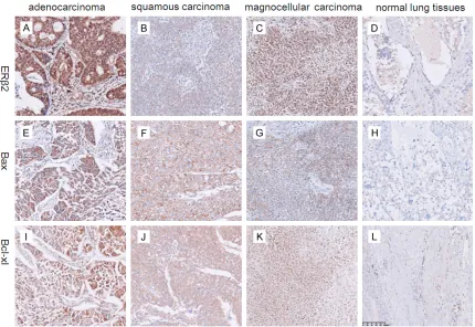
To assess the expression level of ERβ2, Bcl-xl and Bax, immunohistochemistry were used in 108 cases NSCLC and BPL paraffin-embedded tissues. Expression status was dichotomized as negative (Allred score <3) and positive (≥3) based on statistical assessment. Positive ERβ2 expression was observed in 61 out of 108 (44.48%) in NSCLC, whereas positive Bcl-xl and Bax expression was detected in 56 out of 108 (51.85%) and 49 out of 108 (45.37%) in NSCLC respectively. Compared with expression of ERβ2, Bcl-xl and Bax in BPL, it was significantly higher in NSCLC respectively (Figure 1).
Meanwhile, the correlations among ERβ2, Bcl-xl and Bax were analyzed. The analysis result showed that there was a significant positive correlation between ERβ2 and Bcl-xl in NSCLC, and the similar results were observed between ERβ2 and Bax, Bcl-xl and Bax (Table 1).
Expression of ERβ2, Bcl-xl and Bax in relation to clinic and pathologic characteristics
[image:3.612.91.346.97.217.2]The correlations between the expression of ERβ2, Bcl-xl and Bax and clinic and pathologic Table 1. The different analyses of the ERβ2, Bcl-xl and in
Bax NSCLC and BPL expression
Allred score ERβ2 Bcl-xl Bax
NSCLC BPL NSCLC BPL NSCLC BPL
- 47 31 52 27 59 34
+ 24 5 23 7 18 4
++ 21 3 18 4 15 1
+++ 16 1 15 2 16 1
Positive Rate (%) 44.48 22.50 51.85 32.50 45.37 15.00
X2 Value 13.522 4.393 11.529
P Value <0.001 0.027 0.001
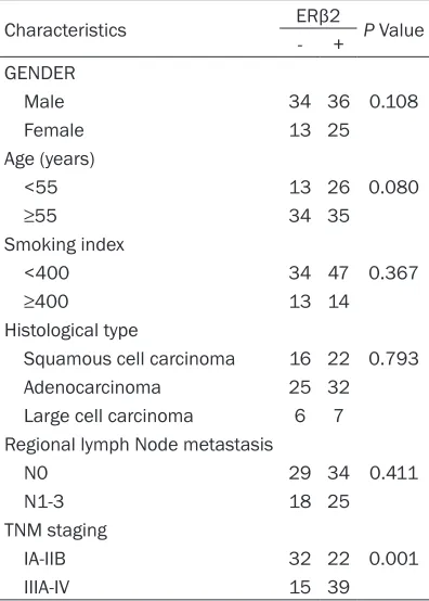
Table 2. Correlation of cytoplasm and nuclear ERβ2 with clinic pathologic parameters
Characteristics ERβ2 P Value
- +
GENDER
Male 34 36 0.108
Female 13 25
Age (years)
<55 13 26 0.080
≥55 34 35
Smoking index
<400 34 47 0.367
≥400 13 14 Histological type
Squamous cell carcinoma 16 22 0.793
Adenocarcinoma 25 32
Large cell carcinoma 6 7 Regional lymph Node metastasis
N0 29 34 0.411
N1-3 18 25
TNM staging
IA-IIB 32 22 0.001
IIIA-IV 15 39
ers. Expression status was dichoto-mized using Allred score >3 as cutoff. Cytoplasmic and nuclear staining was determined, where for negative -; + for weak; ++ for moderate; +++ for strong staining.
Statistical analysis
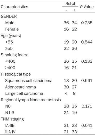
[image:3.612.91.289.263.540.2]characteristics were analyzed. The expression of ERβ2 was not correlated with gender, age, smoking index, histological type, regional lymph node metastasis, whereas it was correlated with TNM staging of patients (Table 2). The sim-ilar results of were Bcl-xl and Bax obtained simultaneously, the expression of Bcl-xl and Bax was not associated with any other known clinic pathologic indicators except TNM staging of patients (Tables 3, 4). Spearman correlation analysis showed that the expression of ERβ2, Bcl-xl and Bax were correlative (Table 5).
Correlation of the expression of ERβ2, Bcl-xl and Bax with OS
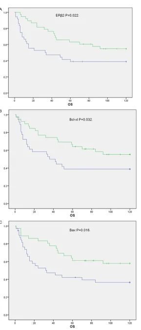
Kaplan-Meier survival analysis was used to assess survival with respect to ERβ2, Bcl-xl and Bax expression. Kaplan-Meier survival analysis showed that ERβ2 higher expression was si- gnificantly associated with the better OS (P= 0.022), and the results of Bcl-xl and Bax expres-sion were similar (P=0.032 and 0.016) (Figure 2).
Discussion
Lung cancer is the most malignancy in many countries and it has become one cause of can-cer death in world, surpassing gastric cancan-cer [22, 23]. Lung cancer has two types, one is small lung cancer, the other is non-small lung cancer (NSCLC). As we know, NSCLC is now the predominant type of lung cancer, which includes lung adenocarcinoma, lung squamous carcino-ma and large cell carcinocarcino-ma. In the previous articles, we have reported the role of estrogen receptor β2 (ERβ2) in the progress of non-small lung cancer, but we have not found the associa-tions between ERβ2 and other proteins [24].
[image:4.612.91.288.96.375.2]In this study, we mainly discuss the associa-tions ERβ2 and the other two proteins, and this is firstly aimed at elucidating the expression and prognostic of ERβ2, bcl-xl and bax in NS- CLC. Our results suggest the importance of evaluating ERβ2, bcl-xl and bax immunoreac- Table 3. Correlation of cytoplasm and nuclear
ERβ2 with clinic pathologic parameters
Characteristics Bax P Value
- +
GENDER
Male 38 32 0.542
Female 21 17
Age (years)
<55 20 19 0.372
≥55 38 30
Smoking index
<400 43 38 0.370
≥400 16 11 Histological type
Squamous cell carcinoma 20 18 0.941
Adenocarcinoma 31 26
Large cell carcinoma 7 5 Regional lymph Node metastasis
N0 35 28 0.569
N1-3 23 19
TNM staging
IA-IIB 35 19 0.026
IIIA-IV 24 30
Table 4. Correlation of cytoplasm and nuclear ERβ2 with clinic pathologic parameters
Characteristics Bcl-xl P Value
- +
GENDER
Male 36 34 0.235
Female 16 22
Age (years)
<55 19 20 0.544
≥55 22 36
Smoking index
<400 36 35 0.133
≥400 16 21 Histological type
Squamous cell carcinoma 18 20 0.561
Adenocarcinoma 30 27
Large cell carcinoma 4 9 Regional lymph Node metastasis
N0 28 35 0.171
N1-3 24 19
TNM staging
IA-IIB 31 23 0.041
[image:4.612.322.521.98.135.2]IIIA-IV 21 33
Table 5. Spearman correlation S(r) between expression of ERβ2, bcl-xl and Bax
Patients ERβ2 vs. Bcl-xl Bcl-xl vs. Bax Bax vs. ERβ2
[image:4.612.91.287.426.703.2]Figure 2. Evaluation of ERβ2, Bcl-xl and Bax as a predictor for OS by the Kaplan-Meier (KM) plot.
tivity as they have important and distinct prognostic impli- cations.
Our data shows that ERβ2, bcl-xl and bax were expressed in NSCLC respectively. Compared with BPL tissues, ERβ2, bcl-xl and bax were over-expressed in NSCLC obviously through the immunohistochemistry. Further statistics by Spearman correlation method, we found there is significant association between ERβ2 and bcl-xl, bax respectively. This phenome-non suggests that ERβ2 not only is a prognostic marker, but also maybe involve in the progress of NSCLC by very complicated mechanisms. In our discovery, it was interest-ing that expression of ERβ2 was associated with both bcl-xl and bax, which were known as anti-apoptotic factors. It suggested that expression of ERβ2 may be associated with anti-apoptosis mechanism of cancer cells, but there was no more clear data about this finding now.
predictors of OS in NSCLC. They were signifi-cant associated with good OS.
In summary, we consider that expression ERβ2, BCL-xl and Bax can provide us help on the role and relationship with other makers of ERβ2. Our data shows that ERβ2, BCL-xl and Bax may be prognostic factors in NSCLC, though there still were many un-revealed questions, in par-ticularity, the mechanism of them. These prog-nostic makers could play an important role in stratifying patients in clinical trials through ana-lyzing more adequate cases.
Acknowledgements
This study was support by the National Natural Science Foundation of China (No.81501964).
Disclosure of conflict of interest
None.
Address correspondence to: Zhaoguo Liu, Depart-ment of General Thoracic Surgery, The First Affiliated Hospital of Sun Yat-sen University, 58 Zhongshan 2nd Road, Guangzhou 510080, People’s Republic of
China. E-mail: [email protected]
References
[1] Pietras RJ, Marquez DC, Chen HW, Tsai E, Weinberg O, Fishbein M. Estrogen and growth
factor receptor interactions in human br- east and non-small cell lung cancer cells. Ste-roids 2005; 70: 372-81.
[2] Goldstraw P, Ball D, Jett JR, Le Chevalier T, Lim E, Nicholson AG, Shepherd FA. Non-small-cell
lung cancer. Lancet 2011; 378: 1727-40. [3] Jemal A, Siegel R, Xu J, Ward E. Cancer
statis-tics, 2010. CA Cancer J Clin 2010; 60: 277-300.
[4] Yang XR, Pfeiffer RM, Garcia-Closas M, Rimm DL, Lissowska J, Brinton LA, Peplonska B, Hewitt SM, Cartun RW, Mandich D, Sasano H, Evans DB, Sutter TR, Sherman ME. Hormonal markers in breast cancer: coexpression, rela -tionship with pathologic characteristics, and
risk factor associations in a population-based
study. Cancer Res 2007; 67: 10608-17. [5] Henschke CI, Yip R, Miettinen OS. Women’s
susceptibility to tobacco carcinogens and
sur-vival after diagnosis of lung cancer. JAMA
2006; 296: 180-4.
[6] Stabile LP, Davis AL, Gubish CT, Hopkins TM, Luketich JD, Christie N, Finkelstein S, Siegfried JM. Human non-small cell lung tumors and
cells derived from normal lung express both estrogen receptor alpha and beta and show biological responses to estrogen. Cancer Res 2002; 62: 2141-50.
[7] Niikawa H, Suzuki T, Miki Y, Suzuki S, Nagasaki S, Akahira J, Honma S, Evans DB, Hayashi S, Kondo T, Sasano H. Intratumoral estrogens
and estrogen receptors in human non-small cell lung carcinoma. Clin Cancer Res 2008; 14: 4417-26.
[8] Hershberger PA, Vasquez AC, Kanterewicz B, Land S, Siegfried JM, Nichols M. Regulation of
endogenous gene expression in human non-small cell lung cancer cells by estrogen recep-tor ligands. Cancer Res 2005; 65: 1598-605. [9] Hammoud Z, Tan B, Badve S, Bigsby RM. Estro
-gen promotes tumor progression in a -
geneti-cally defined mouse model of lung adenocarci -noma. Endocr Relat Cancer 2008; 15: 475-83. [10] Roman-Blas JA, Castaneda S, Largo R, Herre
-ro-Beaumont G. Osteoarthritis associated with estrogen deficiency. Arthritis Res Ther 2009;
11: 241.
[11] Davydov MI, Bogush TA, Polotskii BE, Tiulian -din SA. [Estrogen receptors beta--new target in cellular lung cancer treatment]. Vestn Ross
Akad Med Nauk 2012; 16-22.
[12] Moore JT, McKee DD, Slentz-Kesler K, Moore LB, Jones SA, Horne EL, Su JL, Kliewer SA, Lehmann JM, Willson TM. Cloning and charac
-terization of human estrogen receptor beta isoforms. Biochem Biophys Res Commun
1998; 247: 75-8.
[13] Shaaban AM, Green AR, Karthik S, Alizadeh Y, Hughes TA, Harkins L, Ellis IO, Robertson JF, Paish EC, Saunders PT, Groome NP, Speirs V.
Nuclear and cytoplasmic expression of
ERbe-ta1, ERbeta2, and ERbeta5 identifies distinct
prognostic outcome for breast cancer patients. Clin Cancer Res 2008; 14: 5228-35.
[14] Yan M, Rayoo M, Takano EA, Fox SB. Nuclear
and cytoplasmic expressions of ERbeta1 and ERbeta2 are predictive of response to therapy and alters prognosis in familial breast cancers.
Breast Cancer Res Treat 2011; 126: 395-405.
[15] Chi A, Chen X, Chirala M, Younes M. Differen -tial expression of estrogen receptor beta iso-forms in human breast cancer tissue. Antican-cer Res 2003; 23: 211-6.
[16] Scherr AL, Gdynia G, Salou M, Radhakrishnan P, Duglova K, Heller A, Keim S, Kautz N, Jasso
-wicz A, Elssner C, He YW, Jaeger D, Heiken
-walder M, Schneider M, Weber A, Roth W, Schulze-Bergkamen H, Koehler BC. Bcl-xL is an
oncogenic driver in colorectal cancer. Cell
Death Dis 2016; 7: e2342.
[17] Cory S, Adams JM. The Bcl2 family: regulators
[18] Manne U, Myers RB, Moron C, Poczatek RB, Dillard S, Weiss H, Brown D, Srivastava S, Griz
-zle WE. Prognostic significance of Bcl-2 expres -sion and p53 nuclear accumulation in colorec-tal adenocarcinoma. Int J Cancer 1997; 74: 346-58.
[19] Krajewska M, Krajewski S, Epstein JI, Shabaik
A, Sauvageot J, Song K, Kitada S, Reed JC. Im-munohistochemical analysis of 2, bax, bcl-X, and mcl-1 expression in prostate cancers. Am J Pathol 1996; 148: 1567-76.
[20] Jin-Song Y, Zhao-Xia W, Cheng-Yu L, Xiao-Di L, Ming S, Yuan-Yuan G, Wei D. Prognostic signifi
-cance of Bcl-xL gene expression in human
colorectal cancer. Acta Histochem 2011; 113: 810-4.
[21] Jeong SH, Lee HW, Han JH, Kang SY, Choi JH,
Jung YM, Choi H, Oh YT, Park KJ, Hwang SC,
Sheen SS, Oh YJ, Kim JH, Lim HY. Low
expres-sion of Bax predicts poor prognosis in resected
small cell lung cancer patients with non-squamous histology. Jpn J Clin Oncol 2008; 38: 661-9.
[22] Bae JM, Won YJ, Jung KW, Suh KA, Yun YH, Shin MH, Ahn YO, Lee DH, Shin HR, Ahn DH, Oh DK, Park JG. Survival of Korean cancer pa
-tients diagnosed in 1995. Cancer Res Treat
2002; 34: 319-25.
[23] Spira A, Ettinger DS. Multidisciplinary manage
-ment of lung cancer. N Engl J Med 2004; 350:
379-92.
[24] Liu Z, Liao Y, Tang H, Chen G. The expression of estrogen receptors beta2, 5 identifies and is
associated with prognosis in non-small cell lung cancer. Endocrine 2013; 44: 517-24. [25] Thongon N, Boonmuen N, Suksen K, Wichit P,
Chairoungdua A, Tuchinda P, Suksamrarn A,
Winuthayanon W, Piyachaturawat P. Selective
estrogen receptor modulator (SERM)-like ac -tivities of diarylheptanoid, a phytoestrogen from curcuma comosa, in breast cancer cells, pre-osteoblast cells, and rat uterine tissues. J Agric Food Chem 2017; 65: 3490-3496. [26] Groeger AM, Esposito V, De Luca A, Cassandro
R, Tonini G, Ambrogi V, Baldi F, Goldfarb R, Mineo TC, Baldi A, Wolner E. Prognostic value
of immunohistochemical expression of p53,
bax, Bcl-2 and Bcl-xL in resected non-small-cell