Case Report
Brunner’s gland adenoma of duodenum:
report of two cases
Li Lu, Ruotong Li, Guojing Zhang, Zhicheng Zhao, Weihua Fu, Weidong Li
Institute of General Surgery, Tianjin Medical University General Hospital, Tianjin 300052, China Received March 27, 2015; Accepted May 20, 2015; Epub June 1, 2015; Published June 15, 2015
Abstract: Brunner’s gland adenoma is a rare tumor of the duodenum and might also be an unusual cause of gas-trointestinal bleeding or obstruction. The pathogenesis of Brunner gland hamartoma of the duodenum is unknown. We report two cases of Brunner’s gland adenoma. Surgical resection was carried out because the tumor size was big in both cases and one accompanied with bleeding. Pathological examination revealed submucosal nodular hyperplasia of the Brunner’s glands.
Keywords: Brunner’s gland, adenoma, duodenum
Introduction
Brunner’s gland adenoma, also known as Brunner’s hamartoma, is a rare duodenal lesion that comprises not more than 5% of benign duodenal tumors. Most Brunner’s gland adeno-ma is sadeno-mall size adeno-masses and adeno-many patients are asymptomatic. Occasionally, they may be large in size with clinical manifestations of hemor-rhage or obstruction. According to our experi-ence here are 2 cases of Brunner’s gland ade-noma reported with the literature review. Case report
Case 1
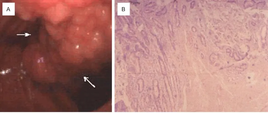
A 43-year-old Chinese man presented with complains of melena for 10-days. He did not have symptoms of haematemesis and epigas-tralgia. There is no history of weight loss and he has no family history of gastrointestinal diseas-es. During the physical examination, he had normal vital signs. No abnormalities detected in the abdominal examination. Laboratory data showed patient was anemic with hemoglobin level of 7.8 g/dL. In routine stool blood test, occult blood was positive. On upper endoscopy, a 2.5 cm large, pedunculated, polypoid mass with ulcerated crater was found in the first por-tion of the duodenum (Figure 1A). EUS showed that tumor was originated from muscularis
pro-pria (Figure 1B). Multiple biopsy specimens were obtained, which later revealed normal mucosa and regenerative changes. Contrast-enhanced computed tomography scan showed a polypoid mass arising from the first portion of the duodenum, extending about 5cm in length to the second portion (Figure 1C and 1D). Following the preoperative check up, explorato-ry laparotomy was done. Duodenum was mobi-lized with Kocher maneuver. Pylorus identified, longitudinal incision made on the pylorus ring for duodenal mucosa exploration. During the exploration a mobile pednculated mass was palpated, with a fixed base in the anterior part of duodenum (Figure 2A) and length 7 cm. Mass was excised from the base and duodenal mucosa repaired. Finally pyloroplasty was done. The gross surgical specimen showed a large duodenal lesion measuring 7.3×3.4×2.9 cm (Figure 2A and 2B). Pathological examination revealed packed Brunner’s glands and ducts admixed with smooth muscle (Figure 2C). No signs of malignancy or dysplasia were found. The postoperative days were uneventful and patient was discharged.
Case 2
com-plaining of fever and leukocytopenia. During the process of routine examination most of her results were found to be within normal limits. But in upper gastrointestinal endoscopy, a large, friable, ulcerated mass of 2 cm was noted incidentally in the second part of duode-nal papilla. There were no signs of active bleed-ing and the biopsy of the lesion was taken. Histological result revealed normal mucosa with no abnormal finding. Then the patient was transferred to Surgery Department.
Patient was planned for exploratory laparotomy and resection of the tumor. With Kocher maneu-ver, first and second portions of duodenum mobilized. Longitudinal incision made at the antimesenteric duodenum wall. Inner mucosa and papilla was exposed. Mass was identified
with its base related with the papilla (Figure 3A). Resection of the mass and sphinteroplasty of the papilla were done. Incised duodenum wall closed transversely. On histological exami-nation, the resected specimen predominantly composed of hyperplasia of Brunner’s glands. Gland hyperproliferation extending beyond muscularis mucosae reaching lower portion of duodenal villi with irregular and squat profile of crowded architecture and features consistent with a Brunner’s gland hamartoma (Figure 3B). She did well and was discharged on the 2 weeks after surgery.
Discussion
[image:2.612.90.525.71.446.2]Primary duodenal tumors are rare, accounting less than 1% among the total gastrointestinal
tumors, and benign tumors are only 16% of all benign tumors of the small intestine [1, 2]. Since Curveilhier [3] described the first case of benign duodenal Brunner’s gland adenoma in 1835, sporadic reports have been recorded (about 100 cases). In 1688, Brunner gave a precise anatomic description of the duodenal submucosal glands and coined the term “pan-creas secundarium”. In 1846, Middeldorpf cor-rected these glands’ name as Brunner’s glands. Brunner’s glands are submucosal mucin-secreting glands. They are predominantly local-ized in the duodenal bulb and proximal duode-num and progressively decrease in size and number in the distal portions. Brunner’s glands secrete an alkaline fluid composed of viscous mucin to protect the duodenal epithelium from acid chyme of the stomach.
The etiology of Brunner’s gland adenoma remains obscure. It has been postulated that
[image:3.612.97.521.72.170.2]an increased gastric acid secretion could stim-ulate these structures to undergo hyperplasia [4]. Franzin et al. [5] have reported an associa-tion between Brunner’s gland adenoma and hyperchlorhydria in patients with chronic gas-tric erosions and duodenal ulcers, but Spellberg et al. [6] have not found regression of the lesion with acid secretion inhibitors. Some scholars think that loss of alkaline protection normally provided by the exocrine pancreas would have led to a compensatory hyperplasia of the Brunner glands with increased production of mucus and alkali. Stolte et al. [7] examined 105 duodenopancreatectomy specimens sh- owing that 75.7% of chronic pancreatitis was associated with diffuse Brunner gland hyper-plasia. In our 2 cases, Brunner gland hyperpla-sia was not associated to chronic pancreatitis. But this mechanism does not explain the hyper-plasia of other mesenchymal components such as smooth muscle, Paneth cells and adipose
Figure 2. A. A photograph of operation field demonstrate a lesion in duodenum, measuring it’s length approximately 7 cm. B. The gross surgical specimen showed a large duodenal lesion measuring 7.3×3.4×2.9 cm. C. Pathological examination revealed packed Brunner’s glands and ducts admixed with smooth muscle.
[image:3.612.90.522.234.416.2]tissue. Another hypothesis suggests that Helicobacter pylori (H pylori) infection may play a role in the pathogenesis of Brunner’s gland adenoma [8]. In a recent study, H pylori infec-tion was found in five out of seven (71%) Brunner’s gland adenoma cases [9]. In our two patients, there were no H pylori infection found. The extreme rarity of Brunner’s gland adenoma and the high prevalence of H pylori infection in general population do not allow us to draw a clear pathogenetic relation. At present, the most accredited pathogenetic hypothesis remains that Brunner’s gland adenoma is a duodenal dysembryoplastic lesion or hamarto-ma [10].
Most patients with Brunner’s gland adenoma are asymptomatic or have nonspecific com-plaints such as nausea, bloating, or vague abdominal pain [11, 12]. In these cases, the lesion is usually an incidental finding detected during endoscopy or imaging studies. The most common presentations in symptomatic pa- tients are gastrointestinal bleeding and ob- structive symptoms. Levine et al. [13] studied the characteristics of a group of 27 patients with a Brunner’s gland adenoma. They found that the majority of patients with tumor-related blood loss had melena and showed evidence of chronic bleeding with ulceration of the majority of these tumors (37%), patients who presented with obstructive symptoms (37%). They also found that asymptomatic patients had smaller lesions (mean, 1.6 cm), patients with obstruc-tive and bleeding symptoms had similar-sized lesions (mean, 2.1 and 2.8 cm, respectively) [13]. On rare occasions, patients can present with gastric outlet obstruction [14]. In our two cases, there were no symptoms of gastrointes-tinal obstruction but one of them with gastroin-testinal bleeding.
At present diagnosis of Brunner’s gland adeno-ma is not always easy before operation. Large adenomas may be detected by ultrasonogra-phy and Computed tomograultrasonogra-phy [15]. CT is also useful to confirm the absence of extra-luminal extension of Brunner’s gland adenoma [16]. Smooth-walled polypoid filling defects may be seen in the duodenal bulb or corresponding portion of the duodenum on upper gastrointes-tinal barium studies [10]. But Radiological find-ing are often non-specific indeed, the duodenal filling defect can mimic several other lesions, such as leiomyoma, lipoma or lymphoma [16].
Endoscopy can localizes the lesion; however, biopsies are usually negative or reveal only Brunner’s gland hyperplasia. Only a deep endo-scopic or a surgical biopsy provides adequate tissue because the Brunner’s gland prolifera-tions may be covered by normal mucosa [17, 18]. In our cases, diagnosis was made on surgi-cal specimen of duodenal mass, because Brunner’s gland hyperproliferation was extend-ed beyond muscularis mucosae reaching lower portion of duodenal villi. Endoscopic ultrasound (EUS) is helpful in assessing the origin, extent, and vascularity of these suspected submuco-sal lesions. Hizawa and coworkers described EUS features seen on 6 cases of Brunner gland adenoma, which were of a heterogeneous solid or cystic mass within the submucosa [19]. On histology, the duodenal cysts invade into the muscularis propria. They were lined by cylindric epithelial cells, and in one of them, the epithe-lium was pseudostratified. There was no evi-dence of malignancy.
Therefore, it is still controversial whether as- ymptomatic small Brunner’s gland adenoma found incidentally needs to be removed. Some people think that it only needs follow-up, where-as others suggested that it should undergo endoscopic excision in order to prevent compli-cations. For symptomatic Brunner’s gland ade-noma they usually need surgical treatment. In our two patients, surgical resection was carried out because size of the tumor was big in both cases and one accompanied with bleeding. In our view, conservative treatment with endo-scopic polypectomy or limited surgical resec-tion is appropriate. Removal of the suspected Brunner’s gland adenoma is recommended to both, confirm the diagnosis as well as to avoid potential complications including obstruction and bleeding. There have been no reports of recurrence after either endoscopic or surgical resection. Generally, they are benign and have a good prognosis [17].
Disclosure of conflict of interest
None.
References
[1] Van de Walle P, Dillemans B, Vandelanotte M, Proot L. The laparoscopic resection of a benign stromal tumour of the duodenum. Acta Chir Belg 1997; 97: 127-9.
[2] Ohba R, Otaka M, Jin M, Odashima M, Matsu-hashi T, Horikawa Y, Hatakeyama N, Mimori N, Kinoshita N, Koizumi S, Takahashi T, Wata-nabe S. Large Brunner’s gland hyperplasia treated with modified endoscopic submucosal dissection. Dig Dis Sci 2007; 52: 170-2. [3] Cruveilhier J. Anatomy of the Human Body.
New York, NY: Harper and Bros; 1844. [4] Peetz ME, Moseley HS. Brunner’s glands
hy-perplasia. Am Surg 1989; 55: 474-7.
[5] Franzin G, Musola R, Ghidini O, Manfrini C, Fratton A. Nodular hyperplasia of Brunner’s glands. Gastrointest Endosc 1985; 31: 374-8. [6] Spellberg MA, Vucelic B. A case of Brunner’s
glands hyperplasia with diarrhoea responsive to cimetidine. Am J Gastroenterol 1980; 73: 519-22.
[7] Stolte M, Schwabe H, Prestele H. Relationship between diseases of the pancreas and hyper-plasia of Brunner’s glands. Virchows Arch A Pathol Anat Histol 1981; 394: 75-87.
[8] Kurella RR, Ancha HR, Hussain S, Lightfoot SA, Harty R. Evolution of Brunner gland hamarto-ma associated with Helicobacter pylori infec-tion. South Med J 2008; 101: 648-50. [9] Kovacevic I, Ljubicic N, Cupic H, Doko M, Zovak
M, Troskot B, Kujundzić M, Banić M. Helico -bacter pylori infection in patients with Brun-ner’s gland adenoma. Acta Med Croatica 2001; 55: 157-60.
[10] Gao YP, Zhu JS, Zheng WJ. Brunner’s gland ad-enoma of duodenum: a case report and litera-ture review. Word J Gastroenterol 2004; 10: 2616-7.
[11] Singla R, Bharti P, Jain R , Kumar S, Ganguly KK, Kar P. Giant Brunner gland adenoma man-ifesting as iron deficiency anaemia and intus -susception. Natl Med J India 2010; 23: 376-7. [12] Yadav D, Hertan H, Pitchumoni CS. A giant
Brunner gland adenoma presenting as gastro-intestinal hemorrhage. J Clin Gastroenterol 2001; 32: 448-50.
[13] Levine JA, Burgart LJ, Batts KP, Wang KK. Brunner’s gland hamartomas: clinical presen-tation and pathologic features of 27 cases. Am J Gastroenterol 1995; 90: 290-4.
[14] El Faleh I, Lutz N, Osterheld MC, Reinberg O, Nydegger A. Gastric outlet obstruction by Brun-ner’s gland hyperplasia in an 8-year-old child. J Pediatr Surg 2009; 44: E21-4.
[15] Bastlein C, Decking R, Voeth C, Ottenjann R. Giant brunneroma of the duodenum. Endos-copy 1988; 20: 154-5.
[16] Merine D, Jones B, Ghahremani GG, Hamilton SR, Bayless TM. Hyperplasia of Brunner gl- ands: the spectrum of its radiographic mani-festations. Gastrointest Radiol 1991; 16: 104-8.
[17] Krishnamurthy P, Junaid O, Moezzi J, Ali SA, Gopalswamy N. Gastric outlet obstruction caused by Brunner’s gland hyperplasia: case report and review of literature. Gastrointest Endosc 2006; 64: 464-7.
[18] Walden DT, Marcon NE. Endoscopic injection and polypectomy for bleeding Brunner gland hamartoma: case report and expanded litera-ture review. Gastrointest Endosc 1998; 47: 403-7.