Original Article
Expression of osteoprotegerin, RNAK and RANKL genes
in femoral head avascular necrosis and
related signaling pathway
Qingtang Miao, Sibin Hao, Hongmei Li, Fang Sun, Xueling Wang
Department of Orthopedics, Zhangqiu People’s Hospital, Jinan 250200, Shandong, China
Received June 22, 2015; Accepted July 27, 2015; Epub September 1, 2015; Published September 15, 2015
Abstract: Femoral head avascular necrosis (AVN) causes the damage of hip joint and related dysfunctions, thus
consisting of a clinical challenge. Osteoprotegerin (OPG), receptor activator of nuclear factor κB (RANK) and its ligand (RANKL) all regulate the formation of bones via gene transcriptional regulation for the balance between osteoblasts and osteoclasts. This study thus investigated the expressional profiles of OPG, RANK and RANKL genes
in AVN patients, and explored related molecular mediating pathways. Real-time qPCR was used to measure the
gene expression of OPG, RANK and RANKL genes in AVN femoral head tissue samples from 42 patients, along with normal tissues. Western blotting analysis was performed to quantify protein levels of OPG and RANKL. There was a trend but not statistically significant elevation of mRNA levels of OPG in femoral head AVN tissues compared to
normal tissues (P>0.05). The expression of RNAK and RNAKL, however, was significantly elevated in necrotic tissues
(P<0.05). No significant difference in protein levels of OPG or RANKL between groups. The expression of OPG, RANK and RANKL genes exert a crucial role in the progression of AVN, suggesting their roles in mediating bone homeosta -sis and potential effects on bone destruction.
Keywords: Femoral head avascular necrosis, osteoprotegerin, receptor activator of nuclear factor κB, ligand for receptor activator of nuclear factor κB, gene expression regulation
Introduction
Femoral head avascular necrosis (AVN) is a complicated and common disease in orthope-dics. Current intervention methods had limited treatment efficacy, thus frequently causing the damage of femoral head and degradation of hip joints [1]. AVN can be further aggravated by the use of steroid, alcoholic intake, systemic lupus erythematosus, sickle cell anemia, meta-bolic disorders, tumors or trauma [2, 3]. The body’s repaired mechanism may function as the vessel proliferation and temporally active osteoclasts at the necrotic lesion and adjacent tissues at the early stage of AVN. Such self-repair, however, had limited effects as the bone formation by osteoblasts will be inhibited, along with the persistent activation of osteoclasts, which finally lead to the degradation of chon -drocytes and breakage of joint surface [4-6]. The dynamic balance between osteoblasts and osteoclasts is mediated by various molecular
Materials and methods
Patient information
Tissue samples were collected from a total of 44 femoral head AVN patients, all of which received the total hip arthroplasties (THA) in our hospital. Among all patients, two individu-als were excluded due to the lower protein con-tents in their samples. The condition of femoral head AVN was determined to be grade III or grade IV based on association research circula-tion osseous (ARCO) standards. This study has been approved by the ethical committee of our hospital. Written consents have been obtained from patients and their families.
Sample collection
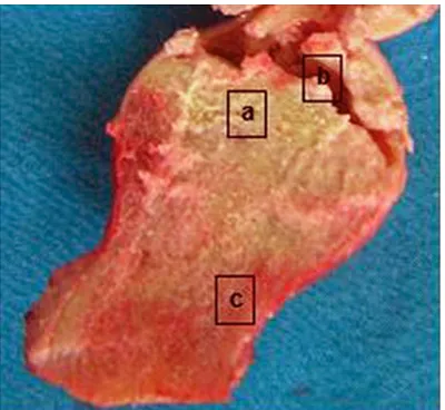
The femoral head sample collected from the surgery was longitudinally divided into two parts. Total RNA was extracted from both necrotic and normal tissues at comparable sites. The morphology of sectioning surface was recorded during the surgery for the deter-mination of the borderline between necrotic and normal tissues (Figure 1). Necrotic bone tissues were samples from inferior chondro-cytes (about 1~3 mm below the cartilage). Normal tissues were collected from the head/ neck of the femoral, with more than 1 cm away from the borderline. Collected tissues were rinsed in 0.9% saline for 5 min to clear bone marrow and residual blood. RNA and proteins were then extracted as reported previously [1].
In brief, fresh tissue samples were lysed in Trizol reagents (Invitrogen, UK) for total RNA extraction following manual instructions. RNA was then purified by RNeasy Mini-kit (Qiagen, Germany) and analyzed by 1% agarose gel elec-trophoresis and UV spectrophotometer. The optical density (OD) ratio at 260/280 nm was between 1.8 and 2.0, suggesting highly purified RNA. We further extracted and cultured cells from seven necrotic tissues along with normal controlled tissues following established proto-cols [12]. The staining of cultured cells by 85L-2 kit (Sigma-Aldrich, UK) also revealed the exis -tence of living osteoblasts even in necrotic tis-sues (Figure 2).
Real-time qPCR
To quantify the gene expression of OPG, RANK and RANKL genes, we employed fluorescent qPCR technique. The in vitro reverse transcrip-tion was performed using RNA as the template. PCR system contained 2 μL cDNA template, 0.5 μM specific primers, 1 mM MgCl2 and 2 μL poly -merase. The parameters were: initial denature (95°C, 10 min); 45 cycles containing denature (95°C, 15 sec), annealing (54°C, 10 sec) and elongation (72°C, 16 sec). House-keeping gene with stable transcription levels such as h-PBGD gene was used as the reference to quantify the expression level of target genes. A cycle thresh-old (Ct) value for cDNA copy number was deter-mined from the standard curve of each gene and the reference. The relative expression level was determined by 2-ΔΔCt method.
DNA oligonucleotide primers and hybridization probes were synthesized by TIB Molbiol (Germany). Fluorescent labeling was conjugat-ed on the adjacent sites of hybridization probe. In brief, recipient fluorescent molecule LC Red 640 labeled the 5’-end of the first probe, while fluorescein (FITC, 3FL) was used to label the 3’-end in the second probe. The probe wit 5’-label prevents the polymerase elongation during 3’-phosphrylation in PCR. Sequences of primers and hybridization probes were listed in Table 1. The lengths of PCR products for OPG, RANKL and RANK genes were 147 bp, 164 bp and 132 bp, respectively.
Western blotting
Both necrotic and normal tissues were firstly rinsed by PBS and were then lysed using NET-Figure 1. Femoral head tissue sample. a. Necrotic
[image:2.612.90.290.70.254.2]Triton lysis buffer (containing 0.01 M Trsi-Cl, 0.1 M NaCl, 1 mM EDTA pH 7.4, 1% Triton X-100, 10% glycerol, 0.1% SDS, 0.5% proteinase inhib-itor). Proteins were separate by electrophoresis on Nu-PAGE-Tris aceate gel (Invitrogen, CA) and were transferred to PVDF membrane (BioRad, UK). Non-specific binding sites were blocked by 5% defatted milk powder, mouse anti-RANKL monoclonal antibody (1:100, Santa-Cruz, US) or human anti-OPG monoclonal antibody (1:250, Abcam, UK) was added to detect the protein expression. Human anti-actin monoclo-nal antibody (1:500, Serotec, UK) was used as an internal reference. After overnight incuba-tion, anti-mouse/human IgG conjugated with
horseradish peroxidase (HRP) (1:500, Abcam, UK) was added to amplify the signal. ELC detec -tion reagent was then used to visualize the positive protein bands. Radioactive autography was performed under the exposure of XAR-5 films (Kodak, Japan). Protein bands were quan -tified by a laser scanner (Epson, Japan). The relative expression of proteins was calculated as the ratio of OD values of target protein bands against actin.
Statistical analysis
[image:3.612.90.524.71.496.2]SPSS 16.0 software package was used to ana-lyze all collected data, which were presented as Figure 2. Cultured cells from femoral head samples. A and B. Normal tissues; C and D. Necrotic bone tissues. Ar
mean ± standard deviation (SD) or medians with minimal/maximal values. Wilcoxon rank-sum test was used in between-comparison, a statistical significance was defined when P<0.05.
The other four patients had no identified risk factor for AVN.
mRNA levels of OPG, RANKL and RANK
The ratio of OPG/h-PBGD mRNA in necrotic bone tissues was higher than normal tissues but with no statistical significance (Figure 3, P>0.05). The expression of RANK and RANKL was significantly higher in necrotic and normal tissues (P<0.05 in both cases).
Western blotting
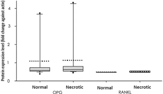
[image:4.612.89.525.84.274.2]Total proteins from both necrotic and normal tissues were analyzed for expressions of OPG and RANKL. As shown in Figures 4 and 5, OPG Table 1. Primer and probe sequences used in qPCR
Target gene Primer/probe name Oligonucleotide sequence (5’->3’) OPG Sense primer 5’-gaagc tggaa cccca gag-3’
Antisense primer 5’-gtgtt gcatt tcctt tctga gtta-3’ 1st probe 5’-caatt tgtgt gtttt ctaca gggtg tt-FL
2nd probe 5’-LC640-agatg acgtc tcatt tgaga agaac ccat-PH
RANKL Sense primer 5’-gcaaa aggaa ttaca acata tcgtt-3’
Antisense primer 5’-acttt atggg aacca gatgg g-3’ 1st probe 5’-ccaga tctaa ccatg agcca tccac c-FL
2nd probe 5’-LC640-tcgct ttctc tgctc tgatg tgctg tg-PH
RANK Sense primer 5’-aggga aagca ctcac agcta at-3’ Antisense primer 5’-acatg ctccc tgctg acc-3’
1st probe 5’-agtgg agata aggag tcctc aggtg aca-FL
2nd probe 5’-LC640-ttgtg tcagt acaca cacgg caaac tt-PH
Figure 3. mRNA levels of OPG, RANKL and RANK genes. The box-plot dis -played 25% quartile, mean and 75% quartile, in addition to 10% and 90% error bar. Medians were given by dashed lines while outraged data points
showed minimal/maximal values. The expression of RANK/RANKL in ne
[image:4.612.85.387.95.451.2]-crotic tissues was significantly increased compared to controlled ones.
Figure 4. Western blotting for OPG and RANKL pro -teins. Actin was used as the internal control protein. AVN, necrotic tissues; NL, normal tissues.
Results
Patient information
[image:4.612.90.287.533.607.2]protein was expressed in both normal and necrotic tissues with similar levels (P>0.05). The RANKL protein also had similar levels in both tissues (P>0.05). RANK protein, however, was not identified in either tissue.
Correlation analysis between gene expression and risk factors for AVN
We firstly divided all patients into three age groups: (1) between 20 and 35 years old; (2) between 36 and 50 years old; (5) older than 50 years. The expressional profiles in each group were studied and found no significant correla -tions between any gene expression level and age (P>0.05). We also compared the gene expression level between women before meno-pause and those after menomeno-pause. The RANK level in necrotic tissues was significantly elevat -ed in menopaus-ed women (3.36 vs. 0.95, P<0.05). Moreover, RANK gene expression was also significantly potentiated in smokers com -pared to non-smoking patients (2.23 vs. 1.23, P<0.05).
Discussion
The pathogenesis of AVN in femoral heads involves multiple factors, of which the imbal-ance between osteoblasts and osteoclasts are crucial due to their mediation on bone forma-tion [13]. The focal ischemia in AVN leads to the necrosis of bones and marrows. Although
reabsorption. OPG, RANKL and RANK have been reported to be important factors in main-taining balances between those two cells [17] as the dysregulation of those factors may lead to the occurrence of various orthopedics dis-eases [12, 18, 19]. In this study, we investigat-ed the gene expressions of OPG, RANKL and RANK in femoral head samples of AVN patients. Our results showed elevated but not statisti-cally significant difference of OPG mRAN in necrotic tissues. The mRNA of RANKL and RANK, however, were highly expressed in necrotic tissues when compared to control tis-sues. Protein levels of either OPG or RANKL, were not significantly changed between necrot -ic and normal tissues. In summary, inconsis-tent patterns between mRNA and protein expressions existed for those genes, suggest-ing further post-translational regulation in necrotic tissues.
[image:5.612.92.377.74.239.2]This study, for the first time, suggested relative -ly elevated OPG mRNA in necrotic bone tissues, although with no statistical significance. This may be caused by decreased activity of osteo-clast, which can disrupt the balance between osteoblasts and osteoclast, further compro-mising bone reformation. There have been studies reported the elevated bone density and osteopetrosis in mice with OPG-overexpression. In contrast, the gene knockout of OPG may lead to early onset osteoporosis. A recent clinical Figure 5. Quantified protein levels of OPG and RANKL. Protein levels were
expressed as fold changes against actin in the same tissue and were plotted as the box plot showing 25% quartile, mean and 75% quartile, in addition to 10% and 90% error bar. Medians were given by dashed lines while outraged
data points showed minimal/maximal values. No significant has been identi
-fied between normal and necrotic tissues in either protein.
survey showed significant correlation between gene expressions of OPG, RANKL and RANK with the risks of bone fracture, suggesting the crucial role of OPG in postnatal bone develop-ment. Other studies focused on mutation of RANK or OPG and diseases such as osteolysis, Paget’s disease and progressive bone deformi-ty, further supporting the interaction among those three factors in maintain normal bone volumes [20-23]. In our study, the elevation of OPG mRNA in necrotic tissues was consistent with imaging results and mouse model with higher bone density [24]. Whether the elevate OPG is the result of AVN or just one etiological factor for ostesopetrosis, however, needs to be elucidated.
In a comparison between RANKL/OPG ratios between necrotic and normal tissues, no sig-nificant differences have been identified. However, both gene expression levels were elevated in necrotic tissues, reflecting the requirement of osteoblasts in bone reforma-tion. Genes responsible for osteoblasts matu-ration and functions, such as bone morphologi-cal proteins (BMPs), related transcriptional factors (RUNX2) and other signaling molecules have been demonstrated to be related with bone volume abnormality and bone diseases [25, 26]. For example, recent studies showed the decreasing bone volume by active osteo-clasts, which are induced by BMPs via RANKL-OPG pathway, making BMPs as key regulators for osteoclasts [27, 28].
In summary, the expression of OPG, RANK and RANKL genes are closely related with progres -sion of femoral head AVN. Those genes may affect bone homeostasis and may exert certain roles in bone reabsorption and long-term hip joint breakage.
Disclosure of conflict of interest
None.
Address correspondence to: Dr. Qingtang Miao, Department of Orthopedics, Shandong Zhangqiu People’s Hospital, 1920 Huiquan Road, Zhangqiu, Jinan 250200, Shandong, China. Tel: +86-531-83259635; Fax: +86-531-+86-531-83259635; E-mail: lmtridverymuch@yeah.net
References
[1] Baelde HJ, Cleton-Jansen AM, van Beerendonk H, Namba M, Bovée JV, Hogendoorn PC. High
quality RNA isolation from tumours with low cellularity and high extracellular matrix compo-nent for cDNA microarrays: application to chondrosarcoma. J Clin Pathol 2001; 54: 778-82.
[2] Bjorkman A, Svensson PJ, Hillarp A, Burtscher IM, Rünow A, Benoni G. Factor V leiden and
prothrombin gene mutation: risk factors for os-teonecrosis of the femoral head in adults. Clin Orthop Relat Res 2004; 168-72.
[3] Browner WS, Lui LY and Cummings SR. Asso -ciations of serum osteoprotegerin levels with diabetes, stroke, bone density, fractures, and mortality in elderly women. J Clin Endocrinol Metab 2001; 86: 631-7.
[4] Chen D, Zhao M and Mundy GR. Bone morpho -genetic proteins. Growth Factors 2004; 22: 233-41.
[5] Canalis E, Economides AN and Gazzerro E.
Bone morphogenetic proteins, their antago -nists, and the skeleton. Endocr Rev 2003; 24: 218-35.
[6] Celik A, Tekis D, Saglam F, Tunali S, Kabakci N,
Ozaksoy D, Manisali M, Ozcan MA, Meral M, Gülay H, Camsari T. Association of corticoste-roids and factor V, prothrombin, and MTHFR gene mutations with avascular osteonecrosis in renal allograft recipients. Transplant Proc 2006; 38: 512-6.
[7] Chong B, Hegde M, Fawkner M, Simonet S, Cassinelli H, Coker M, Kanis J, Seidel J, Tau C, Tüysüz B, Yüksel B, Love D; International Hy -perphosphatasia Collaborative Group.
Idio-pathic hyperphosphatasia and TNFRSF11B
mutations: relationships between phenotype
and genotype. J Bone Miner Res 2003; 18:
2095-104.
[8] Cundy T, Hegde M, Naot D, Chong B, King A,
Wallace R, Mulley J, Love DR, Seidel J, Fawkner
M, Banovic T, Callon KE, Grey AB, Reid IR, Mid -dleton-Hardie CA, Cornish J. A mutation in the
gene TNFRSF11B encoding osteoprotegerin
causes an idiopathic hyperphosphatasia phe-notype. Hum Mol Genet 2002; 11: 2119-27. [9] Estrada K, Styrkarsdottir U, Evangelou E, Hsu
YH, Duncan EL, Ntzani EE, Oei L, Albagha OM, Amin N, Kemp JP, Koller DL, Li G, Liu CT, Min -ster RL, Moayyeri A, Vandenput L, Willner D,
Xiao SM, Yerges-Armstrong LM, Zheng HF, Alonso N, Eriksson J, Kammerer CM, Kaptoge SK, Leo PJ, Thorleifsson G, Wilson SG, Wilson JF, Aalto V, Alen M, Aragaki AK, Aspelund T,
Center JR, Dailiana Z, Duggan DJ, Garcia M, Garcia-Giralt N, Giroux S, Hallmans G, Hocking
LJ, Husted LB, Jameson KA, Khusainova R, Kim GS, Kooperberg C, Koromila T, Kruk M,
Laaksonen M, Lacroix AZ, Lee SH, Leung PC,
Lewis JR, Masi L, Mencej-Bedrac S, Nguyen TV,
Scol-len S, Siggeirsdottir K, Smith AV, Svensson O,
Trompet S, Trummer O, van Schoor NM, Woo J,
Zhu K, Balcells S, Brandi ML, Buckley BM,
Cheng S, Christiansen C, Cooper C, Dedoussis G, Ford I, Frost M, Goltzman D,
González-Macías J, Kähönen M, Karlsson M, Khusnutdi
-nova E, Koh JM, Kollia P, Langdahl BL, Leslie
WD, Lips P, Ljunggren Ö, Lorenc RS, Marc J,
Mellström D, Obermayer-Pietsch B, Olmos JM, Pettersson-Kymmer U, Reid DM, Riancho JA,
Ridker PM, Rousseau F, Slagboom PE, Tang NL, Urreizti R, Van Hul W, Viikari J, Zarrabeitia
MT, Aulchenko YS, Castano-Betancourt M,
Grundberg E, Herrera L, Ingvarsson T,
Johanns-dottir H, Kwan T, Li R, Luben R, Medina-Gómez
C, Palsson ST, Reppe S, Rotter JI, Sigurdsson
G, van Meurs JB, Verlaan D, Williams FM, Wood AR, Zhou Y, Gautvik KM, Pastinen T, Raychaud -huri S, Cauley JA, Chasman DI, Clark GR, Cum-mings SR, Danoy P, Dennison EM, Eastell R, Eisman JA, Gudnason V, Hofman A, Jackson
RD, Jones G, Jukema JW, Khaw KT, Lehtimäki T, Liu Y, Lorentzon M, McCloskey E, Mitchell BD, Nandakumar K, Nicholson GC, Oostra BA,
Peacock M, Pols HA, Prince RL, Raitakari O, Reid IR, Robbins J, Sambrook PN, Sham PC, Shuldiner AR, Tylavsky FA, van Duijn CM, Ware-ham NJ, Cupples LA, Econs MJ, Evans DM,
Har-ris TB, Kung AW, Psaty BM, Reeve J, Spector
TD, Streeten EA, Zillikens MC, Thorsteinsdottir
U, Ohlsson C, Karasik D, Richards JB, Brown MA, Stefansson K, Uitterlinden AG, Ralston SH, Ioannidis JP, Kiel DP, Rivadeneira F. Genome-wide meta-analysis identifies 56 bone mineral
density loci and reveals 14 loci associated with risk of fracture. Nat Genet 2012; 44: 491-501. [10] Glueck CJ, Freiberg RA, Fontaine RN, Tracy T,
Wang P. Hypofibrinolysis, thrombophilia, osteo -necrosis. Clin Orthop Relat Res 2001; 19-33. [11] Hadjigeorgiou G, Dardiotis E, Dardioti M,
Karantanas A, Dimitroulias A, Malizos K. Ge -netic association studies in osteonecrosis of the femoral head: mini review of the literature. Skeletal Radiol 2008; 37: 1-7.
[12] Tuli R, Seghatoleslami MR, Tuli S, Wang ML,
Hozack WJ, Manner PA, Danielson KG, Tuan
RS. A simple, high-yield method for obtaining multipotential mesenchymal progenitor cells
from trabecular bone. Mol Biotechnol 2003;
23: 37-49.
[13] Hruska KA, Mathew S and Saab G. Bone mor
-phogenetic proteins in vascular calcification.
Circ Res 2005; 97: 105-14.
[14] Hughes AE, Ralston SH, Marken J, Bell C,
MacPherson H, Wallace RG, van Hul W, Whyte
MP, Nakatsuka K, Hovy L, Anderson DM. Muta -tions in TNFRSF11A, affecting the signal
pep-tide of RANK, cause familial expansile osteoly -sis. Nat Genet 2000; 24: 45-8.
[15] Jorgensen HL, Kusk P, Madsen B, Fenger M, Lauritzen JB. Serum osteoprotegerin (OPG)
and the A163G polymorphism in the OPG pro-moter region are related to peripheral mea-sures of bone mass and fracture odds ratios. J
Bone Miner Metab 2004; 22: 132-8.
[16] Samara S, Dailiana Z, Varitimidis S,
Chassani-dis C, Koromila T, Malizos KN, Kollia P. Bone morphogenetic proteins (BMPs) expression in
the femoral heads of patients with avascular
necrosis. Mol Biol Rep 2013; 40: 4465-72.
[17] Soucacos PN, Beris AE, Malizos K, Koropilias
A, Zalavras H, Dailiana Z. Treatment of avascu-lar necrosis of the femoral head with vascuavascu-lar-
vascular-ized fibular transplant. Clin Orthop Relat Res
2001; 120-30.
[18] Whyte MP, Mills BG, Reinus WR, Podgornik
MN, Roodman GD, Gannon FH, Eddy MC, McAl-ister WH. Expansile skeletal hyperphosphata-sia: a new familial metabolic bone disease. J
Bone Miner Res 2000; 15: 2330-44.
[19] Leibbrandt A and Penninger JM. RANK/RANKL:
regulators of immune responses and bone
physiology. Ann N Y Acad Sci 2008; 1143:
123-50.
[20] Kamiya N. The role of BMPs in bone anabolism and their potential targets SOST and DKK1.
Curr Mol Pharmacol 2012; 5: 153-63. [21] Kearns AE, Khosla S and Kostenuik PJ. Recep
-tor activa-tor of nuclear fac-tor kappaB ligand
and osteoprotegerin regulation of bone remod-eling in health and disease. Endocr Rev 2008; 29: 155-92.
[22] Kostenuik PJ, Osteoprotegerin and RANKL reg -ulate bone resorption, density, geometry and strength. Curr Opin Pharmacol 2005; 5: 618-25.
[23] Zalavras CG, Malizos KN, Dokou E, Vartholo -matos G. The 677C-->T mutation of the methy-lene-tetrahydrofolate reductase gene in the pathogenesis of osteonecrosis of the femoral head. Haematologica 2002; 87: 111-2. [24] Zupan J, Komadina R and Marc J. The relation
-ship between osteoclastogenic and
anti-osteo-clastogenic pro-inflammatory cytokines differs
in human osteoporotic and osteoarthritic bone
tissues. J Biomed Sci 2012; 19: 28.
[25] Wada T, Nakashima T, Hiroshi N, Penninger
JM. RANKL-RANK signaling in osteoclastogen -esis and bone disease. Trends Mol Med 2006; 12: 17-25.
[26] Grewal R, Perey B, Wilmink M, Stothers K. A
randomized prospective study on the treat-ment of intra-articular distal radius fractures:
open reduction and internal fixation with dor -sal plating versus mini open reduction,
percu-taneous fixation, and external fixation. J Hand
[27] Trouvin AP and Goeb V. Receptor activator of
nuclear factor-kappaB ligand and osteoprote -gerin: maintaining the balance to prevent bone loss. Clin Interv Aging 2010; 5: 345-54.
[28] Aubin JE and Bonnelye E. Osteoprotegerin and