Original Article
Long non-coding RNA ANRIL promotes tumorigenesis
in glioma via MAPK signaling pathways
Xuchang Xu, Xuefeng Wan , Zhiren Zhang
Department of Neurosurgery, Zhumadian Central Hospital, Zhumadian 463000, Henan, China
Received August 17, 2016; Accepted August 26, 2016; Epub October 1, 2016; Published October 15, 2016
Abstract: Long non-coding RNA has been proven to be involved in many biological processes in cancer tumorigen-esis and progression. LncRNA ANRIL has various expression patterns in diverse human cancers. However, its clinical
significance and function in glioma has not yet been elucidated. In the present study, we explored the expression
of ANRIL in 33 paired glioma tissues and adjacent non-tumor tissues. We found that the expression of ANRIL was obviously upregulated in glioma tissue compared to adjacent non-tumor tissues and the detected level of ANRIL was associated with tumor grade. In addition, Kaplan-Meier analysis showed that high expression of ANRIL clearly predicted poorer overall survival (OS) in glioma patients. To explore the functional role of ANRIL, siRNA ANRIL and
negative control siRNA were transfected into glioma cells. We found that decreased expression of ANRIL signifi -cantly inhibited cell proliferation in vitro. Further studies indicated that inhibition of ANRIL induced cell cycle arrest at the G1/S transition, and enhanced apoptosis of glioma cells. Finally, Western blot showed that ANRIL may exert its function by targeting the MAPK signaling pathways. In conclusion, our study suggested that ANRIL might act as an oncogenic lncRNA that promotes proliferation of glioma cells and could be regarded as a therapeutic target in human glioma.
Keywords: Glioma, ANRIL, long non-coding RNA, MAPK
Introduction
Glioma is one of the most common types of malignant tumor in central nervous system [1]. Despite recent advances in treatment of sur-gery, radiotherapy and chemotherapy, the prog-nosis of these patients is still poor [2]. There- fore, it is extremely vital for us to elucidate new mechanisms associated with glioma progres-sion and establish potential therapeutic targets for the treatment of glioma.
In the light of recent transcriptomic studies, it has become apparent that the majority of the transcribed genome is non-coding [3]. Among these, there is a kind of non-coding RNA called long non-coding RNA (lncRNA >200 bp in length), which have been proven to regulate many biological processes from nuclear
organi-zation to epigenetic modification of post-tran -scriptional regulation and RNA splicing, espe-cially in cancers [4, 5]. In previous studies, many lncRNAs have been reported to play cru-cial roles in glioma. For example, Hu et al sh-
owed that upregulation of lncRNA AB073614 predicted a poor prognosis in patients with glio-ma [6]. Liu et al suggested that knockdown of lncRNA SPRY4-IT1 suppressed glioma cell pro-liferation, metastasis and epithelial-mesenchy-mal transition [7]. Ke et al showed that knock-down of lncRNA HOTAIR inhibited malignant biological behaviors of human glioma cells via modulation of miR-326 [8]. However, there are little studies about the role of lncRNA ANRIL in glioma.
miR-449a [11]. Chen et al suggested that lncRNA ANRIL inhibited p15 INK4b through the
TGFβ1 signaling pathway in human esophageal
squamous cell carcinoma [12]. However, the relationship between lncRNA ANRIL and glioma is still largely unknown. The objective of our study was to detect the biological behaviors and molecular mechanisms of lncRNA ANRIL in glioma in order to seek a potential therapeutic target for glioma patients.
Materials and methods
Tissue samples
A total of 33 glioma tissues were collected from surgical resections in Zhumadian Central Hospital. The adjacent non-tumor tissues,
which are defined as normal in the results,
were obtained 2 cm away from the glioma tis-sue. 3 patients had grade I (pilocytic ma), 8 patients had grade II (diffuse astrocyto-ma), 13 patients had grade III (anaplastic astro-cytoma) and 9 patients had grade IV (primary brain glioblastoma), according to the 2007
World Health Organization’s classification sys -tem. None of the patients received chemother-apy or radiotherchemother-apy before surgery. Tumor samples and normal tissues were rapidly frozen in liquid nitrogen and kept at -80°C until used. The study was approved by the Research Ethics Committee of Zhumadian Central Hospital, and written informed consent was obtained from all patients.
Cell lines
Human glioma cell lines (U87 and U251) were purchased from ATCC. All glioma cells were cul-tured at 37°C in a CO2 incubator in Dulbecco’s
modified Eagle’s media (DMEM) (Invitrogen)
supplemented with 10% fetal bovine serum (FBS) and 1% penicillin-streptomycin. Cells were used when they were in the logarithmic growth phase.
RNA isolation and quantitative qRT-PCR
Total RNA was extracted from tissues and cell lines using Trizol reagent (Invitrogen) according to the manufacturer’s instructions. The expres-sion levels of ANRIL were determined by quanti-tative real-time polymerase chain reaction (qRT-PCR) using the SYBR® Green master mix
(TaKaRa). The primers are as follows: lnc- RNA ANRIL 5’-GGGGTACCATGTCCGCTGCACT- TT-3’ and 5’-GCAAAGCTTACCCAACCTGGTAAG-
TGG-3’; GAPDH 5’-CGCTCTCTGCTCCTCCTGT- TC-3’ and 5’-ATCCGTTGACTCCGACCTTCAC-3’. The expression of lncRNA was normalized to GAPDH and calculated using the 2-ΔΔCt method.
Small interfering RNA transfection
Small interfering RNAs (siRNAs) specifically tar -geting ANRIL (si-ANRIL) and a scrambled nega-tive control (si-NC) were provided by Gene- Pharma. The sequences of si-ANRIL were as fol-lows: si-ANRIL-1: 5’-GCAAGAAACATTGCTGCTA- GC-3’; si-ANRIL-2: 5’-GCCCAATTATGCTGTGGT- AAC-3’. Transfections were performed using Lipofectamine 2000 (Invitrogen) according to the manufacturer’s protocol. The transfected cells were harvested 48 h after transfection. Cell proliferation assay
24 after transfection, cells were seeded in 96-well plates at 8000 cells per well. The MTT assay was used to determine cell viability at the point of 24, 48 and 72 h after seeding. The absorbance at 570 nm was measured using a Quant Universal Microplate Spectrophotometer (BioTek).
Colony formation assay
Cells were seeded in 6-well plates at a density of 100 cells/well. After incubation for 14 days,
cells were washed twice with PBS, fixed with
methanol and stained with crystal violet. The number of colonies containing >50 cells was counted under a microscope.
Cell cycle and apoptosis assays
For cell cycle analysis, transfected cells were cultured for 24 h and then treated with nocodazole (100 ng/ml) for 16-20 h, at last
cells were collected and were fixed into 70% ethanol at 20°C for 24 h, stained with 50 μg/ml
propidium iodide (Kaiji).
For apoptosis assay, cells were stained with 50
μg/ml propidium iodide and Annexin
V-fluo-rescein isothiocyanate (Kaiji) and following the manufacturer’s instructions. The results were analyzed using ModFit software (BD Bio- science).
Western blot assay
containing 1% protease inhibitors (PMSF); cell protein lysates were separated on 10% SDS PAGE gels and transferred to PVDF membranes.
The membranes were incubated with specific
antibodies. Finally, ECL chromogenic
sub-strates used in our experiment were quantified
by densitometry.
Statistical analysis
All statistical analyses in our experiment were performed using SPSS 18.0 software. Data are shown as the mean ± SD. The differences between groups were analyzed by the Student’s
t-test, Wilcoxon test, or χ2 test. The
Kaplan-Meier method was performed for patients’ overall survival analysis. All experiments were run in triplicate. P<0.05 was considered
statis-tically significant difference.
Results
LncRNA ANRIL is up-regulated in glioma To explore whether lncRNA ANRIL is dysregu-lated in glioma, qRT-PCR was used to
deter-mine the expression level of ANRIL. Among the 33 pairs of glioma tissues, the expression of
ANRIL was significantly up-regulated in glioma
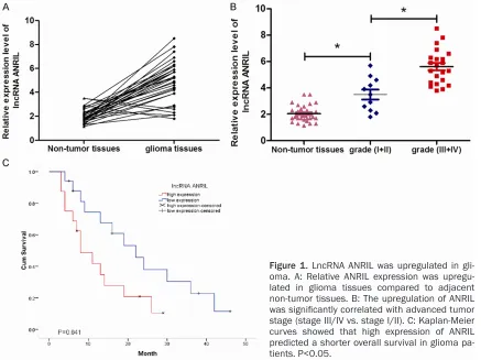
tissues compared to the adjacent non-tumor tissues (Figure 1A, P<0.05). In addition, ANRIL overexpression was closely correlated with advanced tumor stage (III+IV vs. I+II, Figure 1B, P<0.05). We further determined whether ANRIL expression level was associated with the out-come of glioma patients. Kaplan-Meier curve
indicated that high level of ANRIL was signifi -cantly associated with poor overall survival in glioma patients (Figure 1C, P<0.05). These results suggested that ANRIL might play a criti-cal role in glioma progression.
Knockdown of lncRNA ANRIL inhibits glioma cell proliferation
To explore the functional role of lncRNA ANRIL in glioma, the si-ANRIL was transfected into glioma cell lines. qRT-PCR assay revealed that
ANRIL expression was significantly reduced in
[image:3.612.91.528.72.399.2]si-ANRIL transfected glioma cells (Figure 2A, P<0.05). In order to examine the proliferative ability of ANRIL, MTT assay was performed. As Figure 1. LncRNA ANRIL was upregulated in gli-oma. A: Relative ANRIL expression was upregu-lated in glioma tissues compared to adjacent non-tumor tissues. B: The upregulation of ANRIL
was significantly correlated with advanced tumor
Figure 2. Inhibition of lncRNA ANRIL inhibits glioma cells
pro-liferation in vitro. (A) si-ANRIL significantly downregulated the
expression of ANRIL at mRNA levels in glioma cells. (B, C)
De-creased expression of ANRIL in glioma cells significantly re -duced their proliferative capacities as determined by MTT assay (B) and colony formation assay (C). P<0.05.
Figure 3. Inhibition of lncRNA ANRIL induces glioma cells cycle arrest and apoptosis. A: Cell cycle analysis showed that inhibition of ANRIL increased the percentage of G0/G1 phase cells and decreased the percentage of S phase cells. B: Cell apoptosis assay showed that inhibition of the ANRIL promoted glioma cells apoptosis. *P<0.05.
shown in Figure 2B, decreased expression of
ANRIL significantly suppressed cell prolifera -tion in glioma cells compared to the control group. Furthermore, colony formation assay
was performed, and the result similarly
indicat-ed that clonogenic survival was significantly
Knockdown of lncRNA ANRIL induces glioma cell G1 phase arrest and apoptosis
To explore the potential mechanisms
underly-ing cell growth, flow cytometry was used. Cell
cycle distribution analysis revealed that
inhibi-tion of ANRIL resulted in a significant increased
in the cellular population in G0/G1 phase but a sharp decrease in S phase (Figure 3A, P<0.05). Apoptosis analysis showed that silencing of ANRIL induced glioma cell apoptosis compared with control group (Figure 3B, P<0.05). These
findings suggested that ANRIL promoted glio -ma cells proliferation by promoting cell cycle progression and inhibiting cell apoptosis. lncRNA ANRIL activate MAPK signaling path-ways in glioma cells
To further define the underlying molecular
mechanism modulated by ANRIL in the growth of glioma cells, we focused on the Mitogen-activated protein kinase (MAPK) pathway, which including extracellular signal-regulated kinase (ERK), c-Jun N-terminal kinase (JNK) and p38, are crucial molecules involved in path-ways associated with cancer pathogenesis [13]. Western blot analysis was used to detect the activation of MAPK signaling pathways in si-ANRIL transfected glioma cells. Our data showed that inhibition of ANRIL in glioma cells strongly reduced the levels of p-ERK1/2, p-p38 and p-JNK MAPK (Figure 4). Thus, we suggest-ed that lncRNA ANRIL might promote glioma
cell proliferation by regulating MAPK signaling pathway.
Discussion
The sequencing of the human genome revealed that the coding portion of the genome repre-sents less than 2% of the genome. The remain-ing 98% of transcription products of the genome consists of non-coding RNA sequenc-es, including microRNAs and lncRNAs [14]. Recently, numerous pieces of evidence showed that dysregulation in lncRNAs are proved to contribute in tumor development in many can-cer types and can be used to develop as bio-markers and therapy target [15]. For example, Li et al showed that increased expression of lncRNA H19 promotes gastric cancer cell pro-gression and correlated with poor prognosis [16]. Yang et al suggested that lncRNA HULC promoted colorectal carcinoma progression through epigenetically repressing NKD2 expres-sion [17]. Li et al showed that overexpresexpres-sion of lncRNA HOTTIP increased chemoresistance of
osteosarcoma cell by activating the
Wnt/β-catenin pathway [18].
In the present study, we found that the expres-sion of ANRIL was obviously upregulated in glio-ma tissue and the detected levels of ANRIL were associated with tumor grade. Kaplan-Meier analysis suggested that high expression of ANRIL clearly predicted poorer overall sur-vival in glioma. In function assays, we found
that inhibition of ANRIL significantly
suppress-ed cell proliferation in vitro. Further studies revealed that inhibition of ANRIL induced cell cycle arrest at the G1/S transition, and en-
hanced apoptosis of glioma cells. These find -ings demonstrated that ANRIL might act as a tumor oncogene in glioma progression.
ERK/p38/JNK MAPK signaling pathways have been demonstrated to regulate a variety of cel-lular activities, including proliferation, differen-tiation, survival and death [19]. Recent studies suggested that MAPK signaling activation were involved in various human cancers. For exam-ple, Peng et al showed that lncRNA CCHE1 indi-cated a poor prognosis of hepatocellular carci-noma and promoted carcinogenesis via activa-tion of the ERK/MAPK pathway [20]. Wu et al found that lncRNA MALAT1 promoted the prolif-eration and metastasis of gallbladder cancer cells by activating the ERK/MAPK pathway [21]. In this study, we showed that inhibition of ANRIL
[image:5.612.92.290.72.264.2]significantly suppressed p-ERK1/2, p-p38 and
p-JNK MAPK expression in glioma cells. These data indicated that tumor oncogenic effect of ANRIL was at least partly mediated by MAPK pathway in glioma cells, which expanded the function of MAPK signaling pathway in glioma progression.
In conclusion, the present study suggested that lncRNA ANRIL promoted cell proliferation via
MAPK signaling in glioma. Our findings demon -strated that lncRNA ANRIL may become a prom-ising new candidate for glioma prognosis and therapy.
Acknowledgements
The project was supported by the Technology Research and Development Project of Zhu- madian (NO.20604).
Disclosure of conflict of interest None.
Address correspondence to: Zhiren Zhang, De- partment of Neurosurgery, Zhumadian Central Hospital, No.747 West Zhonghua Road, Yicheng District, Zhumadian 463000, Henan, China. E-mail: [email protected]
References
[1] Louis DN, Khaki H, Wiestler OD, Cavenee WK, Burger PC, Jouvet A, Scheithauer BW and
Klei-hues P. The 2007 WHO classification of tu -mours of the central nervous system. Acta Neuropathol 2007; 114: 97-109.
[2] Schwartzbaum JA, Fisher JL, Aldape KD and Wrensch M. Epidemiology and molecular pa-thology of glioma. Nat Clin Pract Neurol 2006; 2: 494-503; quiz 491 p following 516.
[3] Mattick JS and Makunin IV. Non-coding RNA. Hum Mol Genet 2006; 15: R17-29.
[4] Mercer TR, Dinger ME and Mattick JS. Long non-coding RNAs: insights into functions. Nat Rev Genet 2009; 10: 155-159.
[5] Gutschner T and Diederichs S. The hallmarks of cancer: a long non-coding RNA point of view. RNA Biol 2012; 9: 703-719.
[6] Hu L, Lv QL, Chen SH, Sun B, Qu Q, Cheng L, Guo Y, Zhou HH and Fan L. Up-Regulation of Long Non-Coding RNA AB073614 Predicts a Poor Prognosis in Patients with Glioma. Int J Environ Res Public Health 2016; 13: 433. [7] Liu H, Lv Z and Guo E. Knockdown of long
non-coding RNA SPRY4-IT1 suppresses glioma cell proliferation, metastasis and epithelial-mesen-chymal transition. Int J Clin Exp Pathol 2015; 8: 9140-9146.
[8] Ke J, Yao YL, Zheng J, Wang P, Liu YH, Ma J, Li Z, Liu XB, Li ZQ, Wang ZH and Xue YX. Knockdown of long non-coding RNA HOTAIR in-hibits malignant biological behaviors of human glioma cells via modulation of miR-326. Oncotarget 2015; 6: 21934-21949.
[9] Yap KL, Li S, Munoz-Cabello AM, Raguz S, Zeng L, Mujtaba S, Gil J, Walsh MJ and Zhou MM. Molecular interplay of the noncoding RNA ANRIL and methylated histone H3 lysine 27 by polycomb CBX7 in transcriptional silencing of INK4a. Mol Cell 2010; 38: 662-674.
[10] Lin L, Gu ZT, Chen WH and Cao KJ. Increased expression of the long non-coding RNA ANRIL promotes lung cancer cell metastasis and correlates with poor prognosis. Diagn Pathol 2015; 10: 14.
[11] Zhang EB, Kong R, Yin DD, You LH, Sun M, Han L, Xu TP, Xia R, Yang JS, De W and Chen J. Long noncoding RNA ANRIL indicates a poor progno-sis of gastric cancer and promotes tumor growth by epigenetically silencing of miR-99a/ miR-449a. Oncotarget 2014; 5: 2276-2292. [12] Chen D, Zhang Z, Mao C, Zhou Y, Yu L, Yin Y, Wu
S, Mou X and Zhu Y. ANRIL inhibits p15 INK4b
through the TGFβ1 signaling pathway in hu -man esophageal squamous cell carcinoma. Cell Immunol 2014; 289: 91-96.
[13] Johnson GL and Lapadat R. Mitogen-activated protein kinase pathways mediated by ERK, JNK, and p38 protein kinases. Science 2002; 298: 1911-1912.
F, An H, Awe A, Baldwin D, Baden H, Barnstead M, Barrow I, Beeson K, Busam D, Carver A, Center A, Cheng ML, Curry L, Danaher S, Davenport L, Desilets R, Dietz S, Dodson K, Doup L, Ferriera S, Garg N, Gluecksmann A, Hart B, Haynes J, Haynes C, Heiner C, Hladun S, Hostin D, Houck J, Howland T, Ibegwam C, Johnson J, Kalush F, Kline L, Koduru S, Love A, Mann F, May D, McCawley S, McIntosh T, McMullen I, Moy M, Moy L, Murphy B, Nelson K, Pfannkoch C, Pratts E, Puri V, Qureshi H, Reardon M, Rodriguez R, Rogers YH, Romblad D, Ruhfel B, Scott R, Sitter C, Smallwood M, Stewart E, Strong R, Suh E, Thomas R, Tint NN, Tse S, Vech C, Wang G, Wetter J, Williams S, Williams M, Windsor S, Winn-Deen E, Wolfe K, Zaveri J, Zaveri K, Abril JF, Guigo R, Campbell MJ, Sjolander KV, Karlak B, Kejariwal A, Mi H, Lazareva B, Hatton T, Narechania A, Diemer K, Muruganujan A, Guo N, Sato S, Bafna V, Istrail S, Lippert R, Schwartz R, Walenz B, Yooseph S, Allen D, Basu A, Baxendale J, Blick L, Caminha M, Carnes-Stine J, Caulk P, Chiang YH, Coyne M, Dahlke C, Mays A, Dombroski M, Donnelly M, Ely D, Esparham S, Fosler C, Gire H, Glanowski S, Glasser K, Glodek A, Gorokhov M, Graham K, Gropman B, Harris M, Heil J, Henderson S, Hoover J, Jennings D, Jordan C, Jordan J, Kasha J, Kagan L, Kraft C, Levitsky A, Lewis M, Liu X, Lopez J, Ma D, Majoros W, McDaniel J, Murphy S, Newman M, Nguyen T, Nguyen N, Nodell M, Pan S, Peck J, Peterson M, Rowe W, Sanders R, Scott J, Simpson M, Smith T, Sprague A, Stockwell T, Turner R, Venter E, Wang M, Wen M, Wu D, Wu M, Xia A, Zandieh A and Zhu X. The sequence of the hu-man genome. Science 2001; 291: 1304-1351.
[15] Cheetham SW, Gruhl F, Mattick JS and Dinger ME. Long noncoding RNAs and the genetics of cancer. Br J Cancer 2013; 108: 2419-2425. [16] Li H, Yu B, Li J, Su L, Yan M, Zhu Z and Liu B.
Overexpression of lncRNA H19 enhances car-cinogenesis and metastasis of gastric cancer. Oncotarget 2014; 5: 2318-2329.
[17] Yang XJ, Huang CQ, Peng CW, Hou JX and Liu JY. Long noncoding RNA HULC promotes colorectal carcinoma progression through epi-genetically repressing NKD2 expression. Gene 2016; 592: 172-178.
[18] Li Z, Zhao L and Wang Q. Overexpression of long non-coding RNA HOTTIP increases chemo-resistance of osteosarcoma cell by activating
the Wnt/β-catenin pathway. Am J Transl Res
2016; 8: 2385-2393.
[19] Arbabi S and Maier RV. Mitogen-activated pro-tein kinases. Crit Care Med 2002; 30: S74-S79. [20] Peng W and Fan H. Long noncoding RNA
CCHE1 indicates a poor prognosis of hepato-cellular carcinoma and promotes carcinogen-esis via activation of the ERK/MAPK pathway. Biomed Pharmacother 2016; 83: 450-455. [21] Wu XS, Wang XA, Wu WG, Hu YP, Li ML, Ding Q,