Original Article
Apoptosis prediction via inhibition of AKT signaling
pathway by neogrifolin
Yang Chen, Guo-Fang Peng, Xiang-Zhen Han, Wei Wang, Guo-Qiang Zhang, Xiao Li
Department of Orthopedics, Linyi People’s Hospital, 27 Jiefang Road, Linyi City 276003, Shandong Province, P.R. China
Received November 10, 2014; Accepted January 6, 2015; Epub February 1, 2015; Published February 15, 2015
Abstract: Neogrifolin, a natural biologically active substance isolated from the edible bodies of the mushroom Albatrellus confluens, has been shown to possess several pharmacological properties. No studies were investi-gated against osteosarcoma cancer. Hence, in this study, we investiinvesti-gated the apoptosis-inducing effects and the mechanisms of neogrifolin on human osteosarcoma cells. Our results demonstrated that neogrifolin induced con-centration- and time-dependent suppression of proliferation. Further, induction of apoptosis in U2OS and MG63 osteosarcoma cell lines were also observed. Neogrifolin induced the release of cytochrome c accompanied by acti-vation of caspase-9, caspase-3 and cleavage of poly (ADP-ribose) polymerase (PARP). In addition, z-VAD-fmk, a uni-versal inhibitor of caspases, prevented caspase-3 activation and PARP cleavage and inhibited neogrifolin-induced cell growth inhibition. Furthermore, neogrifolin treatment resulted in a reduction of phosphorylated AKT level, FOXO transcription factor, and glycogen synthase kinase 3 (GSK3). Knockdown of GSK3 with siRNA inhibited the apoptotic effects of neogrifolin. On the other hand, neogrifolin treatment also down-regulated the expression of the inhibitor of apoptosis protein (IAP) in both osteosarcoma cells. Collectively, our results suggested that neogrifolin is a potential candidate for osteosarcoma.
Keywords: Neogrifolin, osteosarcoma, AKT signaling pathway
Introduction
Osteosarcoma is an aggressive cancerous neo-plasm arising from primitive transformed cells of mesenchymal origin that exhibit osteoblastic differentiation and produce malignant osteoid [1, 2]. The highest incidence of osteosarcoma is in the second decade of life, which suggests a relationship between bone growth and tumor development [3]. One of the critical steps for normal skeletal development and bone forma-tion is the proliferative expansion of mesenchy-mal cells, osteoprogenitors, and immature ost- eoblasts. Gene and viral therapy, a highly prom-ising strategy for the treatment, has shown some therapeutic effects, but the therapeutic effect in vivo is less obviously than in vitro, still having been a lack of real breakthrough [4]. Osteosarcomatous is due to involve the high heterogeneity caused by the interaction of mul-tifactor [5].
Osteosarcoma is found to be the most common histological form of primary bone cancer
com-prising 2.4% of all malignancies in pediatric patients, and approximately 20% of all primary bone cancers [6, 7]. About 8% of all cases occur in the skull and jaw, and another 8% in the pel-vis [8]. Neoplastic cells undergo osteoblastic differentiation and form tumoral bone, and have the high incidence, the high malignant degree, the high rate of disability and the high-speed migration. There is a preference for origi-nation in the metaphyseal region of tubular long bones with 42% occurring in the femur, 19% in the tibia, and 10% in the humerus [9, 10]. The osteosarcomatous patient must face to resects the trouble bone, even amputation for saving his life. And sometimes amputation is the inevi-table way of the large focal lesion, but the five-year survival rate of these osteosarcomatous amputation is only 17.4% to 21.8% to the early and mid-term patients [11].
poor. In order to ensure a cure, it is necessary to develop more effective adjuvant treatments. The treatment for osteosarcoma consists of neo-adjuvant chemotherapy, delayed wide resection and adjuvant chemotherapy adapted to tumor tissue removed by surgery. The sur-vival rate for patients with bone osteosarcoma has improved mostly thanks to the addition of systemic chemotherapy following surgical removal of the tumor [14]. The primary treat-ment is a combination of surgery and chemo-therapy. The major problems associated with chemotherapy are the cytotoxic effects [15]. Thus, safe and more effective anticancer treat-ments are needed for patients with osteosa- rcoma.
Mushrooms (macrofungi) accumulate a variety of cytotoxic secondary metabolites including polyphenols, terpenoids, and alkaloids [16, 17]. Medicinal mushrooms such as Ganoderma lucidum, Phellinus linteus, and Coriolus versi-color have an established history of use in tra-ditional Asian therapies [18, 19]. Several
[image:2.612.92.522.70.233.2]poly-saccharides and polysaccharide conjugates have been commercialized for the clinical treat-ment of patients undergoing anticancer thera-py. Neogrifolin, a natural biologically active sub-stance isolated from the edible bodies of the mushroom Albatrellus confluens [20]. Two closely related naturally occurring biologically active phenolic compounds, grifolin and neogri-folin were isolated from Albatrellus ssp. (Basi- ciomycetes) [21-23]. Such compounds were derived from tetraketides and belong to the group of polyketide-terpenoids. Grifolin, neogri-folin and their derivatives possess not only anti-microbial properties [24, 25] but also antioxida-tive [26], tyrosinase inhibiting [27] and cytotox-ic activities [28] as well as activity on the vanil-loid receptors [29]. No research reports are available so far in the literature based on osteo-sarcoma studies. Based on the above men-tioned facts, we decided to investigate the mechanism of the chemopreventive effects of neogrifolin in human osteosarcoma cell lines. We examined the involvement of AKT and its substrates FOXO and GSK3 and inhibitor of Figure 1. A. U2OS and MG63 cell lines incubated with 0, 5, 10, 25, 50, or 100 μM of neogrifolin for 24 h. B. U2OS and MG63 cell lines incubated with 50 μM neogrifolin for 6 h, 12 h and 24 h.
[image:2.612.92.518.292.391.2]apoptosis protein (IAP) families in the apoptotic effects of neogrifolin in these cells.
Materials and methods
Materials
Neogrifolin was obtained from reputed chemi-cal suppliers (Netsun USA, California, USA). Anti-phospho-AKT (Ser 473), Anti-phospho-AKT (Thr 308), anti-PARP, anti-caspase-3 antibod-ies, z-VAD-fmk were purchased from Santa Cruz Biotechnology (Santa Cruz, CA USA). Hoechst H33258, Propidium iodide (PI), 3-(4,5-Dime- thylthiazol-2-yl)-2,5-diphenyl tetrazolium bro-mide (MTT), monoclonal mouse anti-β-actin antibody and heat-inactivated fetal bovine serum were obtained from Sigma-Aldrich Co. (St. Louis, MO, USA). Negative control RNA, GSK-3α/βsiRNA, anti-phospho-GSK3, anti-pho-spho-FKHRL1, anti-cytochrome c antibodies were purchased from Cell Signaling Techno- logies (Beverly, MA, USA). Rhodamine dye was purchased from Alexis Corp (San Diego, CA, USA).
Cell culture and methods
Human osteosarcoma cell lines MG63 and U2OS were obtained from American Type Culture Collection, (Rockville, MD, USA). Cells were grown in DMEM with 10% (v/v) fetal calf serum, streptomycin100 μg/ml, and penicillin 100 U/ml. Cultures were maintained at 37°C in a humidified incubator in an O2 atmosphere of 95% and 5% CO2.
Determination of cell viability by MTT assay
The cell viability was assessed by using 3-(4,5-dimethylthiazol-2-yl)-2,5-diphenyl tetra-zolium bromide (MTT) assay. 104 cells was
plat-ed on a 96-well plate in the presence or absence of indicated concentrations of neogri-folin for 6-24 h, then added 100 μl of 0.5 mg/ ml MTT to each well. After 4 h incubation at 37°C, 100 μl of dimethyl sulfoxide (DMSO) was added to each well to allow the formed forma-zan crystals to dissolve. The optical density (OD) value at 565 nm was measured.
Hoechst nuclear staining assessing the per-centage of apoptotic cells
Apoptotic morphology was studied by staining the cells with Hoechst 33258 stain. Cells were seeded on cover slips on a 6-well plate in the
presence or absence of 50 μM neogrifolin. After 24 h incubation, the cover glasses were carefully washed with phosphate-buffered saline (PBS) and stained with 20 μg/ml of Hoechst 33258 for 10 min. Later, the cells were washed in PBS and observed under a fluo-rescence microscope (Leica Microsystems AG, Wetzlar, Germany).
Flow cytometry
After neogrifolin-treatment for 4 h, 8 h and 24 h, cells suspension was prepared by trypsiniza-tion, and was centrifuged at 1,000 rpm for 5 min at 4°C. Pellets were then rinsed with ice-cold PBS and fixed with 70% ethanol for 24 h. Then the samples were stained with staining buffer (PBS containing 50 μg/ml of propidium iodide, 10 μg/ml RNase A, 0.1% sodium citrate and 0.1 Triton X-100) for 30 min at room tem-perature in the dark. DNA content was analyzed by flow cytometry (EPICS XL, BECKMAN CO- ULTER, USA). The populations containing less DNA than G1 populations (sub-G1 peak) were considered as apoptotic cells [30].
Cytochrome c release assay
Cytochrome c release from mitochondria was assayed by following the method as reported previously [31]. In this experiment, cells were treated with and without neogrifolin centrifuged at 1000 g. Cell pellets were suspended in 5 vol-umes of a hypotonic buffer containing 20 mM HEPES-KOH (pH 7.5), 10 mM KCl, 1.5 mM MgCl2, 1 mM EDTA, 1 mM EGTA, 1 mM DTT, 20 μg/ml leupeptin, 10 μg/ml aprotinin, and 250 mM sucrose for 15 min on ice. Cells were homogenized by passing them 15-20 times through a 22-gauge, 1.5-inch-long needle. The lysates were centrifuged at 1000 gfor 5 min at 4°C and the supernatants were collected and centrifuged at 12,000 gfor 15 min. The result-ing mitochondrial pellets were resuspended in lysis buffer. Supernatants were transferred to new tubes and centrifuged again at 12,000 g for 15 min, and the resulting supernatants, which represented cytosolic fractions, were separated. The protein from the cytosolic frac-tion of each sample was used to perform west-ern blotting with anti-cytochrome c antibody.
Mitochondrial membrane potential measure-ment
ously [32]. At first, 1 × 10⁶ MG63 or U2OS cells/
ml were incubated with 10 μM rhodamine 123 for 10 min at 37°C. After incorporation of fluo-rescent probe, the cells were incubated up to 4 h with or without neogrifolin. After incubation, the cells were washed twice with phosphate-buffered saline, harvested by centrifugation, and then resuspended in 1.5 ml phosphate-buffered saline. The fluorescent intensity of each cell suspensions was measured at an excitation wavelength 480 nm and an emission wavelength 530 nm in a Perkin Elmer L15B fluorescence spectrophotometer. The fluores-cence intensity was recorded in arbitrary units representing the mitochondrial transmem-brane potential.
GSK-3 siRNA transfection
GSK-3 siRNA transfection was used to knock down the expression of GSK-3α/β as described in the literature method [33]. In this study, MG63 and U2OS cells were transfected with GSK-3 siRNA or negative control RNA at con-centration of 50 nM using Lipofectamine 2000 according to the manufacturer (Invitrogen, Life Technologies, Inc.). After transfection of about 24 h, the cells were exposed to 50 μM neogrifo-lin for 24 h and then were harvested to perform western blotting and flow cytometry analysis.
Western blot analysis
Western blot experiments were performed by following this reported method [34]. In brief, cells were washed with PBS (mM: NaCl 130, KCl 2.5, Na2HPO4 10, KH2PO4 1.5, pH 7.4), ana-lysed with solubilization buffer (mM: Tris-Cl 50,
NaCl 150, 0.02% NaN3, 1% Nonidet P-40, 0.1% SDS, 0.5% sodiumdeoxycholate, NaVO3, 5 μg/ ml leupeptin and 1 μg/ml aprotinin). After cen-trifugation, the supernatants were collected and equivalent protein concentrations were separated by SDS-PAGE. The separated pro-teins were electrotransferred to PVDF mem-branes (Millipore, Bedford, MA, USA). After blocking with PBST (PBS containing 0.05% Tween 20) containing 5% nonfat milk for 1 h, each membrane was incubated with primary antibodies 1 h at room temperature or over-night at 4°C, and then the membranes were probed with the appropriate secondary peroxi-dase-conjugated antibodies (HRP-linked rabbit secondary antibody and HRP linked anti-biotin antibody, 1 h at room temperature). The immunoblots were visualized by enhanced chemiluminescence.
Statistical analysis
The experimental data was analyzed by SP- SS11.5 statistical software. All the data were expressed in the form of mean ± standard devi-ation. Statistical analysis was performed by applying a one-way analysis of variance fol-lowed by the Student t test. P values of less than 0.05 indicated statistical significance. Each experiment is repeated for three times. Results
Neogrifolin suppressed the proliferation in hu-man osteosarcoma cells
iment, U2OS and MG63 cell lines were incubat-ed with 0, 5, 10, 25, 50, or 100 μM of grifolin for 24 h. In the second experiment, U2OS and MG63 cell lines were incubated with 50 μM neogrifolin for 4 h, 8 h and 24 h. At the end of incubation the cell survival rates were deter-mined by MTT methods. Cell viability is exp- ressed as the percentage of cell survival com-pared with the control. Data were obtained from five independent experiments. *P < 0.05, **P < 0.01 compared to the control group. The results suggested that neogrifolin treatment evoked cells growth inhibition in concentration- and time-dependent manners in both human osteosarcoma cell lines. The result is depicted in Figure 1.
Neogrifolin induced apoptosis in osteosarcoma cells
To determine whether the growth inhibition induced by neogrifolin in human osteosarcoma cells was caused by apoptosis, MG63 and U2OS cell lines were treated with 50 μM neog-rifolin for 24 h, and the morphological changes were examined with Hochest 33258 staining. As shown in Figure 2, the nuclei of cells were round and homogeneously stained in the con-trol group, however, 50 μM neogrifolin treat-ment induced nuclei condensation or apoptotic bodies formation in two cell lines. Furthermore, we analyzed the sub-G1 population of cells
treated with 50 μM neogrifolin for 4, 8 and 24 h. Figure 3 demonstrated that neogrifolin treat-ment induced apoptosis in a time-dependent manner (Figure 3). The apoptotic population was measured as the percentage of total cell populations with sub-G1 DNA content. These results shown are representative of six inde-pendent experiments. All these results sug-gested that neogrifolin treatment suppresses the growth of osteosarcoma cells by inducing apoptosis.
Effects of neogrifolin treatment on mitochon-drial membrane potential and cytochrome c release in osteosarcoma cells
In this experimental study, we examined the effects of neogrifolin treatment on mitochon-drial membrane potential and cytochrome c
[image:6.612.324.525.70.161.2] [image:6.612.90.316.73.312.2]release in osteosarcoma cells. The effect of neogrifolin on the mitochondria membrane potential is shown in Figure 4A. From the Figure 4A, it can be said that neogrifolin treatment induced the loss of the mitochondria mem-brane potential in two tested cells. In addition we tested the cytochrome c release. Release of cytochrome c from mitochondria to cytosol has been suggested to be involved in apoptosis process induced by various apoptotic inducers. Figure 4B showed whether the release of cyto-chrome c is involved in the apoptotic effect of neogrifolin in MG63 and U2OS osteosarcoma Figure 5. A and B. 50 μM Neogrifolin treatment for 24
cells. It can be inferred from Figure 4B, that treatment with 50 μM neogrifolin for 24 h induced the translocation of cytochrome c from the mitochondria to cytosol in two osteosarco-ma cell lines. The results shown are represen-tative of five independent experiments and the data are expressed as mean ± SEM of 5 deter-minations, *P < 0.01 and **P < 0.001 com-pared with control group.
Neogrifolin induced caspases-dependent apoptosis in osteosarcoma cells
Further downstream in the apoptotic pathway, we investigated the effects of neogrifolin treat-ment on the activation of caspases-9 and cas-pase-3 and PARP. As shown in Figure 5A and 5B, neogrifolin treatment activated caspases-9 and caspase-3 and PARP as determined by the cleavage of caspases-9 and caspase-3 and PARP. The cleavage of caspase-3 and PARP was abolished by the pre-treatment with univer-sal caspases inhibitor z-VAD-fmk. Moreover, z-VAD-fmk pre-incubation blocked the neogrifo-lin induced cell death in MG63 and U2OS osteosarcoma cells (Figure 5C). All these results suggested that neogrifolin activated caspase 9/3 and induced PARP cleavage.
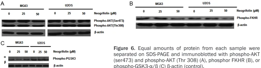
Dephosphorylation of AKT and its substrates induced by neogrifolin
In order to determine whether the Akt activity is associated with apoptotic effects of neogrifo-lin, we carried out the protein expression and phosphorylation level of Akt after neogrifolin treatment. MG63 and U2OS cells were treated with various doses of neogrifolin as indicated for 24 h. Neogrifolin treatment decreased the phosphorylation level of Akt at both Thr308 and Ser473 sites (Figure 6A). In both the cells, Akt is found to be constitutively activated. We then examined the effect of neogrifolin on the phosphorylation level of two Akt downstream
targets: Fork head transcription factors (FKHR) and GSK3 in MG63 and U2OS osteosarcoma cell lines. As shown in Figures 6B and 6C, con-stitutive phosphorylation of FKHR and GSK3 was seen in two osteosarcoma cell lines and the phosphorylation level was decreased by the treatment with neogrifolin. All these results suggested that neogrifolin inhibited constitu-tively active AKT signaling pathway in osteosar-coma cell lines. The results shown are repre-sentative of five independent experiments.
Knockdown of GSK3 with siRNA inhibited neogrifolin-mediated apoptosis
To further prove the role of GSK3 in the neogri-folin-induced apoptosis, MG63 and U2OS cells were transiently transfected with GSK3 siRNA or negative control RNA for 24 h. Cell were then harvested to perform western blot and after transfection, MG63 and U2OS cells were treat-ed with neogrifolin for another 24 h. The cell viability and apoptotic rates were measured by MTT and flow cytometry methods, respectively. As shown in Figure 7A, GSK3 siRNA transfec-tion significantly knocked down the expression of GSK3. Knockdown of GSK3 inhibited the apoptotic effects of neogrifolin in the two test-ed osteosarcoma cells (Figure 7B and 7C). The results shown are representative of five (A) or six (B and C) independent experiments.
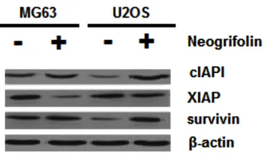
Neogrifolin reduced the expression of IAP pro-tein family
[image:7.612.92.524.71.184.2]Inhibitors of apoptosis proteins (IAP) have been shown to be able to inhibit cell apoptosis and have direct effects on caspase-9 and cas-pase-3 [35]. Therefore, we also determined the effects of grifolin treatment on the expression of IAP family members. As shown in Figure 8, 50 μM grifolin treatment for 24 h decreased the expression of cIAP1, XIAP and survivin in MG63 and U2OS osteosarcoma cells.
Figure 6. Equal amounts of protein from each sample were separated on SDS-PAGE and immunoblotted with phospho-AKT (ser473) and phospho-AKT (Thr 308) (A), phosphor FKHR (B), or
pase-3 activation resulting in the cleavage of PARP [36, 37].
In this study, the release of cytochrome c, acti-vation of caspase-9 and caspase-3, and the cleavage of PARP were observed after neogrifo-lin treatment. In addition, z-VAD-fmk, a univer-sal inhibitor of caspases, preventedcaspase-3 activation and PARP cleavage and inhibited neogrifolin-induced cell growth inhibition. These data indicated that release of cyto-chrome c mediated caspases activation and PARP cleavage is involved in the apoptotic effects of neogrifolin in osteosarcoma cells. The Akt/mTOR signal transduction is critical to control processes integral in cancer develop-ment, such as protein translation, growth, Discussion
In this study, our results demonstrated that neogrifolin decreased cell viability in both MG63 and U2OS osteosarcoma cell lines in dose and time-dependent fashions. The pre-dominant mode of cell death in these cells was found to be apoptosis. This was determined by characteristic changes in cell morphology with Hochest 33258 staining and by the presence of sub-G1 peak with flow cytometry method. During apoptosis, the permeability of mitochon-drial membrane increased, leading to the loss of membrane potential and release of cyto-chrome c to cytosol and the released cyto-chrome c binds to Apaf-1, and then this com-pound activates caspase-9, triggering
[image:8.612.89.524.68.479.2]metabolism, therapeutic resistance, and sur-vival. This pathway is frequently over-expressed or over-activated in osteosarcoma, providing a strong rationale to target it in cancer therapy [38, 39]. Our present results showed that Akt is constitutively phosphorylated at both Ser473 and Thr 308 in the two tested osteosarcoma cell lines.
Neogrifolin inhibited AKT phosphorylation at these two sites. As the downstream targets of Akt, GSK3 and FKHR family of transcription fac-tors have been reported to be involved the reg-ulation of the cells survival. Akt promotes cells survival by phosphorylating GSK3 and FKHR family of transcription factors, which inacti-vates them and prevents the apoptosis [40, 41]. Therefore, we next examined the phos-phorylation levels of GSK3 and FKHR in neogri-folin treated and untreated osteosarcoma cells. The results showed that the constitutive phos-phorylation of GSK3 and FKHR were observed in the two osteosarcoma cell lines and the phosphorylation levels were inhibited by neogri-folin treatment. To further confirm the role of GSK3 in neogrifolin-mediated apoptosis, we knock down the expression of GSK3 by siRNA transfection. We found the GSK3 knockdown significantly inhibited the apoptotic effects of neogrifolin in two osteosarcoma cell lines. Our data suggested that Akt signaling pathway play critical role in regulating the growth and surviv-al of osteosarcoma cells. Neogrifolin treatment inactivates Akt signaling pathway thereby inhib-iting these cells growth. One mechanism of Akt promoting cells survival is by up-regulating many survival genes including inhibitors of apoptosis proteins (IAPs) [42]. IAPs have been demonstrated to be able to inhibit the activity
of caspases and prevent the apoptosis induced by various stimuli.
Survivin is important for osteosarcoma cell sur-vival, growth, and apoptosis resistance, and down-regulating survivin induces osteosarco-ma cell apoptosis and growth inhibition [43, 44]. Survivin is a structurally unique member of the inhibitor of apoptosis protein family, which is important for mitotic progression, cancer survival, and apoptosis inhibition. Its marked expression in cancers versus normal tissues and its association with unfavorable disease outcome have made survivin a promising new target for anti-cancer interventions [45, 46]. Our present study showed that osteosarcoma cell lines expressed IAPs including cIAP1, XIAP and survivin. Neogrifolin treatment decreased the expression of these proteins. It suggested that the down-regulation of IAPs is also involved in the apoptosis process induced by neogrifolin treatment.
In conclusion, research report showed that AKT and its downstream targets FKHR and GSK3 are constitutively active in MG63 and U2OS osteosarcoma cell lines. Neogrifolin-induced inhibition of Akt signaling pathway leads to apoptosis in osteosarcoma cells through cyto-chrome c release from the mitochondria and activated caspases and down-regulation of IAPs (cIAP1, XIAP and survivin). Collective results suggested that neogrifolin may act as a promising antitumor agent against human os- teosarcoma.
Disclosure of conflict of interest
None.
Address correspondence to: Dr. Xiao Li, Department of Orthopedics, Linyi People’s Hospital, 27 Jiefang Road, Linyi 276003, Shandong Province, P.R. China. Tel: +86-0539-8038762; Fax: +86-539-8296088; E-mail: xiaolixiao28@gmail.com
References
[1] Bramwell VH. The role of chemotherapy in the management of non-metastatic operable ex-tremity osteosarcoma. Semin Oncol 1997; 24: 561-571.
[image:9.612.95.287.71.183.2][2] Haydon RC, Luu HH, He TC. Osteosarcoma and osteoblastic differentiation: a new perspective Figure 8. U2OS and MG63 cells treated with and
without 50 μM neogrifolin against cIAP1, XIAP, sur
E, Steiner HH. Immunization with
virus-modi-fied tumor cells. Semin Oncol 1998; 25:
677-696.
[6] Meyers PA, Gorlick R. Osteosarcoma. Pediatr Clin North Am 1997; 44: 973-989.
[7] Bao PP, Zheng Y, Wang CF, Gu K, Jin F, Lu W. Time trends and characteristics of childhood cancer among children age 0-14 in Shanghai. Pediatr Blood Cancer 2009; 53: 13-16. [8] Kamath AT, Feng CG, Macdonald M, Briscoe H,
Britton WJ. Differential protective efficacy of
DNA vaccines expressing secreted proteins of Mycobacterium tuberculosis. Infect Immun 1999; 67: 1702-1707.
[9] Tsuchiya H, Kanazawa Y, Abdel-wanis ME, Asa-da N, Abe S, Isu K, Sugita T, Tomita K. Effect of
timing of pulmonary metastases identification
on prognosis of patients with osteosarcoma: the Japanese Musculoskeletal Oncology Group study. J Clin Oncol 2002; 20: 3470-3477. [10] Corradi D, Wenger De, Bertoni F, Bacchini P,
Bosio S, Goldoni M, Unni KK, Sim FH, Inwards CY. Multicentric osteosarcoma: clinicopatho-logic and radiographic study of 56 cases. Am J Clin Pathol 2011; 136: 799-807.
[11] Afonso CL, Tulman ER, Lu Z, Zsak L, Kutish GF, Rock DL. The genome of fowlpox virus. J Virol 2000; 74: 3815-3831.
[12] Goorin AM, Schwartzentruber DJ, Devidas M, Gebhardt MC, Ayala AG, Harris MB, Helman LJ, Grier HE, Link MP. Presurgical Chemotherapy Compared With Immediate Surgery and Adju-vant Chemotherapy for Nonmetastatic Osteo-sarcoma: Pediatric Oncology Group Study POG-8651. J Clin Oncol 2003; 21: 1574-1580. [13] Ritter J, Bielack SS. Osteosarcoma. Ann Oncol
2010; 21: vii320-vii325.
[14] Gobin B, Moriceau G, Ory B, Charrier C, Brion R, Blanchard F, Redini F, Heymann D. Imatinib mesylate exerts anti-proliferative effects on os-teosarcoma cells and inhibits the tumour growth in immune competent murine models. PLoS One 2014; 9: e90795.
[15] McAllister TW, Ahles TA, Saykin AJ, Ferguson RJ, McDonald BC, Lewis LD, Flashman LA, Rhodes CH. Cognitive effects of cytotoxic can-cer chemotherapy: predisposing risk factors
mide from Pholiota spumosa (Basidiomycetes) inhibits cell growth of human prostate cancer cells. Phytomedicine 2007; 14: 185-191. [18] Moradali MF, Mostafavi H, Ghods S,
Hedja-roude GA. Immunomodulating and anticancer agents in the realm of macromycetes fungi (macrofungi). Int Immunopharmacol 2007; 7: 701-724.
[19] Zhang M, Cui SW, Cheung PCK, Wang Q. Antitu-mor polysaccharides from mushrooms: a re-view on their isolation process, structural char-acteristics and antitumor activity. Trends Food Sci Technol 2007; 18: 4-19.
[20] Ding ZH, Dong ZJ, Liu JK. Albaconol: a novel prenylated resorcinol (=benzene-1,3-diol) from
Basidiomycetes Albatrellus confluens. Helv
Chim Acta 2001; 84: 259-262.
[21] Hirata Y, Nakanishi K. Grifolin, an antibiotic from a Basidiomycete. J Biol Chem 1950; 184: 135-143.
[22] Vrkoč J, Buděšinský M, Dolejš L. Phenolic me -roterpenoids from the Basidiomycete Albatrel- lus ovinus. Phytochemistry 1977; 16: 1409-1411.
[23] Besl H, Hoefle G, Jendrny B, Jägers E, Steglich
W. Pilzpigmente, XXXI. Farnesylphenole aus Albatrellus-Arten (Basidiomycetes). Chem Ber 1977; 110: 3770-3776.
[24] Zechlin L, Wolf M, Steglich W, Anke T.
Antibio-tika aus Basidiomyceten, XII. Cristatsäure, ein modifiziertes Farnesylphenol aus Fruchtkör -pern von Albatrellus cristatus. Liebigs Ann Chem 1981; 12: 2099-2105.
[25] Hashimoto T, Quang DN, Nukada M, Asakawa Y. Isolation, synthesis and biological activity of grifolic acid derivatives from inedible mush-room Albatrellus dispansus. Heterocycles 2005; 65: 2431-2439.
[26] Nukata M, Hashimoto T, Yamamoto I, Iwasaki N, Tanaka M, Asakawa Y. Neogrifolin deriva-tives possessing anti-oxidative activity from the mushroom Albatrellus ovinus. Phytochem-istry 2002; 59: 731-737.
[27] Misasa H, Matsui Y, Uehara H, Tanaka H, Ishi-hara M, Shibata H. Tyrosinase inhibitors from
[28] Yang XL, Qin C, Wang F, Dong ZJ, Liu JK. A new meroterpenoid pigment from the
Basidiomy-cete Albatrellus confluens. Chem Biodivers
2008; 5: 484-489.
[29] Hellwig V, Nopper R, Mauler F, Freitag J, Ji-Kai L, Zhi-Hui D, Stadler M. Activities of prenylphe-nol derivatives from fruitbodies of Albatrellus ssp. on the human and rat vanilloid receptor 1 (VR1) and characterisation of the novel natural
product, confluentin. Arch Pharm 2003; 336:
119-126.
[30] Zhang HN, Zhou JG, Qiu QY, Ren JL, Guan YY. ClC-3 chloride channel prevents apoptosis in-duced by thapsigargin in PC12 cells. Apoptosis 2006; 11: 327-336.
[31] Uddin S, Hussain AR, Al-Hussein KA, Manoga-ran PS, Wickrema A, Gutierrez MI, Bhatia KG. Inhibition of Phosphatidylinositol 3-kinase/ AKT-signaling promotes apoptosis of primary effusion lymphoma cells. Clin Cancer Res 2005; 11: 3102-3108.
[32] Lin HI, Lee YJ, Chen BF, Tsai MC, Lu JL, Chou CJ, Jow GM. Involvement of Bcl-2 family, cyto-chrome c and caspase 3 in induction of apop-tosis by beauvericin in human non-small cell lung cancer cells. Cancer Lett 2005; 230: 248-259.
[33] Pang RP, Zhou JG, Zeng ZR, Li XY, Chen W, Chen MH, Hu PJ. Celecoxi induces apoptosis in
COX-2 deficient human gastric cancer cells through Akt/GSK3β/NAG-1 pathway. Cancer
Lett 2006; 251: 268-277.
[34] Zhou JG, Ren JL, Qiu QY, He H, Guan YY. Regu-lation of intracellular Cl-concentration through volume-regulated ClC-3 chloride channel in A10 vascular smooth muscle cells. J Biol Chem 2005; 280: 7301-7308.
[35] Kornacker M, Verneris MR, Kornacker B, Scheffold C, Negrin RS. Survivin expression correlates with apoptosis resistance after lym-phocyte activation and is found preferentially in memory T cells. Immunol Lett 2001; 76: 169-173.
[36] Chan DW, Son SC, Block W,Ye R, Khanna KK, Wold MS, Douglas P, Goodarzi AA, Pelley J, Taya Y, Lavin MF, Lees-Miller SP. Purification
and characterization of ATM from human pla-centa. A manganese-dependent, wortmannin-sensitive serine/threonine protein kinase. J Biol Chem 2000; 275: 7803-7810.
[37] Slee EA, Harte MT, Kluck RM, Wolf BB, Casiano CA, Newmeyer DD, Wang HG, Reed JC, Nichol-son DW, Alnemri ES, Green DR, Martin SJ. Or-dering the cytochrome c-initiated caspase cas-cade: hierarchical activation of caspases-2, -3, -6, -7, -8, and -10 in a caspase-9-dependent manner. J Cell Biol 1999; 144: 281-292. [38] Vivanco I, Sawyers CL. The
phosphatidylinosi-tol 3-Kinase AKT pathway in human cancer. Nat Rev Cancer 2002; 2: 489-501.
[39] Hennessy BT, Smith DL, Ram PT, Lu Y, Mills GB. Exploiting the PI3K/AKT pathway for cancer drug discovery. Nat Rev Drug Discov 2005; 4: 988-1004.
[40] Romashkova JA, Makarov SS. NF-kappa B is a target of AKT in anti-apoptotic PDGF signaling. Nature 1999; 401: 86-90.
[41] Brunet A, Park J, Tran H, Hu LS, Hemmings BA, Greenberg ME. Protein kinase SGK mediates survival signals by phosphorylating the fork-head transcription factor FKHRL1 (FOXO3a). Mol Cell Biol 2001; 21: 952-965.
[42] Altieri DC, Marchisio PC. Survivin apoptosis: An interloper between cell death and cell prolifer-ation in cancer. Lab Invest 1999; 79: 1327-1333.
[43] Wu YF, Liang XJ, Liu YY, Gong W, Liu JX, Wang XP, Zhuang ZQ, Guo Y, Shen HY. Antisense oli-gonucleotide targeting surviving inhibits gro- wth by inducing apoptosis in human osteosar-coma cells MG-63. Neoplasma 2010; 57: 501-506.
[44] Zou J, Gan M, Mao N, Zhu X, Shi Q, Yang H. Sensitization of osteosarcoma cell line SaOS-2 to chemotherapy by downregulating survivin. Arch Med Res 2010; 41: 162-169.
[45] Altieri DC. Survivin, versatile modulation of cell division and apoptosis in cancer. Oncogene 2003; 22: 8581-8589.