Original Article
Diagnostic value of circulating miR-155, miR-21,
and miR-10b as promising biomarkers
in human breast cancer
Jingquan Zhang*, Cen Jiang*, Xinming Shi, Hua Yu, Han Lin, Yibing Peng
Department of Laboratory Medicine, Ruijin Hospital, Shanghai Jiaotong University School of Medicine, Shanghai, China. *Equal contributors.
Received June 5, 2016; Accepted August 23, 2016; Epub October 1, 2016; Published October 15, 2016
Abstract: Background: Breast cancer (BC) remains one of the top threats to the health of women and so far, there are no powerful and convenient methods to diagnose BC. In this study, we selected 3 microRNAs, miR-155, miR-21, and miR-10b, to assess their diagnostic value in BC screening. Methods: From March, 2014 to March, 2015, 106 BC patients and 106 age-match healthy participants were recruited in our study. Blood samples were collected from the total 206 participants. MicroRNAs were extracted from plasma and quantified by RT-qPCR, which their relative expressions were normalized by external control, cel-miR-39. An new miRNA Score including miR-155, miR-21 and miR-10b, was constructed using multivariate logistic regression. Statistical analyses were conducted to compare microRNAs level as well as other clinical characteristics between two groups. Results: The levels of circulating miR-155, miR-21, and miR-10b were significantly up-regulated in BC patients compared with healthy participants. ROC curve analyses revealed that the AUC (95% CI, sensitivity, specificity) value for miR-10b, miR-29c, and miR-205 were 0.692 (95% CI: 0.625-0.754; sensitivity=66.0%, specificity=68.9%), 0.748 (95% CI: 0.684-0.805; sensitiv-ity=77.4%, specificity=67.9%), 0.794 (95% CI: 0.733-0.846; sensitivity=68.9%, specificity=75.3%), respectively. The new miR-score had the best performance with AUC (95% CI, sensitivity, specificity) of 0.860 (95% CI: 0.806-0.903; sensitivity=83.0%, specificity=77.3%). Conclusions: The 3 selected miRNAs, miR-155, miR-21, and miR-10b, were significantly up-regulated in BC patients and may be an ideal, noninvasive screening tool for BC detection.
Keywords: microRNA, breast cancer, diagnose
Introduction
Breast cancer (BC) is the second most common cancer in the world, and by far, it remains one of the top threats to the health of women, con-tributing to an estimated 25% of all new can-cers or cases diagnosed in 2012 [1]. Early detection of breast cancer is vital to reduce the mortality of this disease [2, 3]. Several biologi-cal features are routinely used for the diagno-sis and prognodiagno-sis of patients with BC and for determining the therapy, such as histological grade, lymph node status, hormone receptor status, and human epidermal growth factor receptor type 2 status [4]. However, some patients, with a similar combination of BC fea-tures, may have different clinical outcomes. And, even with the most acceptable methods, such as mammography, ultrasonography and
magnetic resonance imaging, for breast cancer detection, concerns remain for rates of misdi-agnosis, missed diagnosis and the overdiagno-sis [2, 3].
New affordable methods are therefore needed to help diagnosis and to suggest the most appropriate treatment for patients with BC on an individual basis. As a solution, microRNAs (miRNAs) have been proposed as promising bio-markers of BC because they can be readily detected in tumor biopsies and are also stably
serum/plasma miRNAs levels in breast cancer. Lodes et al. [9] used a panhuman microarray platform to evaluate serum miRNAs expression
patterns of five types of cancer, including breast
cancer, and found that miRNAs expression pat-terns could discriminate normal and breast cancer patients. Zhao et al. [10] performed a pilot study to compare the levels of plasma miRNAs from early-stage breast cancer patients
and healthy controls and also confirm a differ -ence expression of plasma miRNAs between
two groups. Thus it may be now clear that miR -NAs have the potential to provide new diagnos-tic, prognosdiagnos-tic, and predictive biomarkers for BC, with a great impact on the clinical manage-ment of patients with BC.
In present study, we selected 3 candidate circu-lating miRNAs (miR-155, miR-21, and miR-10b) to assess their diagnostic values in BC scr- eening by comparing their expression level in serum between BC patients and healthy controls.
Materials and methods
Subjects and sample collection
From March, 2014 to March, 2015, a total of 106 breast cancer (BC) participants from the Ruijin Hospital, Shanghai Jiaotong University School of Medicine, were enrolled in our study. 106 age-matched normal subjects from Medical Examination Center in the same period were included. Each enrolled patient has to meet the following criteria: 1) diagnosis of
pri-mary BC was clinically confirmed by histopa -thology or biopsy; 2) patients have no severe infection, active clinical comorbidities, or a his-tory of any other malignancy; 3) patients have not received chemotherapy, radiotherapy, or
operation. The stage of tumor was determined according to the tumor-node-metastasis (TNM) staging system. The research was conducted
in strict accordance with the protocol approved by the Ethics Committee of Ruijin Hospital, Shanghai Jiaotong University School of Med- icine, and a written informed consent was obtained from each subject before their partici-pation in the study.
Up to 8 mL of whole blood were collected from each participant in an ethylenediamine tet-raacetic acid tube. Blood samples were centri-fuged at 1200 rpm for 10 min at 4°C to
sepa-rate the blood cells. The supernatant was then
centrifuged at 12000 rpm for 15 min at 4°C to
completely remove cellular contaminants. Then
plasma were aliquoted into microcentrifuge tubes, marked and stored at -80°C until use. Blood samples were processed and plasma was frozen within 2 hours of collection.
RNA extraction
Total RNA was extracted from 0.2 ml plasma samples using the Norgen RNA Purification Kit (Norgen Biotek Corporation, Thorold, Ontario,
Canada) following the manufacturer’s protocol.
Briefly, lysis solution was added to 200 μL of plasma, and then ethanol was added. The
lysates were then loaded onto the provided col-umn, and most of the contaminating cellular
proteins were removed as they flowed through it. The column was then washed 3 times with 400 μL of wash solution. The purified total RNA was eluted into as much as 50 μL of elution buffer. The concentration and purity of the RNA
solution was measured by detecting its absor-bance at 260/280 and 260/230 nm with NanoDrop 1000A spectrophotometer. (Nano-
Drop Technologies, Wilmington, DE). All the purified RNA samples were stored at -80°C for
further processing.
Reverse transcription and real-time quantita-tive PCR (RT-qPCR)
Reverse transcription for total RNA was
per-formed by the TaqMan MicroRNA Reverse Transcription kit (Applied Biosystems, Darm-stadt, Germany). The 30 μl-reverse transcrip
-tion reac-tion contained: 1 μL Oligo(dt)18, 1 μL Random Hexamer primer, 1 μL stem loop prim
-er, 1 μL RNase Inhibitor, 1 μL M-MLV Rtase, 4 μL Reaction Buffer, 2 μl 10 mM dNTP Mix, 10 μl total RNA, 9 μL ddH2O (DNase-free). The reac
-tion was performed on a MJ Research PTC-200 Peltier Thermal Cycler (Global Medical
Inst-rumentation) at 16°C for 30 min, 42°C for 40 min, and 85°C for 5 min.
Then RT-qPCR was performed to quantify the
expression level of 155, 21 and
miR-10b with SYBR Green PCR Master Mix (Thermo)
following the manufacturer’s instructions.
Cel-mir-39 was used as an extrinsic control. The 25 μl-amplifications reaction contained: 10 μL SYBR Green Mix, 2 μL miR-specific forward-primer, 2 μL miR-specific revere-forward-primer, 2 μl total cDNA and 14 μl ddH2O (DNase-free). The
(3.8% vs. 8.5%, P=0.152). Among 106 BC patients, 11 were diagnosed as Stage I, 43 as
stage II, 32 as stage III, and 20 as stage IV.
MicroRNAs level and BC susceptibility
Three microRNAs (155, 21, and
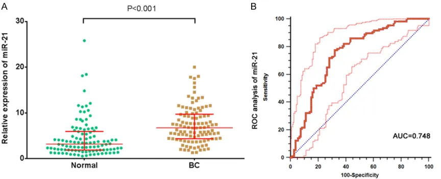
miR-10b) were examined in serum samples from 106 BC patients and 106 healthy participants. Serum relative levels of miR-155, miR-21, and miR-10b were plotted in the form of scatter dots in Figures 1A-3A. As shown, relative expression of miR-155, miR-21, and miR-10b in BC patients were 2.87 (1.43, 4.97), 6.74 (4.36, 9.71), 0.78 (0.34, 1.42) respectively. And the relative expression of 3 miRNAs were 1.44 (0.69, 3.05), 3.19 (1.86, 5.94), 0.25 (0.11, 0.47) in healthy participants. Statistically
sig-nificant difference can be observed between
BC patients and healthy participants (all P<0.001). Hence, these 3 miRNAs were able to discriminate BC patients from healthy controls based on their aberrant expression patterns.
Then we performed multivariate logistic
reg-ression to construct an new miRNA score: Score=0.25*miR-155+0.2*miR-21+1.59*miR-10b. As shown in Figure 4A, the score of BC patients was also higher than that of healthy min and in 40 cycles at 95°C for 15 s and 60°C
for 60 s on a ABI 7500 thermocycler (Applied
Biosystems). The relative expression of each
miRNA was calculated from the following equa-tion: relative expression 2-∆Ct, where Ct is the
threshold cycle for a sample, and ∆Ct=mean
CtmiRNA-mean Ctcel-miR-39. Each blood sample was performed in duplicate wells and repeated 3 times.
Statistical analysis
Continuous variables of normal distributions are expressed as mean ± standard deviation (SD) and skewed distributions are expressed as median (interquartile range). Categorical val-ues were expressed by absolute and relative frequencies. Differences in variables were ana-lyzed using Student t tests (for normally
distrib-uted data), Wilcoxon signed rank test (for
skewed distributed data) or the chi-square test (for categorical data). Receiver operating char-acteristic (ROC) curves were constructed based on the miRNAs levels between groups. Area under the ROC curve (AUC) was generated to assess the diagnostic values of the candidate
microRNAs. The cutoff values for microRNA lev -els were determined by Youden index. In
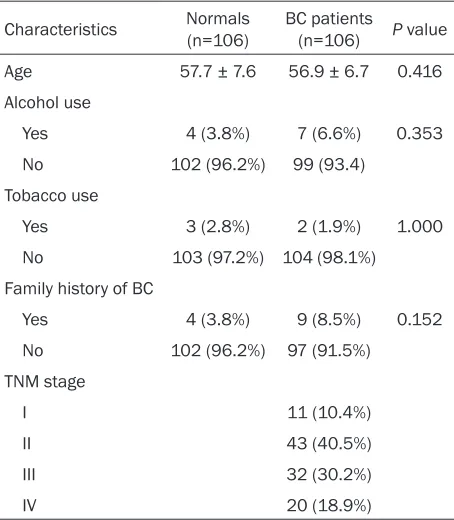
addi-Table 1. Characteristics of normals and breast can-cer patients
Characteristics Normals (n=106) BC patients (n=106) P value
Age 57.7 ± 7.6 56.9 ± 6.7 0.416
Alcohol use
Yes 4 (3.8%) 7 (6.6%) 0.353
No 102 (96.2%) 99 (93.4)
Tobacco use
Yes 3 (2.8%) 2 (1.9%) 1.000
No 103 (97.2%) 104 (98.1%)
Family history of BC
Yes 4 (3.8%) 9 (8.5%) 0.152
No 102 (96.2%) 97 (91.5%)
TNM stage
I 11 (10.4%)
II 43 (40.5%)
III 32 (30.2%)
IV 20 (18.9%)
tion, we performed multivariate logistic regression to construct an new miRNA Score including 155, 21 and miR-10b, which may have an more powerful diagnosability. A P value less than 0.05
was considered as statistically significant.
All statistical analysis were performed
by STATA version 12.0 software (Stata Corp, College Station, TX), and the graphs
were obtained from GraphPad Prism 5.0 (GraphPad Software Inc., CA).
Results
Clinical characteristics of study population
Serum samples were acquired from 216 subjects (106 BC patients, 106 healthy par-ticipants). As presented in Table 1, no
sig-nificant difference in age (57.7 ± 7.6 vs.
56.9 ± 6.7, P=0.416), alcohol use (3.8% vs. 6.6%, P=0.353) as well as tobacco use (2.8% vs. 1.9%, P=1.00) were observed between healthy participants and BC pati- ents group. A higher family history of BC was observed in BC patients, however the
[image:3.612.92.319.96.356.2]Figure 1. Diagnostic performance of serum miR-155 in differentiating breast cancer (BC) patients from participants. A. Relative expression levels of serum 155 in BC patients and participants. B. ROC curve analysis of serum miR-155 in differentiating BC patients from participants.
Figure 2. Diagnostic performance of serum miR-21 in differentiating breast cancer (BC) patients from participants. A. Relative expression levels of serum 21 in BC patients and participants. B. ROC curve analysis of serum miR-21 in differentiating BC patients from participants.
participants [3.86 (2.67, 5.60) vs. 1.76 (1.27, 2.46), all P<0.001].
Diagnostic performance of MicroRNAs in BC detection
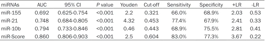
We further evaluated the diagnostic value of 3
selected miRNAs and the new miRNA score by ROC curves and AUC values. Figures 1B-4B shows the ability of the 3 miRNAs and miR-score to distinguish between BC patients and
healthy participants. The AUC of 155,
miR-21, and miR-10b were 0.692 (0.625-0.754), 0.748 (0.684-0.805), 0.794 (0.733-0.846), respectively. miR-Score had a highest AUC of 0.860 (0.806-0.903). More information about
the diagnosability were shown in Table 2. Among 3 selected miRNAs, miR-21 had a er sensitivity of 77.4% and miR-10b had a
high-er specificity of 75.5%. We also can obshigh-erve
that 10b had a higher+LR of 2.81 and miR-21 had a lower-LR of 0.33. However, compared with these 3 miRNAs, miR-Score showed the best diagnosability, with the highest sensitivity
of 83.0%, specificity of 77.3%, +LR of 3.67 and
lowest-LR of 0.22. Discussion
Currently, the identification of cancer-specific miRNAs profiles in the circulation is an emerg
[image:4.612.93.525.301.476.2]ideal diagnosability of 3 selected miRNAs, with AUCs of 0.692, 0.748 and 0.794, respectively. Furthermore, we constructed a new score, named miR-score, which show greater diagnos-ability than 3 selected miRNAs. Collectively, our study provides evidences that serum level of results suggested that plasma levels of
miR-155, miR-21 and miR-10b in BC patients were higher than those in healthy participants. Receiver operating characteristic (ROC) curves were used to assess the diagnostic values of
[image:5.612.94.519.73.237.2]the candidate microRNAs. We can observe
[image:5.612.92.518.305.469.2]Figure 3. Diagnostic performance of serum miR-10b in differentiating breast cancer (BC) patients from participants. A. Relative expression levels of serum 10b in BC patients and participants. B. ROC curve analysis of serum miR-10b in differentiating BC patients from participants.
Figure 4. Diagnostic performance of miR-score in differentiating breast cancer (BC) patients from participants. A. Relative expression levels of miR-score in BC patients and participants. B. ROC curve analysis of miR-score in dif-ferentiating BC patients from participants. miR-score=0.25 miR-155+0.2 miR-21+1.59 miR-10b.
Table 2. The diagnosis value of microRNAs normal and breast cancer patients
miRNAs AUC 95% CI P value Youden Cut-off Sensitivity Specificity +LR -LR
miR-155 0.692 0.625-0.754 <0.001 2.2 0.321 66.0% 68.9% 2.03 0.53
miR-21 0.748 0.684-0.805 <0.001 4.32 0.453 77.4% 67.9% 2.41 0.33
miR-10b 0.794 0.733-0.846 <0.001 0.46 0.443 68.9% 75.5% 2.81 0.41
miR-Score 0.860 0.806-0.903 <0.001 2.5 0.604 83.0% 77.3% 3.67 0.22
[image:5.612.91.525.548.616.2]regulates SOCS1 in breast cancer, in turn
lead-ing to persistent activation of STAT3 signallead-ing, which significantly promoted the proliferation
of cancer cells [21]. MiR-21 has been reported to promote oncogenesis and progression of various carcinomas via direct targeting of tumor suppressing phosphatase and tensin homolog
(PTEN) [22]. Then Liu et al. [23] found miR-21 is
up-regulated in breast cancer bell lines, clinical specimens, and serum samples and demon-strated miR-21 could promote oncogenesis by
miR-34b/c through affecting PTEN/PI3K/AKT/
FOXO3a signaling. A correlation between ele-vated miR-10b expression and poor prognosis was recent reported in gastric cancer, renal cancer, colorectal tumors, and bladder tumors [24-26]. miR-10b is a particularly interesting candidate given its close correlation with meta-static behaviors [27]. Moreover, higher miR-10b expression level was recently detected in serum from metastatic breast cancer patients [28], consisting with our result.
Obviously, the studies on circulating miRNA
pro-files offer an exciting expectation. A reliable
miRNA biomarker in circulation will dramatically facilitate the management of BC. However, the development of miRNA biomarker is cumber-some. A major concern is the different normal-ization strategies. Internal controls, like miR-16, may not be ideal for they are not always consistent across BC patients and controls [29, 30]. In present study, we took advantage of spiked-in cel-miR-39 for normalization. However, these synthetic miRNAs are less
sta-ble than endogenous miRNAs. Thus, finding an
ideal miRNA for normalization should be imper-ative. Also, other certain limitations in our
study should be mentioned. The population of
enrolled patients and healthy controls was rela-tively small. Further study on a larger sample is
needed to confirm our results. Secondly, the
plasma has been stored at -80°C for average 7 months before use. Immediate analysis should be taken after blood samples are collected for
a better reflection of real condition.
Taken together, this study extended the find -ings of previous studies about the serum levels of miR-155, miR-21 and miR-10b in breast can-cer patients. Our data provided complementary information on its diagnostic value, indicating new insights into the diagnosability of serum level of the 3 selected miRNAs. However, whe- ther this correlation is exactly proportional requires carefully scrutiny.
miR-155, miR-21 and miR-10b have great clini-cal value as promising biomarkers in BC pre-liminary screening.
Advanced technologies, such as microarray expression data, have shown that aberrant miRNAs expression is the rule rather than the exception in BC [11, 12]. BC-relative miRNAs, which have an important role in the pathophysi-ology of the disease, facilitating invasion,
epi-thelial to mesenchymal transition (EMT), and
maintenance of BC stem cells, have become an interesting topic in BC management [13, 14]. Recently, some attempts have been made to identify affordable BC signatures for diagnosis, and prediction of the therapeutic response.
With respect to diagnosis, Iorio et al. [15] found
that miRNAs aberrantly expressed in human breast cancer could clearly separate normal versus cancer tissues with 100% accuracy and
the conclusion were confirmed by microarray
and Northern blot analyses. Blenkiron et al.
[16] identified 133 miRNAs that displayed aber -rant expression levels in breast tumor tissues compared with normal breast tissues and
bead-based flow cytometric miRNAs expres
-sion profiling might be a suitable platform to
classify breast cancer into prognostic
molecu-lar subtypes. With respect to prediction, Rothé
F et al. [17] study showed that expression of miR-210 is linked to tumor proliferation and appears to be a strong potential biomarker of clinical outcome in BC.
Although direct measurements of tissue gene biomarkers have greatly improved BC diagno-sis, the invasive and unpleasant nature of the diagnostic procedures limits their application. Isolation and subsequent characterization of circulating miRNAs provide the opportunity to bypass the problems associated with tissue biopsy. Gloria Bertoli et al. [18] have depicted an overview of circulating miRNAs that can already be considered as BC biomarkers, such as 195, 16, 25, 222, miR-324-3p and so on. In the present study, we screened the level of circulating 155, miR-21 and miR-10b in BC patients and healthy
par-ticipants by using RT-qPCR. Mir-155 is a robust
[12] Shi M and Guo N. MicroRNA expression and its implications for the diagnosis and therapeutic strategies of breast cancer. Cancer Treat Rev 2009; 35: 328-334.
[13] Sotiropoulou G, Pampalakis G, Lianidou E and Mourelatos Z. Emerging roles of microRNAs as molecular switches in the integrated circuit of the cancer cell. RNA 2009; 15: 1443-1461. [14] Weber B, Stresemann C, Brueckner B and Lyko
F. Methylation of human microRNA genes in normal and neoplastic cells. Cell Cycle 2007; 6: 1001-1005.
[15] Iorio MV, Ferracin M, Liu CG, Veronese A, Spiz-zo R, Sabbioni S, Magri E, Pedriali M, Fabbri M, Campiglio M, Menard S, Palazzo JP, Rosenberg A, Musiani P, Volinia S, Nenci I, Calin GA, Quer-zoli P, Negrini M and Croce CM. MicroRNA gene expression deregulation in human breast can-cer. Cancer Res 2005; 65: 7065-7070. [16] Blenkiron C, Goldstein LD, Thorne NP, Spiteri I,
Chin SF, Dunning MJ, Barbosa-Morais NL, Te-schendorff AE, Green AR, Ellis IO, Tavare S, Caldas C and Miska EA. MicroRNA expression profiling of human breast cancer identifies new markers of tumor subtype. Genome Biol 2007; 8: R214.
[17] Rothe F, Ignatiadis M, Chaboteaux C, Haibe-Kains B, Kheddoumi N, Majjaj S, Badran B, Fayyad-Kazan H, Desmedt C, Harris AL, Piccart M and Sotiriou C. Global microRNA expression profiling identifies MiR-210 associated with tu-mor proliferation, invasion and poor clinical outcome in breast cancer. PLoS One 2011; 6: e20980.
[18] Bertoli G, Cava C and Castiglioni I. MicroRNAs: New Biomarkers for Diagnosis, Prognosis, Therapy Prediction and Therapeutic Tools for Breast Cancer. Theranostics 2015; 5: 1122-1143.
[19] Wang J and Wu J. Role of miR-155 in breast cancer. Front Biosci (Landmark Ed) 2012; 17: 2350-2355.
[20] Mitchell PS, Parkin RK, Kroh EM, Fritz BR, Wyman SK, Pogosova-Agadjanyan EL, Peter-son A, Noteboom J, O’Briant KC, Allen A, Lin DW, Urban N, Drescher CW, Knudsen BS, Stire-walt DL, Gentleman R, Vessella RL, Nelson PS, Martin DB and Tewari M. Circulating microR-NAs as stable blood-based markers for cancer detection. Proc Natl Acad Sci U S A 2008; 105: 10513-10518.
[21] Jiang S, Zhang HW, Lu MH, He XH, Li Y, Gu H, Liu MF and Wang ED. MicroRNA-155 functions as an OncomiR in breast cancer by targeting the suppressor of cytokine signaling 1 gene. Cancer Res 2010; 70: 3119-3127.
[22] Wickramasinghe NS, Manavalan TT, Dough-erty SM, Riggs KA, Li Y and Klinge CM. Estra-diol downregulates miR-21 expression and in-creases miR-21 target gene expression in Acknoweledgements
This study was supported by the scientific
research fund project of Shanghai jiaotong uni-versity school of medicine (12XJ10012). Disclosure of conflict of interest
None.
Address correspondence to: Yibing Peng, Depart- ment of Laboratory Medicine, Ruijin Hospital, Shanghai Jiaotong University School of Medicine, 197 Ruijin Second Road, Shanghai 20025, China. E-mail: pyb9861@sina.com
References
[1] Ferlay J, Soerjomataram I, Dikshit R, Eser S, Mathers C, Rebelo M, Parkin DM, Forman D and Bray F. Cancer incidence and mortality worldwide: sources, methods and major pat-terns in GLOBOCAN 2012. Int J Cancer 2015; 136: E359-386.
[2] Rim A and Chellman-Jeffers M. Trends in breast cancer screening and diagnosis. Cleve Clin J Med 2008; 75 Suppl 1: S2-9.
[3] Nattinger AB. In the clinic. Breast cancer screening and prevention. Ann Intern Med 2010; 152: ITC41.
[4] Viale G. The current state of breast cancer classification. Ann Oncol 2012; 23 Suppl 10: x207-210.
[5] Jiang D and Zhao N. A clinical prognostic pre-diction of lymph node-negative breast cancer by gene expression profiles. J Cancer Res Clin Oncol 2006; 132: 579-587.
[6] Shen J, Stass SA and Jiang F. MicroRNAs as potential biomarkers in human solid tumors. Cancer Lett 2013; 329: 125-136.
[7] Qu H, Xu W, Huang Y and Yang S. Circulating miRNAs: promising biomarkers of human can-cer. Asian Pac J Cancer Prev 2011; 12: 1117-1125.
[8] Bartel DP. MicroRNAs: target recognition and regulatory functions. Cell 2009; 136: 215-233.
[9] Lodes MJ, Caraballo M, Suciu D, Munro S, Ku-mar A and Anderson B. Detection of cancer with serum miRNAs on an oligonucleotide mi-croarray. PLoS One 2009; 4: e6229.
[10] Zhao H, Shen J, Medico L, Wang D, Ambrosone CB and Liu S. A pilot study of circulating miR-NAs as potential biomarkers of early stage breast cancer. PLoS One 2010; 5: e13735. [11] Andorfer CA, Necela BM, Thompson EA and
MCF-7 breast cancer cells. Nucleic Acids Res 2009; 37: 2584-2595.
[23] Liu X, Feng J, Tang L, Liao L, Xu Q and Zhu S. The regulation and function of miR-21-FOXO3a-miR-34b/c signaling in breast cancer. Int J Mol Sci 2015; 16: 3148-3162.
[24] Zaravinos A, Radojicic J, Lambrou GI, Volanis D, Delakas D, Stathopoulos EN and Spandidos DA. Expression of miRNAs involved in angio-genesis, tumor cell proliferation, tumor sup-pressor inhibition, epithelial-mesenchymal transition and activation of metastasis in blad-der cancer. J Urol 2012; 188: 615-623. [25] Li X, Zhang Y, Zhang Y, Ding J, Wu K and Fan D.
Survival prediction of gastric cancer by a sev-en-microRNA signature. Gut 2010; 59: 579-585.
[26] Heinzelmann J, Henning B, Sanjmyatav J, Posorski N, Steiner T, Wunderlich H, Gajda MR and Junker K. Specific miRNA signatures are associated with metastasis and poor progno-sis in clear cell renal cell carcinoma. World J Urol 2011; 29: 367-373.
[27] Ma L, Teruya-Feldstein J and Weinberg RA. Tu-mour invasion and metastasis initiated by mi-croRNA-10b in breast cancer. Nature 2007; 449: 682-688.
[28] Zhao FL, Hu GD, Wang XF, Zhang XH, Zhang YK and Yu ZS. Serum overexpression of microR-NA-10b in patients with bone metastatic pri-mary breast cancer. J Int Med Res 2012; 40: 859-866.
[29] McDonald JS, Milosevic D, Reddi HV, Grebe SK and Algeciras-Schimnich A. Analysis of circulat-ing microRNA: preanalytical and analytical challenges. Clin Chem 2011; 57: 833-840. [30] Creemers EE, Tijsen AJ and Pinto YM.