Original Article
Inhibition of IL17A promotes bufalin-induced
apoptosis in colon cancer cells via miR-96/DDIT3
Honghua Cao1, Wei Zhang1, Junlin Liang2
1Department of Gastrointestinal Surgery, Guigang City People’s Hospital, Guigang, Guangxi, China; 2Department
of Gastrointestinal Surgery, The First Affiliated Hospital of Guangxi Medical University, Nanning, Guangxi, China
Received November 29, 2015; Accepted January 26, 2016; Epub April 1, 2016; Published April 15, 2016
Abstract: Bufalin is used clinically to treat patients with many solid malignant tumors. However, the mechanisms remain to be further elucidated. Our study focused on IL17A involved in bufacin inducing apoptosis of colon cancer cells. The data showed that bufalin could induce colon cancer cell apoptosis via inhibiting IL17A. Ectopic expression of IL17A promoted the proliferation and induced anti-apoptosis of colon cancer cells by MTT and flow cytometry analysis. Further study verified bufalin inhibiting IL17A induced apoptosis was through miR-96-DDIT3, our study demonstrated that bufalin may inhibit the proliferation and promote the apoptosis of colon cancer cells. Bufalin-associated IL-17A inhibition may indirectly be involved in cell proliferation and apoptosis by miR-96 targeting DDIT3, pointing to use as a potential molecular target of bufacin in colon cancer therapy.
Keywords: Bufalin, IL17A, DDIT3, colon cancer, miR-96
Introduction
Colon cancer is one of the common cancers in the world and has higher cancer mortality and morbidity in developed countries [1]. Many studies indicated that the malignant transfor-mation and progression of cancer due to epi-genetic changes and oncogenic signaling acti-vation. In recent years, the epigenetic altera-tions, in particular, the aberrant expression of microRNAs (miRNAs), have been shown critical roles in cancer formation, metastasis, and response to cancer therapy [1-3]. However, the interaction between immune cells, inflamma-tory cytokines, and cancer evolution is still largely unknown. Recently, several inflammato-ry cytokines have been shown to promote colon cancer progression [4].
IL-17 (IL-17A), initially termed as cytotoxic T-ly- mphocyte-associated antigen (CTLA)-8, is the member of IL-17 cytokine family consisting of six homologous proteins (from IL-17A to IL-17F) [5, 6]. A large body of evidence suggests that IL-17 is an essential proinflammatory cytokine due to inducing a mass of cytokines and che-mokines secretion by distinct cell types, such as mesenchymal cells and myeloid cells, which recruit monocytes and neutrophils into the site
of inflammation [7]. IL-17 promotes the expres-sion of antimicrobial peptides from epithelial cells and facilitates host defense against infec-tions [6-8]. This evidence indicates that IL-17 is an important inflammatory cytokine which links innate and adaptive immunity. Recently, several studies have shown that IL-17 has either a pro-tumor or antipro-tumor role in different cancer mod-els. However the majority of studies consider that IL-17 acts as a promoter in tumor initiation and progression in colon [9].
Bufalin, one of Chinese medicine, inhibits cell proliferation and induces apoptosis in various tumor cell lines [10, 11], including colon cancer [12]. However, the precise molecular mecha-nisms of the bufalin induced apoptosis of col-on cancer cells are still unclear. At the present study, we will investigate mechanism of bufalin induced colon cancer cell apoptosis. We found that bufalin could promote cell apoptosis via inhibiting IL-17A-miR-96-DDIT3 in colon cancer. Material and methods
Cell culture
SW620 were obtained primarily from the Ame-rican Type Culture Collection (ATCC, Manassas, VA, USA). The cells were cultured in DMEM sup-plemented with 10% fetal bovine serum, 100 U/mL of penicillin and 100 µg/mL of streptomy-cin. Cells were cultured at 37°C in a humidified atmosphere of 5% CO2.
MiRNA and siRNA transfection
The lentiviral vectors mediated miR-96, IL-17A and DDIT3 were constructed. Lentivirus was producted using 293T cells transfected with lentivirus vectors and packaging plasmids with Lipofectamine 2000 (Invitrogen, Carlsbad, CA, USA) according to the manufacturer’s instruc-tions. The expression of miR-96 was examin- ed by qRT-PCR. IL-17A and DDIT3 siRNAs were ordered from Sigma (Sigma-Aldrich, Saint Lo- uis, MO, USA).
ELISA assay
Cytokine levels were measured in the superna-tant of colon cancer cells with bufalin
treat-siRNA and then counted and re-seeded to 96-well plates with 5×10³ cells per well. 10 μl MTT solution (5 mg/ml) was added to each well at day 1, 3 and 5, and then the cells were cul-tured for 4 h. After the incubation, the crystal was dissolved with 0.1% SDS and finally the ELISA reader was used to measure the absor-bance at 570 nm.
Colony forming assay
Colon cancer cells were transfected the miR-NAs, seeded in 6 well plates (200 cells in every well) and incubated at 37°C, 5% CO2 for 24 h. Non-adherent cells were removed. Cell growth medium was changed every 3-5 days. The colo-nies with more than 50 cells were counted after 14 days.
Cell apoptosis
[image:2.629.101.382.79.351.2]Colon cancer cells transfected with plasmids were seeded in 6-well plates and harvested after 48 h. The cells were stained with 5 μl of Annexin V and 5 μl of propidium iodide (BD
Figure 1.Bufalin inhibited IL-17A expression in colon cancer cells. A. Cyto-kines profile in HT-29 colon cancer cells with exposure to bufalin for 24 h. The conditioned medium was collected for cytokine ELISA assay. B. Colon cancer cells including HCT116, HCT8, HT29, LS174T, LOVO, SW480 and SW620 were treated with bufalin for 24 h, and the conditioned medium was assayed for IL-17A protein by ELISA.
ment. ELISA assays were per-formed with Quantikine kits according to the manufac- turer’s instructions (R&D Sys- tems, Inc., Minneapolis, MN, USA).
QRT-PCR
Total RNA from the cells trans-fected with miRNAs or siRNA was isolated from the cells using Trizol reagent (Invitro- gen, Carlsbad, CA, USA). QRT-PCR was performed to mea-sure mRNA expression. Pri- mers were designed and or- dered from Sangon Biotech (Shanghai, China). The rela-tive expression levels were calculated by comparing Ct values of the samples with those of the reference, all data normalized to the inter-nal control GAPDH or U6 snRNA.
MTT assay
Biosciences, San Jose, CA, USA) for 15 min at room temperature. The apoptosis rate (%) of the stained cells was analyzed using a flow cytometry (BD, USA).
Hoechst staining
Hoechst 33342 dye (Molecular Probes, Eugene, OR, USA) was used to stain the nuclei. Cells
were transfected miRNAs and incubated in the Hoechst labeling solution for 30 mins at room temperature. Images were obtained using a Leica DM IRB inverted fluorescence micro-scope (Wetzlar, Germany) at 400× magnifi- cation.
Western blot analysis
Colon cancer cells were lysed in RIPA lysis buf-fer with protease and phosphatase inhibitors. The protein concentration was determined using Bradford assay (Bio-Rad, Philadelphia, PA).Equivalent protein was performed to SDS-PAGE and transferred to NC membranes (Mi- llipore, Bedford, MA, USA). The NC membranes with protein blotting was blocked in 5% non-fat, incubated with primary antibodies, and then incubated with secondary antibodies conjugat-ed with HRP. Blots were visualizconjugat-ed on X-ray films using an ECL detection system (Pierce, IL).
Statistical analysis
Data were analyzed by SPSS 13.0 software and presented as mean ± SEM of at least three independent experiments. Two-tailed Student’s t test was used for comparisons of two inde-pendent groups. Gene expression was ana-lyzed by Mann-Whitney U test. P values of <0.05 were considered statistically significant. Results
Bufalin inhibits IL-17A expression in colon can-cer cells
[image:3.629.98.296.79.447.2]Cytokines play important roles in cancer pro-gression. To investigate whether bufalin influ-ence cytokines in colon cancer, the colon can-cer cells were treated with bufalin and condi-tioned medium was collected for ELISA analy-sis. The results showed that bufalin could induce changes of many cytokines’ expression (Figure 1A). IL-17A decreased significantly in HT-29 cell. Cytokines such as IL-6 decreased, but it was research widely. In our research, we focused on IL-17A. To verify the result, HCT116, HCT8, HT29, LS174T, LOVO, SW480 and SW620 cells were treated with bufalin and IL-17A expression was evaluated by ELISA. We found IL-17A protein was reduced in most of the cell lines with bufalin treatment (Figure 1B). These gave us the evidence that IL-17A inhibition co- uld have a suppressive effect on colon cancer.
Inhibition of IL-17A promotes bufalin-induced apoptosis
Bufalin could induce apoptosis in various can-cer cells. To answer whether IL-17A deletion induces colon cancer cell apoptosis, HT-29 and SW480 cells were transfected with IL-17A siR-NAs or siRNA control, IL-17A mRNA and protein levels was decreased (Figure 2A). Cell apopto-sis was examined by flow cytometry and it was shown that in the cells with IL-17A down-regula-tion, apoptosis rate increased compared with the control in HT-29 and SW480 cells (Figure 2B). Apoptosis associated protein was ana-lyzed by western blot, and it was found that DDIT3 protein level was higher in HT-29 and SW480 cells with IL-17A inhibition (Figure 2C). Bcl-2 decreased and Bax increased. These data indicated that inhibition of IL-17A could promote cell apoptosis in HT-29 and SW480 cells. It is interesting that DDIT3 increases in the colon cells with IL-17A inhibition.
IL-17A down-regulates DDIT3 expressions via miR-96
[image:4.629.98.531.78.440.2]It is interesting that DDIT3 increases in the colon cells with IL-17A inhibition. To investigate the potential molecule that regulates DDIT3 up-regulation in colon cancers, we next checked whether IL-17A down-regulates DDIT3 expres-sion in colon cancer cells through miRNAs. DDIT3 may be regulated by many miRNAs including miR-96. HT-29 cells were treated with IL-17A, and the predicted miRNAs were ana-lyzed and the data showed that miR-96 increased (Figure 3A). Bioinformatic analysis showed that DDIT3 was directly suppressed by miR-96 (Figure 3B). As shown in Figure 3C, the luciferase activity of wide type of DDIT3 3’UTR in HT29 cells was much lower than in control cells. The luciferase activity of mutation of DDIT3 3’UTR was rescued in the cells. Compared with control, endogenous DDIT3 mRNA levels (Figure 3D) were down-regulated
when HT29 and SW480 cells were transfected with miR-96. DDIT3 protein was down-regulat-ed in the cells with miR-96 (Figure 3E). So, DDIT3 is involved in bufalin induced colon can-cer cell apoptosis by miR-96.
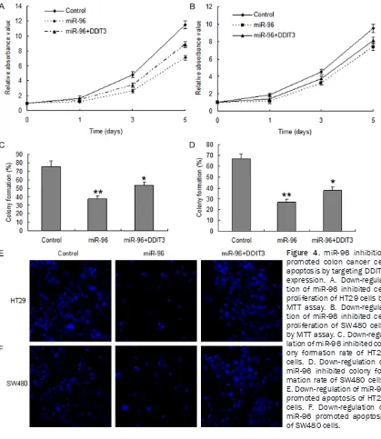
MiR-96 inhibition promotes colon cancer cell apoptosis by targeting DDIT3 expression
To further research the mechanism of DDIT3 regulation, colon cancer cells were transfected with miR-96 inhibitor for cellular function analy-sis. HT29 and SW480 cells were transfected with miR-96 inhibitor in or the control
(miRNA-control), the results of MTT assay displayed that miR-96 inhibited cell proliferation in HT29 cells (Figure 4A) and SW480 cells (Figure 4B). The colony formation rate in the two cells with miR-96 overexpression increased compared with the controls (Figure 4C and 4D). Cell apop-tosis was increased in the HT29 and SW480 cells with miR-96 inhibition (Figure 4E and 4F).
Correlation of miR-96 and DDIT3 in colon can-cer tissues
[image:5.629.103.522.81.564.2]Above data suggested that miR-96 inhibits colon cancer cell apoptosis by targeting DDIT3.
In order to investigate miR-96 expression in colon cancer tissues, the data from real time RT-PCR showed that miR-96 was up-regulated in colon cancer tissues than their normal tis-sues (Figure 5A and 5B). The clinic data showed that miR-96 was negatively associated with DDIT3 protein in colon cancer tissues (Figure 5C).
Discussion
Bufalin, a class of toxic steroids purified from Chinese traditional medicine, has therapeutic effect on cancer, but its molecular mechanism on cancer is unclear. In the present study, we found that bufalin could suppress IL-17A expres-sion in colon cancer cells. Further study showed that deletion of IL-17A could lead to cell apopto-sis and DDIT3 expression. DDIT3 was identified as a target gene of miR-96 stimulated by IL-17A in colon cancer cells.
MiRNAs are a class of small noncoding RNAs that typically inhibit the translation and stability of messenger RNAs (mRNAs) by binding to the 3’-untranslated regions (3’-UTR) of their target
mRNAs. MiRNAs have 19-22 nucleotides and are found in all multi-cellular eukaryotic cells. MiRNAs have important roles in various biologi-cal and pathologibiologi-cal processes, such as devel-opment, cell proliferation, differentiation, apop-tosis, inflammation, stress response and migra-tion. Recent studies showed that the differen-tial expression of miR-96 in many type of can-cers, such as esophageal cancer, colorectal cancer, pancreatic cancer, glioma, breast can-cer and etc. The target genes of miR-96 that identified such as RECK, FOXO1, FOXO3a, NU- AK1, HERG1 and HBP [13-26]. Previous study suggested that miR-96 acted as a tumor sup-pressor miRNA. Our data show that miR-96 is up-regulated in advanced and metastatic colon cancer by targeting DDIT3. Taken together with previous studies, it suggests that roles of miR-96 can be complicated by regulating different downstream effectors in different cancer con-texts and it functions mainly as an oncogen in colon cancer.
[image:6.629.98.530.80.362.2]DNA-damage-inducible transcript 3 (DDIT3) is also known as CHOP, growth arrest- and DNA damage-inducible gene 153 (GADD153), and
C/EBPg [27]. DDIT3 protein is composed of two known functional domains, an N-terminal tran-scriptional activation domain and a C-terminal basic-leucine zipper (bZIP) domain consisting of a basic amino-acid-rich DNA-binding region followed by a leucine zipper dimerization motif [27-29]. There are reports showed that bZIP domain is important for CHOP-induced apopto-sis [27, 30, 31]. Re-expression of DDIT3 could promote cell apoptosis of colon cancer cells. However, in mouse model studies, re-expres-sion of DDIT3 could significantly but not com-pletely reverse the miR-96-imposed promotion on tumor growth, which suggests that other potential targets of miR-96 may have other genes.
Our present study has identified bufalin could inhibit IL-17A expression in colon cancer cells. IL-17A inhibits cell apoptosis via targeting DDIT3, an apoptosis inhibitor. DDIT3 is regulat-ed by miR-96 which is negatively correlatregulat-ed in colon cancer tissues. Our finding suggested that IL-17A and miR-96 are therapeutic targets for colon cancer.
Disclosure of conflict of interest
None.
Address correspondence to: Junlin Liang, De- partment of Gastrointestinal Surgery, The First Affiliated Hospital of Guangxi Medical University, 6 Shuangyong Road, Nanning 53000, Guangxi, China. E-mail: ljunlin15@sina.com
References
[1] Siegel R, Ma J, Zou Z, Jemal A. Cancer statis-tics, 2014. CA Cancer J Clin 2014; 64: 9-29. [2] Giovannucci E. Modifiable risk factors for colon
cancer. Gastroenterol Clin North Am 2002; 31: 925-943.
[3] Itzkowitz SH and Yio X. Inflammation and can-cer IV. Colorectal cancan-cer in inflammatory bowel disease: the role of inflammation. Am J Physiol Gastrointest Liver Physiol 2004; 287: G7-17. [4] Chiba T, Marusawa H, Ushijima T.
Inflammation-associated cancer development in digestive organs: mechanisms and roles for genetic and epigenetic modulation. Gastroenterology 2012; 143: 550-63.
[5] Wang K, Kim MK, Di Caro G, Wong J, Shalapour S, Wan J, Zhang W, Zhong Z, Sanchez-Lopez E, Wu LW, Taniguchi K, Feng Y, Fearon E, Grivennikov SI, Karin M. Interleukin-17 recep-tor a signaling in transformed enterocytes
pro-motes early colorectal tumorigenesis. Immu- nity 2014; 41: 1052-63.
[6] Chin CC, Chen CN, Kuo HC, Shi CS, Hsieh MC, Kuo YH, Tung SY, Lee KF, Huang WS. Interleukin-17 induces CC chemokine receptor 6 expression and cell migration in colorectal cancer cells. J Cell Physiol 2014; 230: 1430-7. [7] Omrane I, Marrakchi R, Baroudi O, Mezlini A, Ayari H, Medimegh I, Stambouli N, Kourda N, Bouzaienne H, Uhrhammer N, Bougatef K, Bignon YJ, Benammar-Elgaaied A. Significant association between interleukin-17A polymor-phism and colorectal cancer. Tumour Biol 2014; 35: 6627-32.
[8] Chung AS, Wu X, Zhuang G, Ngu H, Kasman I, Zhang J, Vernes JM, Jiang Z, Meng YG, Peale FV, Ouyang W, Ferrara N. An interleukin-17-me-diated paracrine network promotes tumor re-sistance to anti-angiogenic therapy. Nat Med 2013; 19: 1114-23.
[9] Li L, Boussiotis VA. The role of IL-17-producing Foxp3+ CD4+ T cells in inflammatory bowel disease and colon cancer. Clin Immunol 2013; 148: 246-53.
[10] Zhu Z, Li E, Liu Y, Gao Y, Sun H, Ma G, Wang Z, Liu X, Wang Q, Qu X, Liu Y, Yu Y. Inhibition of Jak-STAT3 pathway enhances bufalin-induced apoptosis in colon cancer SW620 cells. World J Surg Oncol 2012; 10: 228.
[11] Xie CM, Chan WY, Yu S, Zhao J, Cheng CH. Bufalin induces autophagy-mediated cell de- ath in human colon cancer cells through reac-tive oxygen species generation and JNK activa-tion. Free Radic Biol Med 2011; 51: 1365-75. [12] Hu Q, Liang B, Sun Y, Guo XL, Bao YJ, Xie DH,
Zhou M, Duan YR, Yin PH, Peng ZH. Preparation of bufalin-loaded pluronic polyetherimide na- noparticles, cellular uptake, distribution, and effect on colorectal cancer. Int J Nanomedicine 2014; 9: 4035-41.
[13] Xia H, Chen S, Chen K, Huang H, Ma H. MiR-96 promotes proliferation and chemo-or radiore-sistance by down-regulating RECK in esopha-geal cancer. Biomed Pharmacother 2014; 68: 951-8.
[14] Gao F, Wang W. MicroRNA-96 promotes the proliferation of colorectal cancer cells and tar-gets tumor protein p53 inducible nuclear pro-tein 1, forkhead box propro-tein O1 (FOXO1) and FOXO3a. Mol Med Rep 2015; 11: 1200-6. [15] Ma Y, Yang HZ, Dong BJ, Zou HB, Zhou Y, Kong
XM, Huang YR. Biphasic regulation of autopha-gy by miR-96 in prostate cancer cells under hypoxia. Oncotarget 2014; 5: 9169-82. [16] Huang X, Lv W, Zhang JH, Lu DL. miR--96
func-tions as a tumor suppressor gene by targeting NUAK1 in pancreatic cancer. Int J Mol Med 2014; 34: 1599-605.
functions as an oncogene in pancreatic cancer and is downregulated by miR-96. Oncotarget 2014; 5: 5832-44.
[18] Yan Z, Wang J, Wang C, Jiao Y, Qi W, Che S. miR-96/HBP1/Wnt/β-catenin regulatory circuitry promotes glioma growth. FEBS Lett 2014; 588: 3038-46.
[19] Yu JJ, Wu YX, Zhao FJ, Xia SJ. miR-96 promotes cell proliferation and clonogenicity by down-regulating of FOXO1 in prostate cancer cells. Med Oncol 2014; 31: 910.
[20] Zhang J, Kong X, Li J, Luo Q, Li X, Shen L, Chen L, Fang L. miR-96 promotes tumor proliferation and invasion by targeting RECK in breast can-cer. Oncol Rep 2014; 31: 1357-63.
[21] Guo H, Li Q, Li W, Zheng T, Zhao S, Liu Z. MiR-96 downregulates RECK to promote growth and motility of non-small cell lung cancer cells. Mol Cell Biochem 2014; 390: 155-60. [22] Fendler A, Jung M, Stephan C, Erbersdobler A,
Jung K, Yousef GM. The antiapoptotic function of miR-96 in prostate cancer by inhibition of FOXO1. PLoS One 2013; 8: e80807.
[23] Haflidadóttir BS, Larne O, Martin M, Persson M, Edsjö A, Bjartell A, Ceder Y. Upregulation of miR-96 enhances cellular proliferation of pros-tate cancer cells through FOXO1. PLoS One 2013; 8: e72400.
[24] Wang Y, Huang JW, Calses P, Kemp CJ, Taniguchi T. MiR-96 downregulates REV1 and RAD51 to promote cellular sensitivity to cispla-tin and PARP inhibition. Cancer Res 2012; 72: 4037-46.
[25] Xu XM, Qian JC, Deng ZL, Cai Z, Tang T, Wang P, Zhang KH, Cai JP. Expression of 21, miR-31, miR-96 and miR-135b is correlated with the clinical parameters of colorectal cancer. Oncol Lett 2012; 4: 339-345.
[26] Lin H, Dai T, Xiong H, Zhao X, Chen X, Yu C, Li J, Wang X, Song L. Unregulated miR-96 induces cell proliferation in human breast cancer by downregulating transcriptional factor FOXO3a. PLoS One 2010; 5: e15797.
[27] Oyadomari S, Mori M. Roles of CHOP/GADD- 153 in endoplasmic reticulum stress. Cell Death Differ 2004; 11: 381-9.
[28] Ron D and Habener JF. CHOP, a novel develop-mentally regulated nuclear protein that dimer-izes with transcription factors C/EBP and LAP and functions as a dominant-negative inhibi- tor of gene transcription. Genes Dev 1992; 6: 439-453.
[29] Ubeda M, Wang XZ, Zinszner H, Wu I, Habener JF and Ron D. Stress-induced binding of the transcriptional factor CHOP to a novel DNA control element. Mol Cell Biol 1996; 16: 1479-1489.
[30] Matsumoto M, Minami M, Takeda K, Sakao Y and Akira S. Ectopic expression of CHOP (GADD153) induces apoptosis in M1 myelo-blastic leukemia cells. FEBS Lett 1996; 395: 143-147.