Original Article
The roles of phenotypic transformation of vascular
smooth muscle cells regulated by AGEs in the
thoracic aortic dissection in neonatal rats
Huaping Wu1,2, Jun Cheng1, Wen Huang1, Fenghe Li1, Li Zhang2, Xiang Li2, Yu Zhao1
1Department of Vascular Surgery, The First Affiliated Hospital of Chongqing Medical University, Chongqing, PR
China; 2Department of Vascular Surgery, Dazhou Central Hospital, Dazhou, PR China
Received December 2, 2015; Accepted February 13, 2016; Epub March 1, 2016; Published March 15, 2016
Abstract: Objective: This study aims to observe the expression of advanced glycation end products (AGEs) and its receptor (RAGE) and explore the roles of phenotypic transformation of vascular smooth muscle cells (VSMCs) regulated by AGEs in the thoracic aortic dissection (TAD) in neonatal rats. Methods: The thoracic aortic dissection in neonatal rat model was established. The expression levels of AGEs and RAGE in thoracic aortic dissection tissues were detected. VSMCs phenotype transformation model was established by recombinant platelet derived growth factor BB (PDGF-BB) stimulation and starvation culture methods. The expression of RAGE was silenced by siRNA to explore the roles of phenotypic transformation of VSMCs regulated by AGEs in the thoracic aortic dissection in neonatal rats. The expression changes of SM α-actin and calponin were detected by western blotting method. The expression changes of AGEs and RAGE were detected by ELISA and RT-PCR methods. Results: The expression levels of AGEs and its receptor RAGE in thoracic aortic dissection were significantly higher than that in normal thoracic aorta (P<0.01). The expression of AGEs and RAGE were negatively correlated with that of SM α-actin and calponin. The regulation of AGEs on phenotypic transformation of VSMCs was blocked after silencing RAGE. Conclusions: AGEs can promote the transformation of VSMCs from the contractile type to the synthetic type in the thoracic aortic dissection and aortic aneurysm tissue.
Keywords: Thoracic aortic dissection, advanced glycation end products, receptor for advanced glycation end prod-ucts, vascular smooth muscle cells
Introduction
Thoracic aortic dissection (TAD) refers to the lesions that longitudinal separation between the aortic intima and the middle occurred and true and false lumen were formed in the arter-ies, which was caused by a sudden pressure increase within the vascular lumen after the lesion in the tunicae media occurred, and then a direct, retrograde, or bidirectional dissecting aneurysm formed or ruptured. Its mortality is very high [1, 2]. At present, the main treatment methods include endovascular graft replace-ment surgery and traditional graft replacereplace-ment surgery, the mortality and postoperative com-plication rates were high [3].
The aortic wall has contractibility and elasticity. Vascular smooth muscle cells (VSMCs) play an important role in the maintenance of vascular
homeostasis and repair of vascular injury. The phenotype of VSMCs was divided into two types: synthetic VSMCs and contractile VSMCs. Synthetic VSMCs have strong ability of migra-tion, proliferation and secretion. Contractile VSMCs highly expressed smooth muscle spe-cific genes and had strong contractibility [4, 5]. The mature VSMCs were able to convert between the two types. VSMCs could transform from the contractile type to the synthetic type in the thoracic aortic dissection and the middle membrane tissue of aneurysm [6]. Smooth muscle alpha-actin (SMα-actin) and calponin can be used to evaluate the phenotypic trans-formation of VSMCs.
tis-sues or cells to destroy them. The biological effect of AGEs is related to the expression of RAGE (Receptor for AGE) [9]. Interactions be- tween AGEs and RAGE can activate a variety of signaling pathways related to cell prolifera-tion and apoptosis, which is associated with the occurrence and development of diabetes, Alzheimer’s disease, atherosclerosis and other diseases [10-13]. Vascular endothelial cells, smooth muscle cells, fibroblasts, macrophages and even neurons express RAGE or have AGEs binding site. At present, the roles of AGEs in the transformation of the arterial VSMCs pheno-type in patients with TAD remain unclear. In this study we observe the expression of AGE and RAGE and explore the roles of phenotypic trans-formation of VSMCs regulated by AGEs in the thoracic aortic dissection in neonatal rats, which could provide theoretical basis for the prevention, diagnosis and treatment of the disease.
Materials and methods
Cell culture
USMC cell lines were purchased from American type culture collection (ATCC). They were cul-tured with DMEM medium containing 10% fetal calf serum at 37°C with 5% CO2.
natal rat thoracic aortic dissection model was established in pregnant rats (n=25). Semicar- bazlde (25 mg/kg.d) was casted to the implan- table capsule osmotic pump and implanted inside the abdomen of pregnant 14 d rats. Neonatal mice were sacrificed after anesthesia and thoracic aortic tissues were taken out [14]. All experimental procedures were approved by the Care of Experimental Animals Committee of our hospital.
HE staining
The thoracic aortic tissue was cut into small pieces and fixed in 10% formalin and embed-ded in paraffin routinely. The paraffin blocks of specimen were cut into continuous sections with 4 μm respectively. The sections were de- waxed with xylene and washed with ethanol and water. They were stained with hematoxylin after that and then differentiated, washed and stained with eosin, then dehydrated, hyalinized and finally mounted on slides and observed under microscope, pictures were taken.
Preparation and detection of VSMCs pheno-type transformation model
[image:2.629.98.370.95.182.2]VSMCs phenotype transformation model was established by platelet derived growth factor BB (PDGF-BB) stimulation and starvation cul-ture methods respectively. Different concentra-tion of PDGF-BB (0 ng/ml, 2 ng/ml, 5 ng/ml and 7 ng/ml) was added into DMEM medium and culture for 48 h and then detection. VSMCs were detected after starvation treatment for 0 h, 24 h, 48 h and 72 h respectively. Successfully established model cells were performed AGEs stimulation experiment, they were divided into control group, 40 μg/mL AGEs group, 20 μg/mL AGEs group, 10 μg/mL AGEs group and 5 μg/ mL AGEs group respectively.
Table 2. The synthetic system of inverse transcription
Components Volume per Reaction dNTP Mix 4 μl Primer Mix 2 μl RNA Template 4 μl 5×RT Buffer 4 μl
DTT 2 μl
[image:2.629.100.300.239.345.2]RAGE siRNA
SiRNA oligonucleotides corresponding to RAGE (5’-CGAGAATCACGCTGC ATGACCATGT-3’) were used for transfection of VSMCs cells using lipo-fectamine 2000 (Life Technologies) according to the manual. The cells were divided into con-trol group, AGEs 40 μg/mL group and RAGE siRNA+AGEs 40 μg/mL group.
Enzyme linked immunosorbent assay (ELISA)
AGEs was detected after grinding the tissue samples with ELISA kit according to the manu-al. OD values at 450 nm were determined by ELISA detector. Standard curve was drawn to calculate the concentration.
Protein extraction and western blotting
The tissues were lysed with RIPA lysis buffer and total proteins were extracted and analyzed with SDS-PAGE electrophoresis. Then it was electrotransferred to the PVDF membrane. After the transmembrane, PVDF membrane was rinsed with TBS for 10 to 15 min, placed in TBS/T blocking buffer containing 5% (w/v) skimmed milk powder and shook at room tem-perature for one hour. It was incubated at room temperature for two hours after added with appropriate dilution degree of primary antibody (diluted with TBST containing 1% (w/v) skimmed milk powder). Then the membrane was rinsed with TBST for three times (5 to 10 minutes one time). The membrane was incubated at room temperature for one hour with HRP labeled sec-ondary antibody (1:10000) diluted with TBST
containing 0.05% (w/v) skimmed milk powder and rinsed for three times with TBST (5 to 10 minutes at a time). The protein bands were scanned and quantified as a ratio to β-actin.
RNA extraction and real-time PCR
The harvested cells were washed with RNase free PBS. Total RNA was extracted using RNeasy Mini Kit (Qiagen) according to the man-ufacturer’s protocol. Their concentration and purity were detected with Qubit Fluorometer. 1 μg RNA was subjected to reverse transcription using reverse transcription kit (Promega). Real-time PCR were performed using SYNBR Green PCR Master Mix (Qigen). The primers used in this study were shown in Table 1, the synthetic system of PCR were shown in Table 2. β-actin gene was used as an internal control for nor-malization of RNA quantity and quality differ-ences in all samples.
Statistical analysis
The results are expressed as mean ± S.D. and analyzed with SPSS 16.0 software, student t-test was used to evaluate the differences between groups. A value of P<0.05 and P<0.01 were taken to denote statistical significance.
Results
Establishment of thoracic aortic dissection model
[image:3.629.99.533.78.269.2]As shown in Figure 1, the histopathological fea-tures of thoracic aortic dissection were middle
arterial degeneration, lamellar necrosis with smooth muscle cell loss. We established tho-racic aortic dissection model successfully.
Expression of AGEs in thoracic aortic tissues
[image:4.629.100.295.80.215.2]The expression levels of AGEs were shown in
Figure 2. It showed that the expression levels of AGEs in thoracic aortic dissection tissues were significantly higher than that of normal thoracic aortic dissection tissues (7350.97± 31.01 ng/ml vs 6830.88±35.46 ng/ml, P< 0.01).
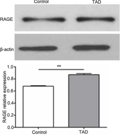
Expression of RAGE in thoracic aortic tissues
Western blotting results were shown in Figure 3. It showed that the expression levels of RAGE
in thoracic aortic dissection tissues were sig-nificantly higher than that of normal thoracic aortic dissection tissues (P<0.01).
Establishment of VSMCs phenotype transfor-mation model by PDGF-BB stimulation
[image:4.629.334.529.83.219.2]As shown in Figure 4, the concentration of AGEs gradually decreased with the increase of PDGF-BB concentration. Western blotting re- sults showed that the expression levels of SMα-actin and Calponin increased with the increase of PDGF-BB concentration (Figure 5). RT-PCR results showed that the expression levels of RAGE decreased with the increase of PDGF-BB concentration (Figure 6).
[image:4.629.101.295.269.489.2]Figure 2.Expression of AGEs in thoracic aortic tis-sues.
Figure 3.Expression of RAGE in thoracic aortic tis-sues.
Figure 4. AGEs expression changes with different concentrations of PDGF-BB.
[image:4.629.336.528.271.503.2]Establishment of VSMCs phenotype transfor-mation model by starvation treatment
ELISA results of AGEs were shown in Figure 7. It showed that the concentration of AGEs gradu-ally increased with the time of starvation treat-ment. Western blotting results showed that the expression levels of SMα-actin and Calponin decreased with the time of starvation treat-ment (Figure 8). RT-PCR results showed that the expression levels of RAGE increased with the time of starvation treatment.
Phenotype transformation of VSMCs in differ-ent concdiffer-entration of AGEs groups
Western blotting results were shown in Figure 9. It showed that the expression levels of SMα-actin and Calponin decreased with the increase of AGEs concentration. RT-PCR results showed that the expression levels of RAGE increased with the increase of AGEs concentration.
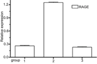
RAGE siRNA results
Western blotting results were shown in Figure 10. It showed that AGEs could down-regulate the expression levels of SMα-actin and Cal- ponin, while the expression levels of SMα-actin and Calponin increased after silencing RAGE.
[image:5.629.332.531.78.275.2]Figure 11 showed that RAGE expression de- creased in RAGE siRNA group.
[image:5.629.99.297.81.223.2]Figure 6.RT-PCR results of RAGE expression under different concentrations of PDGF-BB.
[image:5.629.100.294.280.414.2]Figure 7.AGEs gradually increased with the time of starvation treatment.
Figure 8.Western blotting results of SMα-actin and Calponin expression on different starvation treat-ment time.
[image:5.629.333.531.337.554.2]Discussion
TAD is a common serious cardiovascular dis-ease and its prognosis is poor, the incidence rate was about 3 cases/100,000 [14, 15]. It was found that the interaction of RAGE and AGEs is related to the development of a variety of diseases [11-13]. In this study we found that AGEs levels in TAD tissues were higher than that of normal tissue, which suggested that AGEs may play an important role in the
develop-Increased SMα-actin and Calponin predicated that VSMCs transformed to synthetic type, oth-erwise they transformed to contractile type. PDGF-BB is an important promoting mitogenic factor; it could stimulate VSMCs transform to contractile type. Starvation treatment could stimulate VSMCs transform to synthetic type [18]. We found that the expression levels of SMα-actin and Calponin increased with the increase of PDGF-BB concentration, while the expression levels of RAGE decreased with the increase of PDGF-BB concentration. The ex- pression levels of SMα-actin and Calponin decreased with the time of starvation treat-ment, while the expression levels of RAGE increased with the time of starvation treat-ment. These results suggested that the expres-sion level of RAGE was negatively correlated with the expression of SMα-actin and Calponin. AGEs expression was also negatively correlated with the expression of SMα-actin and Calponin. Therefore, we thought that AGEs and RAGE could promote the transformation of VSMCs from the contractile type to the synthetic type in the thoracic aortic dissection.
We analyzed the interaction of AGEs and RAGE and related pathway through STRING database (http://string-db.org) (Figure 12). We found that Saa1/2, Hmgb1, S100b and other proteins got higher credibility. These proteins abnormally ex- pressed in diabetes, rheumatoid arthritis and other diseases, they played important roles in the development of the diseases. These sug-gested that AGEs was involved in the occur-rence and progression of the disease through its receptor and signaling pathway, AGEs had complex biological effects.
[image:6.629.102.296.80.314.2]In a word, AGEs and RAGE could promote the transformation of VSMCs from the contractile type to the synthetic type in TAD, its specific molecular mechanism need further study.
Figure 10. Western blotting results of SMα-actin and Calponin expression under AGEs and RAGE siR-NA treatment. 1: Control group; 2: AGEs 40 μg/mL group; 3: RAGE siRNA+AGEs 40 μg/mL group.
[image:6.629.100.296.388.519.2]Disclosure of conflict of interest
None.
Address correspondence to: Yu Zhao, Depart- ment of Vascular Surgery, The First Affiliated Hos- pital of Chongqing Medical University, Chongqing, PR China. E-mail: yuzhaocq@sina.com
References
[1] Shalhub S, Dua A and Brooks J. Biomarkers in descending thoracic aortic dissection. Semin Vasc Surg 2014; 27: 196-199.
[2] Roselli EE. Thoracic endovascular aortic repair versus open surgery for type-B chronic dissec-tion. J Thorac Cardiovasc Surg 2015; 149: S163-167.
[3] Olsson C, Thelin S, Stahle E, Ekbom A and Granath F. Thoracic aortic aneurysm and dis-section: increasing prevalence and improved outcomes reported in a nationwide population-based study of more than 14,000 cases from 1987 to 2002. Circulation 2006; 114: 2611-2618.
[4] Yu X and Li Z. MicroRNAs regulate vascular smooth muscle cell functions in atherosclero-sis (review). Int J Mol Med 2014; 34: 923-933. [5] Mao N, Gu T, Shi E, Zhang G, Yu L and Wang C.
Phenotypic switching of vascular smooth mus-cle cells in animal model of rat thoracic aortic aneurysm. Interact Cardiovasc Thorac Surg 2015; 21: 62-70.
[6] El-Hamamsy I and Yacoub MH. Cellular and molecul-ar mechanisms of thoracic aortic an-eurysms. Nat Rev Cardiol 2009; 6: 771-786.
[11] Abe R, Shimizu T, Sugawara H, Watanabe H, Nakamura H, Choei H, Sasaki N, Yamagishi S, Takeuchi M and Shimizu H. Regulation of hu-man melanoma growth and metastasis by AGE-AGE receptor interactions. J Invest Der- matol 2004; 122: 461-467.
[12] Abe R and Yamagishi S. AGE-RAGE system and carcinogenesis. Curr Pharm Des 2008; 14: 940-945.
[13] Mittal SR. Presystolic flow in ascending aorta in a case of left ventricular diastolic dysfunc-tion. Indian Heart J 2015; 67: 152-155. [14] Elefteriades JA. Thoracic aortic aneurysm:
reading the enemy’s playbook. Yale J Biol Med 2008; 81: 175-186.
[15] Yu SY, Lee CH, Hsieh HC, Chou AH and Ko PJ. Treatment of primary infected aortic aneurysm without aortic resection. J Vasc Surg 2012; 56: 943-950.
[16] Chen S, Liu B, Kong D, Li S, Li C, Wang H and Sun Y. Atorvastatin calcium inhibits phenotypic modulation of PDGF-BB-induced VSMCs via down-regulation the Akt signaling pathway. PLoS One 2015; 10: e0122577.
[17] Zhang J, Wang L, Fu W, Wang C, Guo D, Jiang J and Wang Y. Smooth muscle cell phenotypic diversity between dissected and unaffected thoracic aortic media. J Cardiovasc Surg (To- rino) 2013; 54: 511-521.
[image:7.629.102.384.80.264.2][18] Li PC, Sheu MJ, Ma WF, Pan CH, Sheu JH and Wu CH. Anti-Restenotic Roles of Dihydroaus- trasulfone Alcohol Involved in Inhibiting PDGF-BB-Stimulated Proliferation and Migration of Vascular Smooth Muscle Cells. Mar Drugs 2015; 13: 3046-3060.
Figure 12.Bioinformatics analysis of RAGE. Hmgb1: High mobility group box 1; S100b: S100 protein beta polypeptide; Nfkbib: Nuclear factor of kappa light polypeptide gene enhancer in B cells inhibitor beta; Capza1: Capping protein (actin filament) muscle Z-line alpha 1; Saa2: Serum amyloid A 2; Mapk3: Mitogen-activated protein kinase 3.
[7] Stirban A and Tschope D. Vascular Effects of Dietary Advanced Glycation End Products. Int J Endocrinol 2015; 2015: 836498. [8] Tessier FJ. The Maillard
re-action in the human body. The main discoveries and factors that affect glyca-tion. Pathol Biol (Paris) 2010; 58: 214-219.
[9] Fritz G. RAGE: a single re-ceptor fits multiple ligands. Trends Biochem Sci 2011; 36: 625-632.