Original Article
Prognostic value of CD147 and HIF-2α expression
in localized clear cell renal cell carcinoma
Hao Li1,2*, Tian Zhang1,2*, Yang Zhang1,2, Pei Wang1,2, Huijie Bian1,2, Zhi-Nan Chen1,2
1National Translational Science Center for Molecular Medicine, Xi’an, China; 2Department of Cell Biology, Fourth
Military Medical University, Xi’an, China. *Equal contributors.
Received February 19, 2016; Accepted June 11, 2016; Epub September 1, 2016; Published September 15, 2016
Abstract: Objective: To identify the prognostic significance of CD147 and HIF-2α expressions in localized clear cell renal cell carcinoma (ccRCC). Methods: Tissue microarrays of 74 cases of ccRCC without distant metastasis or invasion were performed by CD147 and HIF-2α immunohistochemical staining. The expressions were qualified by intensity and percentage of tumor cells stained. Results: Increased expression of CD147 was positively correlated
with high pT stages (P = .003) and high grade (P = .008). A positive correlation between CD147 and HIF-2α expres -sions (P = .002, rho = .359) was observed. Kaplan-Meier analysis indicated that ccRCC patients with high CD147 expression and high HIF-2α expression were significantly associated with poor overall survival (P = .001 and P =
.002, respectively). Moreover, a significant difference in overall survival was found among the four coexpression subgroups, low CD147 & low HIF-2α, low CD147 & high HIF-2α, high CD147 & low HIF-2α, and high CD147 & high HIF-2α, of which patients with both high expressions of CD147 and HIF-2α had the poorest prognosis (P = .001).
Multivariate survival analysis suggested that the combined expression of CD147 and HIF-2α, instead of CD147 and HIF-2α individually, was an independent prognostic factor. Conclusions: The expressions of CD147 and HIF-2α were significantly correlated with poor survival of patients with localized ccRCC, and combined expression of CD147 and HIF-2α may serve as an independent prognostic factor in localized ccRCC.
Keywords: CD147, HIF-2α, localized ccRCC, prognosis
Introduction
Renal cell carcinoma (RCC) is a universal dis-ease in adults, with a higher incidence of 11.8 per 100,000 in males than 5.8 in females [1]. Patients with RCC were more than 350,000 worldwide in 2013, with more than 140,000 deaths per year, which made it the seventh most common site for tumors [2]. The principle treatment for localized RCC is nephrectomy, which benefits RCC patients limitedly with locally advanced or metastatic RCC [3]. Given the situation that the current management of RCC mainly depends on imaging technologies, identifying the safe and accurate biomarkers to distinguish patients with poor prognosis in the early stage of RCC is urgently needed [2]. Clear cell renal cell carcinoma (ccRCC), repre-senting about 80% of RCC subtypes, is charac-terized as the inactivation of the von Hippel-Lindau tumor-suppressor gene and the reduced
degradation of hypoxia inducible factor (HIF), of which HIF-2α, but not HIF-1α seems to be nec -essary and sufficient for tumor growth [4].
CD147, also known as extracellular matrix me-talloproteinase inducer (EMMPRIN) or basigin, has been reported to be associated with tu- mor progression and prognosis in various can-cers such as hepatocellular carcinoma, breast cancer, and RCC [5, 6]. Previous study has validated the prognostic value of CD147 and vascular endothelial growth factor (VEGF) in advanced RCC [7]. However, the prognostic role of CD147 in localized ccRCC patients is not fully established.
positively correlated with high pT stages and high grade. High expressions of CD147 and HIF-2α, especially combined expression of CD147 and HIF-2α, were positively associated with poor prognosis.
Materials and methods
Tissue microarrays (TMAs) and immunohistochemical staining
The TMAs were purchased from Shanghai Bio- chip Company, Ltd. (Shanghai, China), which contained a total of 90 pairs of ccRCC tissues and matched adjacent tissues. The patients underwent nephrectomy between July 2006 and February 2008, and were followed up to September 2013. The median follow-up period was 70.5 months (range 2-83).
Immunohistochemical staining was conducted for detection of CD147 and HIF-2α. Briefly, TMA
slides (4 μm thickness) were deparaffinaged and dehydrated by xylene and alcohol infusion followed by antigen retrieval and blocking endogenous peroxidase activity. Primary anti-bodies including anti-CD147 HAb18 antibody (produced in our laboratory, 1:100 dilution), anti-HIF-2α antibody (Bioss, Beijing, China; 1:300 dilution) were incubated at 4°C over-night. Following incubation of immunoperoxi-dase, the staining was conducted by a strepta-vidin-peroxidase kit (ZSGB-BIO, Beijing, China). And the slides were visualized by 3,3’-diamino -benzidine (ZSGB-BIO) and counterstained with hematoxylin.
Immunohistochemical scores
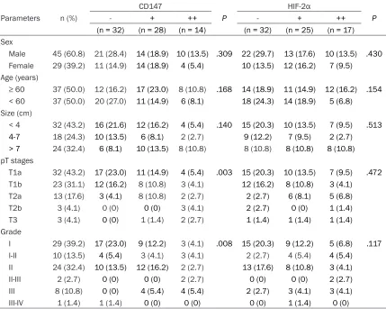
[image:2.612.92.521.97.440.2]Each TMA slide was evaluated by two inde- pendent pathologists. Staining was scored by the intensity and percentage of tumor cells stained. The staining intensity was graded as Table 1. The correlation between CD147 and HIF-2α expressions and clinicopathological parameters in localized ccRCC*
Parameters n (%)
CD147
P
HIF-2α
P
- + ++ - + ++
(n = 32) (n = 28) (n = 14) (n = 32) (n = 25) (n = 17) Sex
Male 45 (60.8) 21 (28.4) 14 (18.9) 10 (13.5) .309 22 (29.7) 13 (17.6) 10 (13.5) .430
Female 29 (39.2) 11 (14.9) 14 (18.9) 4 (5.4) 10 (13.5) 12 (16.2) 7 (9.5) Age (years)
≥ 60 37 (50.0) 12 (16.2) 17 (23.0) 8 (10.8) .168 14 (18.9) 11 (14.9) 12 (16.2) .154
< 60 37 (50.0) 20 (27.0) 11 (14.9) 6 (8.1) 18 (24.3) 14 (18.9) 5 (6.8) Size (cm)
< 4 32 (43.2) 16 (21.6) 12 (16.2) 4 (5.4) .140 15 (20.3) 10 (13.5) 7 (9.5) .513
4-7 18 (24.3) 10 (13.5) 6 (8.1) 2 (2.7) 9 (12.2) 7 (9.5) 2 (2.7) > 7 24 (32.4) 6 (8.1) 10 (13.5) 8 (10.8) 8 (10.8) 8 (10.8) 8 (10.8)
pT stages
T1a 32 (43.2) 17 (23.0) 11 (14.9) 4 (5.4) .003 15 (20.3) 10 (13.5) 7 (9.5) .472 T1b 23 (31.1) 12 (16.2) 8 (10.8) 3 (4.1) 12 (16.2) 8 (10.8) 3 (4.1)
T2a 13 (17.6) 3 (4.1) 8 (10.8) 2 (2.7) 2 (2.7) 6 (8.1) 5 (6.8)
T2b 3 (4.1) 0 (0) 0 (0) 3 (4.1) 2 (2.7) 0 (0) 1 (1.4)
T3 3 (4.1) 0 (0) 1 (1.4) 2 (2.7) 1 (1.4) 1 (1.4) 1 (1.4)
Grade
I 29 (39.2) 17 (23.0) 9 (12.2) 3 (4.1) .008 15 (20.3) 9 (12.2) 5 (6.8) .117 I-II 10 (13.5) 4 (5.4) 3 (4.1) 3 (4.1) 2 (2.7) 4 (5.4) 4 (5.4)
II 24 (32.4) 10 (13.5) 12 (16.2) 2 (2.7) 13 (17.6) 8 (10.8) 3 (4.1)
II-III 2 (2.7) 0 (0) 0 (0) 2 (2.7) 0 (0) 0 (0) 2 (2.7) III 8 (10.8) 0 (0) 4 (5.4) 4 (5.4) 2 (2.7) 3 (4.1) 3 (4.1)
III-IV 1 (1.4) 1 (1.4) 0 (0) 0 (0) 0 (0) 1 (1.4) 0 (0)
9396 Int J Clin Exp Pathol 2016;9(9):9394-9400 0 (no staining), 1 (weak), 2 (moderate), and 3
(strong). The percentage of positive staining was graded as 0 (0-5%), 1 (6-30%), 2 (31-70%), and 3 (71-100%). Then these two scores were summed, giving a resultant score of 0-6. Finally, the levels of staining were classified into four groups by “-” (score 0-1), “+” (score 2-3), “++” (score 4-5), and “+++” (score 6).
Statistical analysis
The correlations between the patients’ clini-copathological parameters and the expres-sions of CD147 and HIF-2α were analyzed by Pearson’s chi-squared test (χ2). Spearman cor-relation analysis was used to analyze the cor -relation between CD147 and HIF-2α expres -sions. Overall survivals (OS) were analyzed using Kaplan-Meier curves and prognostic sig-nificance was assessed by the log-rank test. Multivariate survival analysis was done by using the Cox regression methods. All statisti-cal analyses were performed using IBM SPSS 19.0 (SPSS, Inc., Chicago, IL, USA). Statistical significance was defined as two-sided (P < .05).
Results
Patients
Seventy-four of 90 samples were involved in the analysis except for patients with metasta-sis (3 cases), loss of follow-ups (9 cases), and absence of information of TNM phase (4 cases). The clinical features of patients were summa-rized in Table 1. The study involved 74 patients who were diagnosed as ccRCC without metas-tasis, including 45 men (60.8%) and 29 women (39.2%), with a median age of 69.5 years (range, 29-82 years). Tumor size was divided into 3 groups, of which 32 patients were less than 4 cm (43.2%), 18 were 4-7 cm (24.3%), and 24 were over 7 cm (32.4%). Furthermore, 32 patients were diagnosed as T1a (43.2%), 23 were T1b (31.1%), 13 were T2a (17.6%), 3 were T2b (4.1%), and 3 were T3 (4.1%). For pathology grade, 29 patients were diagnosed as grade I (39.2%), 10 were grade I-II (13.5%), 24 were grade II (32.4%), 2 were grade II-III (2.7%), 8 were grade III (10.8%), and only one patient was grade III-IV (1.4%).
Expressions of CD147 and HIF-2α in localized ccRCC and adjacent tissues
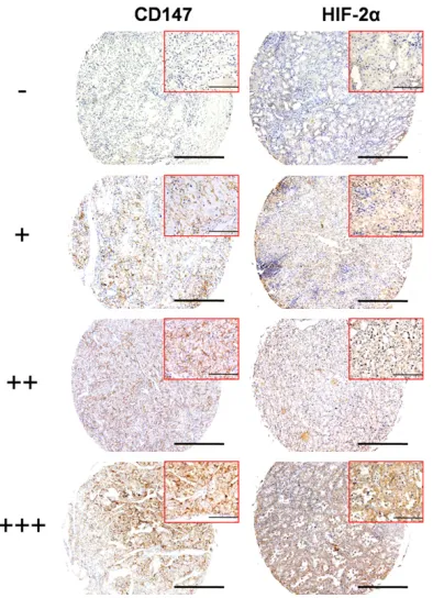
The representative images of CD147 and HIF-2α expressions in ccRCC tissue array were shown in Figure 1. Immunohistochemical stain-ing displayed that CD147 localized in the cyto -plasm and membrane of RCC cells, but not found in stromal cells, with a positive rate of 56.8%. Whereas the positive rate of CD147 was 10.8% in the tissues adjacent to cancer. The expression of CD147 in ccRCC was signifi -cantly higher than that in tissues adjacent to cancer (P < .001). HIF-2α staining was detect-ed in the cytoplasm and nuclear of ccRCC cells, with positive rates of 56.8% and 58.1% in ccRCC and adjacent tissues, respectively. Spearman correlation analyses indicated a positive correlation between CD147 and HIF-2α expressions (P = .002, rho = .359).
Association of expression of CD147 and
HIF-2α with clinicopathological parameters in localized ccRCC
[image:3.612.95.292.73.345.2]The expression of CD147 in ccRCC tissues was associated with pT stage (P = .003) and grade (P = .008), whereas HIF-2α did not show signifi -cant correlation with these clinicopathological Figure 1.Representative immunohistochemical sta-
9398 Int J Clin Exp Pathol 2016;9(9):9394-9400 features (Table 1). Patients with higher pT
stag-es and gradstag-es had significantly higher exprstag-es -sion of CD147.
Correlation of CD147 and HIF-2α expressions
with prognosis in localized ccRCC
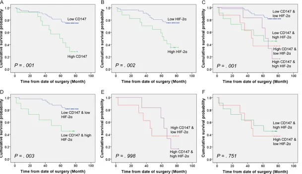
Based on the different levels of CD147 and HIF-2α expressions in the tumor tissues, the OS analysis were conducted by Kaplan-Meier curves and analyzed by the log-rank test. As shown in Figure 2A and 2B, patients with high CD147 expression (P = .001) and high HIF-2α expression (P = .002) were significantly associ -ated with poor OS. The cumulative 5-year OS rates of patients with low and high expressions of CD147 were 81.7% and 50.0%, respectively. Similarly, patients with low and high expres-sions of HIF-2α were 80.7% and 58.8%, respectively.
According to the positive correlation between CD147 and HIF-2α expressions, we wonder whether combined expression of CD147 and HIF-2α was more accurate to predict the prog -nosis than CD147 alone. Thus, patients were categorized into four groups: 1) low expression of CD147 and low expression of HIF-2α (low CD147 & low HIF-2α); 2) low expression of CD147 and high expression of HIF-2α (low
of patients with high CD147 & low HIF-2α and high CD147 & high HIF-2α were not statistically different (Figure 2E). Additionally, patients with low CD147 & high HIF-2α and high CD147 & low HIF-2α showed no statistical difference (Figure 2F).
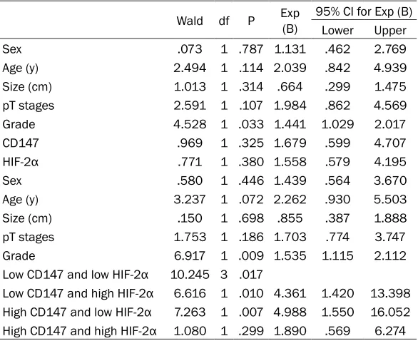
To further evaluate the prognostic significance of CD147 and HIF-2α in localized ccRCC, we carried out Cox regression analysis. Covariates included age, sex, tumor size, pT stages, grade, CD147 expression, and HIF-2α expression. Only grade was found an independent prognostic factor for patients with ccRCC (Table 2). How- ever, when combined expression of CD147 and 2α was added, grade, low CD147 & low HIF-2α expression, low CD147 & high HIF-HIF-2α pression, and high CD147 & low HIF-2α ex-pression were independent prognostic factors (Table 2). These results indicated that com-pared with CD147 and HIF-2α expression indi -vidually, combined expression of CD147 and HIF-2α was more accurate in prediction of ccRCC prognosis.
Discussion
[image:5.612.91.384.108.347.2]In our study, we observed that expression of CD147 in ccRCC tissues was significantly high -er than that in adjacent tissues. Increased Table 2. Cox multivariate analysis of the prognostic significance of
CD147 and HIF-2α expressions and clinicopathological parameters in patients with localized ccRCC
Wald df P Exp (B) 95% CI for Exp (B) Lower Upper Sex .073 1 .787 1.131 .462 2.769
Age (y) 2.494 1 .114 2.039 .842 4.939
Size (cm) 1.013 1 .314 .664 .299 1.475
pT stages 2.591 1 .107 1.984 .862 4.569 Grade 4.528 1 .033 1.441 1.029 2.017
CD147 .969 1 .325 1.679 .599 4.707
HIF-2α .771 1 .380 1.558 .579 4.195
Sex .580 1 .446 1.439 .564 3.670
Age (y) 3.237 1 .072 2.262 .930 5.503
Size (cm) .150 1 .698 .855 .387 1.888
pT stages 1.753 1 .186 1.703 .774 3.747
Grade 6.917 1 .009 1.535 1.115 2.112
Low CD147 and low HIF-2α 10.245 3 .017
Low CD147 and high HIF-2α 6.616 1 .010 4.361 1.420 13.398
High CD147 and low HIF-2α 7.263 1 .007 4.988 1.550 16.052
High CD147 and high HIF-2α 1.080 1 .299 1.890 .569 6.274
expression of CD147 was positively correlated with clinicopathological features including high pT stages and high grade. In the survival ana- lysis, higher expressions of CD147 and HIF-2α were positively associated with poor OS. Moreover, combined expression of CD147 and HIF-2α further demonstrated their prognostic value in ccRCC. These results indicate that high expressions of CD147 and HIF-2α, espe -cially combined expression of CD147 and HIF-2α, is significantly correlated with poor progno -sis of patients with ccRCC.
We discovered that high CD147 expression was associated with clinicopathological features including high pT stages and high grade. This finding was comparable with previous studies showing that CD147 expression was correlated with clinicopathological parameters and poor survival rates in RCC [8, 9]. Moreover, Liang et al. explained the prognostic role of CD147/ VEGF coexpression in patients with advanced RCC [7]. Here, we attempted to prove the prog -nostic role of CD147 in localized ccRCC and found that patients with high CD147 expres -sion were significantly associated with poor OS.
Numerous researches in the prognostic role of HIF family made it clear that cytoplasmic ex- pression of HIF-2α indicated unfavorable prog -nosis in RCC [10, 11]. Several studies have demonstrated the tight correlation between CD147 and HIF family. CD147 promotes mela -noma cell malignant properties through a HIF-2α mediated upregulation of VEGFR-2 [12, 13]. Besides, hypoxia upregulates CD147 expres -sion through a combined effect of HIF-1α and Sp1 to promote glycolysis and tumor progres-sion in epithelial solid tumors [14]. However, the relationship between CD147 and HIF-2α in localized ccRCC is still yet unknown. In the pres -ent study, we revealed the correlation between CD147 and HIF-2α in localized ccRCC. Either CD147 or HIF-2α is reported as a good marker of ccRCC individually, thus we analyzed the combination of CD147 and HIF-2α expression in prognosis of ccRCC. Compared with CD147 and HIF-2α expression individually, combined expression of CD147 and HIF-2α indicated more accurate prediction in ccRCC prognosis. However, whether CD147 and HIF-2α coexpres -sion can serve as an independent prognosis factor in localized ccRCC needs a further research involved in more patients. The
specif-ic role of the crosstalk between CD147 and HIF-2α also needs to be further investigated.
Our results indicate that high expression of CD147 is identified as a poor prognostic fac-tor in localized ccRCC. Expression of CD147 and HIF-2α, especially combined expression of CD147 and HIF-2α, may guide us in predict -ing the prognosis of ccRCC. CD147 may be a practical prognostic factor and therapeutic target in ccRCC.
Acknowledgements
This work was supported by the National Natural Science Foundation of China (3157-1434) and National Basic Research Program of China (2015CB553701).
Disclosure of conflict of interest
None.
Address correspondence to: Zhi-Nan Chen and Hui- jie Bian, Department of Cell Biology, Fourth Military
Medical University, No. 169, Changle West Road,
Xi’an 710032, China. Tel: +86-29-84774480; Fax: +86-29-83293906; E-mail: [email protected] (ZNC); [email protected] (HJB)
References
[1] Jemal A, Bray F, Center MM, Ferlay J, Ward E
and Forman D. Global cancer statistics. CA
Cancer J Clin 2011; 61: 69-90.
[2] Capitanio U and Montorsi F. Renal cancer.
Lancet 2016; 387: 894-906.
[3] Brookman-May S, Langenhuijsen JF, Volpe A, Minervini A, Joniau S, Salagierski M, Roscigno
M, Akdogan B, Vandromme A, Rodriguez-Faba O and Marszalek M. Management of localized
and locally advanced renal tumors. A contem-porary review of current treatment options.
Minerva Med 2013; 104: 237-259.
[4] Li M and Kim WY. Two sides to every story: the HIF-dependent and HIF-independent functions
of pVHL. J Cell Mol Med 2011; 15: 187-195.
[5] Li Y, Xu J, Chen L, Zhong WD, Zhang Z, Mi L,
Zhang Y, Liao CG, Bian HJ, Jiang JL, Yang XM, Li XY, Fan CM, Zhu P, Fu L and Chen ZN. HAb18G
(CD147), a cancer-associated biomarker and
its role in cancer detection. Histopathology
2009; 54: 677-687.
[6] Zhang Q, Zhou J, Ku XM, Chen XG, Zhang L, Xu J, Chen GS, Li Q, Qian F, Tian R, Wen N and
hepatocel-9400 Int J Clin Exp Pathol 2016;9(9):9394-9400
lular carcinoma. Eur J Cancer Prev 2007; 16:
196-202.
[7] Liang YX, He HC, Han ZD, Bi XC, Dai QS, Ye YK, Qin WJ, Zeng GH, Zhu G, Xu CL and Zhong WD. CD147 and VEGF expression in advanced re -nal cell carcinoma and their prognostic value.
Cancer Invest 2009; 27: 788-793.
[8] Jin JS, Hsieh DS, Lin YF, Wang JY, Sheu LF and
Lee WH. Increasing expression of extracellular matrix metalloprotease inducer in renal cell carcinoma: tissue microarray analysis of im-munostaining score with clinicopathological
parameters. Int J Urol 2006; 13: 573-580.
[9] Tsai WC, Sheu LF, Nieh S, Yu CP, Sun GH, Lin YF, Chen A and Jin JS. Association of EMMPRIN and fascin expression in renal cell carcinoma: correlation with clinicopathological
parame-ters. World J Urol 2007; 25: 73-80.
[10] Fan Y, Li H, Ma X, Gao Y, Chen L, Li X, Bao X, Du Q, Zhang Y and Zhang X. Prognostic
Signifi-cance of Hypoxia-Inducible Factor Expression in Renal Cell Carcinoma: A PRISMA-compliant Systematic Review and Meta-Analysis. Medi- cine (Baltimore) 2015; 94: e1646.
[11] Kroeger N, Seligson DB, Signoretti S, Yu H,
Magyar CE, Huang J, Belldegrun AS and Pan- tuck AJ. Poor prognosis and advanced clinico-pathological features of clear cell renal cell carcinoma (ccRCC) are associated with cyto-plasmic subcellular localisation of Hypoxia in-ducible factor-2alpha. Eur J Cancer 2014; 50: 1531-1540.
[12] Bougatef F, Menashi S, Khayati F, Naimi B, Porcher R, Podgorniak MP, Millot G, Janin A, Calvo F, Lebbe C and Mourah S. EMMPRIN promotes melanoma cells malignant proper-ties through a HIF-2 alpha mediated up-regula-tion of VEGF-receptor-2. PLoS One 2010; 5: e12265.
[13] Bougatef F, Quemener C, Kellouche S, Naimi B, Podgorniak MP, Millot G, Gabison EE, Calvo F,
Dosquet C, Lebbe C, Menashi S and Mourah S.
EMMPRIN promotes angiogenesis through hy-poxia-inducible factor-2alpha-mediated regula-tion of soluble VEGF isoforms and their
recep-tor VEGFR-2. Blood 2009; 114: 5547-5556.
[14] Ke X, Fei F, Chen Y, Xu L, Zhang Z, Huang Q, Zhang H, Yang H, Chen Z and Xing J. Hypoxia
upregulates CD147 through a combined effect
of HIF-1alpha and Sp1 to promote glycolysis and tumor progression in epithelial solid