Original Article
MicroRNA-486-5p targets FGF9 and inhibits
colorectal cancer proliferation, migration and invasion
Jun Gan, Cheng-En Hu
Department of General Surgery Huashan Hospital, Fudan University, Shanghai, China
Received January 13, 2016; Accepted March 24, 2016; Epub May 1, 2016; Published May 15, 2016
Abstract: Colorectal cancer (CRC) is one of the most common malignancies worldwide, and microRNAs (miRNAs), which act as tumor suppressors or oncogenes, are involved in CRC development and progression. The purpose of this study was to investigate the function and potential mechanism of miR-486-5p in CRC. The results showed that miR-486-5p was significantly down-regulated in CRC cell lines and tissues. Over-expression of miR-486-5p signifi-cantly inhibited cell proliferation, migration and invasion in vitro and suppressed tumor growth in vivo. Fibroblast growth factor 9 (FGF9) was identified as a direct target of miR-486-5p in CRC cells, and FGF9 expression was negatively correlated with miR-486-5p expression in clinical specimens. Enforced expression of FGF9 significantly reversed the tumor suppressive effects of miR-486-5p. Taken together, these findings revealed a functional and mechanistic link between miR-486-5p and FGF9 in the pathogenesis of CRC and miR-486-5p has potential as a therapeutic target for CRC.
Keywords: Colorectal cancer, miR-486-5p, FGF9, proliferation, invasion
Introduction
Colorectal cancer (CRC) is one of the most com-mon malignancies worldwide with a high inci-dence and mortality rate [1, 2]. Despite advanc-es in therapeutic strategiadvanc-es, the clinical out-come and prognosis of CRC remains poor [3]. Tumor metastasis is a major cause of mortali- ty in the multi-step genetic events involved in CRC [4, 5]. Although many cell growth and metastasis-related genes, including p53, APC and K-ras, have been identified in CRC, the molecular mechanisms that suppress tumor cell growth, migration and invasion are largely unknown. Recent studies have revealed that the non-coding microRNAs (miRNAs) are novel regulators of tumor progression and novel tar-gets for therapy in CRC [6].
miRNAs are endogenous non-coding RNAs con-taining ~22 nucleotides (nt) that can negatively regulate protein expression by inducing the degradation of target mRNAs or impairing their translation or both by specifically binding to the 3’-untranslated regions (3’-UTRs) of target mRNAs [7, 8]. miRNAs participate in multiple cancer cell biological processes, such as
prolif-eration, cell cycle, invasion and apoptosis [9, 10]. Accumulating data point to a central regu-latory role for miRNAs in the initiation and development of CRC, and these miRNAs may function as tumor suppressors or oncogenes [11, 12]. Many miRNAs have been proved to play important roles in human CRC, including miR-224, miR-320a, miR-135, miR-154 and miR-200 [4, 6, 13-15].
miR-486-5p is a recently discovered miRNA and is involved in tumorigenesis. miR-486-5p was reported to be down-regulated in non-small cell lung cancer [16], gastric adenocarcinoma [17], lung cancer [18], breast cancer [19] and hepatocellular carcinoma [20], and acts as a tumor suppressor. In contrast, it has also been shown to be up-regulated in renal cell carcino-ma [21] and functions as an oncogene. However, the role of miR-486-5p in CRC tumorigenesis remains undefined.
MicroRNA-486-5p functions as a tumor suppressor in CRC
suppressed tumor growth in vivo by binding to FGF9 3’ UTR in CRC cells. Our findings showed that the miR-486-5p/FGF9 axis is an important regulator in the development and progression of CRC and may be a candidate target for CRC treatment.
Materials and methods
Cell culture and preparation of clinical sam-ples
Four human CRC cell lines (SW480, HCT116, SW620 and HT29) were obtained from the American Type Culture Collection (ATCC; Ma- nassas, VA, USA) and were cultured in DMEM (Invitrogen, Carlsbad, CA, USA) containing 10% fetal bovine serum (FBS) (Invitrogen) at 5% CO2 humidity and 37°C. The normal colon epithelial cell line FHC was grown in DMEM: F12 supple-mented with 10% FBS (Invitrogen). Twenty CRC tissue samples and matched adjacent normal tissues were obtained from Jinshan Hospital affiliated to Fudan University, and frozen in liq-uid nitrogen and stored at -80°C until use. This study was approved by the Institutional Ethical Review Boards of our institute, and written informed consent was obtained from all pa- tients.
RNA preparation and quantitative RT-PCR analysis
Total RNA was extracted from the cell lines and tissues using TRIzol reagent (Invitrogen) accord-ing to the manufacturer’s protocol. RNA was reverse transcribed to cDNA using the Prime- Script® 1st Strand cDNA Synthesis Kit (TaKaRa, Dalian, China). Quantitative PCR (qRT-PCR) was performed to determine the expression levels of miR-486-5p and FGF9 using SYBR Premix ExTaq (TaKaRa). U6 or β-actin was used as the control for normalization. The relative expres-sion levels of the gene of interest were calcu-lated using the 2-ΔΔCt method. All experiments
were performed in triplicate.
Western blot analysis
Cells were lysed in RIPA buffer (Beyotime, Jiangsu, China) and the protein concentration was determined using the Pierce® BCA Protein
Assay Kit (Thermo Fisher, CA, USA). Total pro-tein was prepared and separated by 10% sodi-um dodecyl sulfate polyacrylamide gel
electro-phoresis (SDS-PAGE), then transferred onto polyvinylidene difluoride membranes and blo- cked with 5% nonfat milk. The target proteins were detected according to standard methods using the following primary antibodies: anti-FGF9 (Santa Cruz, CA, USA), goat anti-peroxi-dase-conjugated secondary antibody (Sigma, St Louis, MO, USA). An anti-β-actin antibody (Sigma) was used as a loading control.
Oligonucleotide and plasmid transfection
miR-486-5p mimic and negative control (miR-NC) were designed and synthesized by RiboBio (Guangzhou, China). Cells were plated in indi-vidual wells of 6-well plates. miR-486-5p mimic or miR-NC was transfected into SW620 and HCT116 cells using Lipofectamine 2000 (In- vitrogen) according to the manufacturer’s pro-tocol. After 48 h, the cells were harvested for the assays described below. The coding region of FGF9 mRNA was amplified by PCR from human genomic DNA and inserted into the mul-tiple cloning site of expression vector pENTER (Invitrogen) and verified by sequencing. SW620 and HCT116 cells were transfected with the FGF9 plasmid (FGF9) or empty pEnter-vector using LipofectamineTM 2000. The cells
were harvested 48 h after transfection for the specified assays. The FGF9 3’ UTR was gener-ated by PCR amplification and subcloned in- to the pGL3-basic luciferase reporter plasmid (Promega).
Cell proliferation assay
Cell viability was detected using the Cell Counting Kit-8 (CCK-8) (Dojindo, Kumamoto, Japan) assay. Cells were seeded on 96-well plates at a density of 3×10³ cells per well. The
viability of cells was determined for five consec-utive days (day 1, 2, 3, 4, and 5) and measured at 450 nm using a microplate reader (Epoch; BioTek, Winooski, VT, USA). All experiments were repeated three times and the averages were calculated.
Cell migration and invasion assays
invasion in vitro according to the manufactur-er’s protocol. 1×10⁵ cells were placed in the top
chamber in triplicate. For the invasion assay, the upper chamber was coated with Matrigel. The cells on the upper surface of the mem-brane were removed following incubation for 24 h, and the cells on the undersurface were fixed, stained with 0.1% crystalviolet and counted under a light microscope. The Transwell migra-tion assay was performed in the same way as the invasion assay, but without the Matrigel coating.
Luciferase reporter assay
Cells were seeded in 24-well plates (1×10⁵/
well) in triplicate and cultured for 24 h. The luciferase reporter gene plasmid pGL3-FGF9-3’-UTR (wild-type, WT) or the control-lucif-erase plasmid (Mutant) were co-transfected into the cells with the control pRL-TK Renilla plasmid (Promega, Madison, WI, USA) using Lipofectamine 2000 Reagent (Invitrogen). Lu- ciferase and Renilla activities were assayed 48 h after transfection using the Dual Luciferase Reporter Assay Kit (Promega) following the manufacturer’s instructions [4].
Tumorigenicity in vivo
Male BALB/c nude mice aged 4-6 weeks were obtained from Shanghai Laboratory Animal Center of China. For the tumor growth assay, SW620 cells transfected with miR-486-5p mimic and miR-NC, were subcutaneously
inject-ed into nude mice. Tumor growth was deter-mined by measuring the tumor volume, V= (mm3, V = tumor length × tumor width2/2) every
3 days using calipers [22]. The animal studies and the experimental protocol were approved by the Institutional Animal Care and Use Com- mittee of Fudan University. All animal experi-ments were performed according to the guide-lines on the care and use of animals for scien-tific use.
Statistical analysis
All data were analyzed using SPSS 16.0 soft-ware (SPSS, Chicago, IL, USA) and expressed as mean ± SD. The mRNA or protein relation-ship between miR-486-5p and FGF9 was ana-lyzed by Pearson’s correlation. The two-tailed Student’s t-test was used to determine the
P-value, and P<0.05 was considered signifi- cant.
Results
miR-486-5p was reduced in CRC cell lines and tissues
[image:3.612.100.520.73.245.2]MicroRNA-486-5p functions as a tumor suppressor in CRC
sues (Figure 1B). These data suggest that miR-486-5p was down-regulated in both CRC cell lines and tissues.
Ectopic expression of miR-486-5p inhibited cell proliferation, migration and invasion and suppressed tumorigenicity
To investigate the biological function of
[image:4.612.93.523.73.540.2]and invasion were inhibited in the miR-486-5p mimic group compared with the negative con-trol group, thus demonstrating the inhibitory effects of miR-486-5p on CRC cells (Figure 2C, 2D).
We next examined the effects of miR-486-5p on the tumorigenicity of CRC cells. SW620 cells transfected with miR-486-5p mimic or miR-NC were subcutaneously injected into the flanks of
[image:5.612.93.519.71.481.2]nude mice, respectively. The tumors were mea-sured until the mice were killed on day 24. Over-expression of miR-486-5p suppressed tu- mor growth compared with the negative control (Figure 2E) and tumor size in the miR-486-5p mimic group was significantly smaller than that in the miR-NC group (Figure 2F). Similar results were observed for tumor weight (Figure 2G). These findings indicate that miR-486-5p func-tions as a tumor suppressor in CRC.
MicroRNA-486-5p functions as a tumor suppressor in CRC
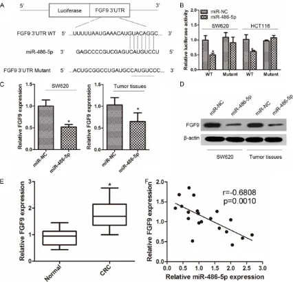
miR-486-5p suppressed the expression of FGF9 by targeting its 3’-UTR
To elucidate the underlying molecular mecha-nisms by which miR-486-5p executes its func-tion, we next identified the target genes of miR-486-5p using the publicly available databases (TargetScan and miRanda). FGF9 was predicted to be a target of miR-486-5p and one putative binding site in the FGF9 3’-UTR is shown in Figure 3A. The luciferase reporter assay was then used to determine whether miR-486-5p directly binds to the 3’-UTR of FGF9. As demon-strated in Figure 3B, compared with miR-NC, miR-486-5p significantly suppressed the lucif-erase activity of the WT3’-UTR, while mutation of the miR-486-5p binding sites blocked this inhibition. In addition, we analyzed the level of FGF9 expression in SW620 cells transfected with miR-486-5p mimic or miR-NC and in tumors isolated from nude mice using qRT-PCR and Western blotting. The results showed that miR-486-5p significantly inhibited the expres-sion of FGF9 levels (Figure 3C, 3D). Next, we determined FGF9 expression in CRC tissues and adjacent normal tissues. qRT-PCR analysis indicated that FGF9 expression was significant-ly increased in CRC tissues compared with the paired normal tissues (Figure 3E). Statistical analysis showed that the level of miR-486-5p was inversely correlated with the level of FGF9 in human CRC tissues (Figure 3F). These re-
sults confirmed that FGF9 is a target of miR-486-5p.
Enforced expression of FGF9 alleviates the ef-fects of miR-486-5p on proliferation, migration and invasion
Given that FGF9 is a direct target of miR-486-5p, we investigated whether over-expression of FGF9 reversed the suppressive effect of miR-486-5p. The FGF9 expressing vector (which encodes the entire coding region of FGF9 wi- thout 3’ UTR) was transiently introduced in SW620 cells co-transfected with miR-486-5p. The results showed that SW620 cells transfect-ed with FGF9 and miR-486-5p grew faster (Figure 4A) and had stronger migration and invasion (Figure 4B) ability than those trans-fected with empty vector and miR-486-5p. These results indicated that over-expression of FGF9 significantly alleviated the suppressive effects of miR-486-5p on CRC cells.
Discussion
It is now widely accepted that miRNAs contrib-ute to cancer development by acting as onco-genes or tumor suppressor onco-genes. Previous studies have shown that miR-486-5p is dysreg-ulated in several cancers and its potential func-tion has also been partly evaluated in several studies [18-21, 23]. For example, Wang et al.
[image:6.612.92.522.74.270.2]contributes to tumor progression and metasta-sis by targeting pro-tumorigenic ARHGAP5 in lung cancer [18]. Zhang et al. found that miR-486-5p expression was significantly down-reg-ulated in breast cancer tissues and cell lines, and over-expression of miR-486-5p markedly suppressed breast cancer cell proliferation in vitro and in vivo, induced G0/G1 arrest, and promoted apoptosis [19]. Huang et al. revealed that miR-486-5p suppressed hepatocellular carcinoma cell proliferation, migration and invasion in vitro and inhibited hepatocellular carcinoma growth in vivo [20]. In contrast, Goto
et al. showed that miR-486-5p was up-regulat-ed in renal cell carcinoma [21]. A previous study reported that miR-486-5p was down-regulated in CRC samples compared with matched non-tumor tissues [23]. However, the function of miR-486-5p in CRC pathogenesis, as well as the molecular mechanisms by which miR-486-5p exerts its function and modulates the malig-nant phenotypes of CRC cells, is not fully understood.
In the present study, miR-486-5p expression was decreased in CRC cell lines and tissues compared with the negative controls. Ectopic expression of miR-486-5p suppressed cell pro-liferation, migration and invasion of CRC cells in vitro and inhibited tumorigenesis in vivo. These findings revealed that miR-486-5p is a poten-tial suppressor of CRC and further work was carried out to characterize the mechanism. TargetScan and miRanda were used to identify potential gene targets of miR-486-5p and revealed that FGF9 was a potential target of miR-486-5p in CRC.
The fibroblast growth factor (FGF) family in- cludes at least 24 distinct polypeptides with molecular masses ranging from 17 to 34 kDa and share 13-71% sequence identity [24]. Many mammalian FGFs are abundantly expressed in a specific spatial and temporal pattern and are involved in many cellular processes, including development [25] and angiogenesis [26]. FGF9, a secretory protein of the FGF family, is report-edly expressed in stromal cells [27-29]. Studies have also demonstrated that FGF9 exhibits mitogenic activity in glioma [30], and in epithe-lial and fibroblast cells [31]. Over-expression of FGF9 has transforming potential in NIH3T3 fibroblasts and stimulates the invasion of epi-thelial and endoepi-thelial cells [32]. This suggests that FGF9 over-expression may result in
uncon-trolled cell proliferation and malignancy. FGF9 has been implicated in various cancers such as ovarian endometrial adenocarcinoma [31], hepatocellular carcinoma [33], prostate carci-noma [34] and gastric cancer [35]. A previous study reported that the expression of FGF9 was strong in a subset of advanced colon cancers, and over-expression was negatively correlated with patient survival [36]. In the present study, we demonstrated that FGF9 was a direct target of miR-486-5p in CRC cells using luciferase reporter assays. FGF9 expression was nega-tively correlated with miR-486-5p expression in CRC tissues. Furthermore, over-expression of FGF9 alleviated the suppressive effects of 5p. These results suggest that miR-486-5p inhibited CRC growth and metastasis partly by repressing FGF9 expression.
In summary, the current study revealed that miR-486-5p is down-regulated in CRC tissues and cell lines. We provide evidence that miR-486-5p is involved in CRC proliferation, migra-tion and invasion in vitro and suppressed tumor growth in vivo, potentially via the direct modula-tion of the downstream target FGF9. These results indicate that miR-486-5p plays an important role during CRC carcinogenesis and may serve as a putative target for CRC diagno-sis and therapy.
Disclosure of conflict of interest
None.
Address correspondence to: Dr. Cheng-En Hu, De- partment of General Surgery Huashan Hospital, Fudan University, 12 Wurumuqizhong Road, Shang- hai 200040, China. E-mail: chengenhu2008@163. com
References
[1] Li T, Yuan G, Zhang L, Ye L, Li S, Fan Y and Sun J. ApoG2 inhibits the antiapoptotic protein, Mcl1, and induces mitochondriadependent apoptosis in human colorectal cancer cells. Mol Med Rep 2015; 12: 6976-84.
[2] Huang S, Jean D, Luca M, Tainsky MA and Bar-Eli M. Loss of AP-2 results in downregulation of c-KIT and enhancement of melanoma tumori-genicity and metastasis. EMBO J 1998; 17: 4358-4369.
MicroRNA-486-5p functions as a tumor suppressor in CRC
[4] Liao WT, Li TT, Wang ZG, Wang SY, He MR, Ye YP, Qi L, Cui YM, Wu P, Jiao HL, Zhang C, Xie YJ, Wang JX and Ding YQ. microRNA-224 promotes cell proliferation and tumor growth in human colorectal cancer by repressing PHLPP1 and PHLPP2. Clin Cancer Res 2013; 19: 4662-4672.
[5] Hanahan D and Weinberg RA. The hallmarks of cancer. Cell 2000; 100: 57-70.
[6] Zhao H, Dong T, Zhou H, Wang L, Huang A, Feng B, Quan Y, Jin R, Zhang W, Sun J, Zhang D and Zheng M. miR-320a suppresses colorec- tal cancer progression by targeting Rac1. Carcinogenesis 2014; 35: 886-895.
[7] Liu J. Control of protein synthesis and mRNA degradation by microRNAs. Curr Opin Cell Biol 2008; 20: 214-221.
[8] Bartel DP. MicroRNAs: genomics, biogenesis, mechanism, and function. Cell 2004; 116: 281-297.
[9] Ambros V. The functions of animal microRNAs. Nature 2004; 431: 350-355.
[10] Baer C, Claus R and Plass C. Genome-wide epi-genetic regulation of miRNAs in cancer. Cancer Res 2013; 73: 473-477.
[11] Calin GA, Sevignani C, Dumitru CD, Hyslop T, Noch E, Yendamuri S, Shimizu M, Rattan S, Bullrich F, Negrini M and Croce CM. Human mi-croRNA genes are frequently located at fragile sites and genomic regions involved in cancers. Proc Natl Acad Sci U S A 2004; 101: 2999-3004.
[12] Asangani IA, Rasheed SA, Nikolova DA, Leupold JH, Colburn NH, Post S and Allgayer H. MicroRNA-21 (miR-21) post-transcriptionally downregulates tumor suppressor Pdcd4 and stimulates invasion, intravasation and metas-tasis in colorectal cancer. Oncogene 2008; 27: 2128-2136.
[13] Nagel R, le Sage C, Diosdado B, van der Waal M, Oude Vrielink JA, Bolijn A, Meijer GA and Agami R. Regulation of the adenomatous pol-yposis coli gene by the miR-135 family in colorectal cancer. Cancer Res 2008; 68: 5795-5802.
[14] Xin C, Zhang H and Liu Z. miR-154 suppresses colorectal cancer cell growth and motility by targeting TLR2. Mol Cell Biochem 2014; 387: 271-277.
[15] Burk U, Schubert J, Wellner U, Schmalhofer O, Vincan E, Spaderna S and Brabletz T. A recipro-cal repression between ZEB1 and members of the miR-200 family promotes EMT and inva-sion in cancer cells. EMBO Rep 2008; 9: 582-589.
[16] Zhu J, Zeng Y, Xu C, Qin H, Lei Z, Shen D, Liu Z and Huang JA. Expression profile analysis of microRNAs and downregulated miR-486-5p and miR-30a-5p in non-small cell lung cancer. Oncol Rep 2015; 34: 1779-1786.
[17] Chen H, Ren C, Han C, Wang D, Chen Y and Fu D. Expression and prognostic value of miR-486-5p in patients with gastric adenocarcino-ma. PLoS One 2015; 10: e0119384.
[18] Wang J, Tian X, Han R, Zhang X, Wang X, Shen H, Xue L, Liu Y, Yan X, Shen J, Mannoor K, Deepak J, Donahue JM, Stass SA, Xing L and Jiang F. Downregulation of miR-486-5p con-tributes to tumor progression and metastasis by targeting protumorigenic ARHGAP5 in lung cancer. Oncogene 2014; 33: 1181-1189. [19] Zhang G, Liu Z, Cui G, Wang X and Yang Z.
MicroRNA-486-5p targeting PIM-1 suppresses cell proliferation in breast cancer cells. Tumour Biol 2014; 35: 11137-11145.
[20] Huang XP, Hou J, Shen XY, Huang CY, Zhang XH, Xie YA and Luo XL. MicroRNA-486-5p, which is downregulated in hepatocellular carci-noma, suppresses tumor growth by targeting PIK3R1. FEBS J 2015; 282: 579-594.
[21] Goto K, Oue N, Shinmei S, Sentani K, Sakamoto N, Naito Y, Hayashi T, Teishima J, Matsubara A and Yasui W. Expression of is a potential prog-nostic factor after nephrectomy in advanced renal cell carcinoma. Mol Clin Oncol 2013; 1: 235-240.
[22] Bambang IF, Xu S, Zhou J, Salto-Tellez M, Sethi SK and Zhang D. Overexpression of endoplas-mic reticulum protein 29 regulates mesenchy-mal-epithelial transition and suppresses xeno-graft tumor growth of invasive breast cancer cells. Lab Invest 2009; 89: 1229-1242. [23] Navon R, Wang H, Steinfeld I, Tsalenko A,
Ben-Dor A and Yakhini Z. Novel rank-based statisti-cal methods reveal microRNAs with differential expression in multiple cancer types. PLoS One 2009; 4: e8003.
[24] Ornitz DM and Itoh N. Fibroblast growth fac-tors. Genome Biol 2001; 2: REVIEWS3005. [25] Goldfarb M. Functions of fibroblast growth
factors in vertebrate development. Cytokine Growth Factor Rev 1996; 7: 311-325.
[26] Gerwins P, Skoldenberg E and Claesson-Welsh L. Function of fibroblast growth factors and vascular endothelial growth factors and their receptors in angiogenesis. Crit Rev Oncol Hematol 2000; 34: 185-194.
[27] Giri D, Ropiquet F and Ittmann M. FGF9 is an autocrine and paracrine prostatic growth fac-tor expressed by prostatic stromal cells. J Cell Physiol 1999; 180: 53-60.
[28] Coffey E, Newman DR and Sannes PL. Ex- pression of fibroblast growth factor 9 in normal human lung and idiopathic pulmonary fibrosis. J Histochem Cytochem 2013; 61: 671-679. [29] Tsai SJ, Wu MH, Chen HM, Chuang PC and
[30] Miyagi N, Kato S, Terasaki M, Aoki T, Sugita Y, Yamaguchi M, Shigemori M and Morimatsu M. Fibroblast growth factor-9 (glia-activating fac-tor) stimulates proliferation and production of glial fibrillary acidic protein in human gliomas either in the presence or in the absence of the endogenous growth factor expression. Oncol Rep 1999; 6: 87-92.
[31] Hendrix ND, Wu R, Kuick R, Schwartz DR, Fearon ER and Cho KR. Fibroblast growth fac-tor 9 has oncogenic activity and is a down-stream target of Wnt signaling in ovarian endo-metrioid adenocarcinomas. Cancer Res 2006; 66: 1354-1362.
[32] Miyamoto M, Naruo K, Seko C, Matsumoto S, Kondo T and Kurokawa T. Molecular cloning of a novel cytokine cDNA encoding the ninth member of the fibroblast growth factor family, which has a unique secretion property. Mol Cell Biol 1993; 13: 4251-4259.
[33] Yang H, Fang F, Chang R and Yang L. MicroRNA-140-5p suppresses tumor growth and metas-tasis by targeting transforming growth factor beta receptor 1 and fibroblast growth factor 9 in hepatocellular carcinoma. Hepatology 2013; 58: 205-217.
[34] Li ZG, Mathew P, Yang J, Starbuck MW, Zurita AJ, Liu J, Sikes C, Multani AS, Efstathiou E, Lopez A, Wang J, Fanning TV, Prieto VG, Kundra V, Vazquez ES, Troncoso P, Raymond AK, Logothetis CJ, Lin SH, Maity S and Navone NM. Androgen receptor-negative human prostate cancer cells induce osteogenesis in mice th- rough FGF9-mediated mechanisms. J Clin In- vest 2008; 118: 2697-2710.
[35] Deng M, Tang HL, Lu XH, Liu MY, Lu XM, Gu YX, Liu JF and He ZM. miR-26a suppresses tumor growth and metastasis by targeting FGF9 in gastric cancer. PLoS One 2013; 8: e72662. [36] Leushacke M, Sporle R, Bernemann C,