Original Article
Centella asiatica inhibits renal interstitial fibrosis
by regulating Smad3 and Smad7 expression
in the TGFβ signaling pathway
Zhu Zhang1,2, Jiwei Ma1, Rui Feng1, Zheng Wang1
1Department of Nephrology, The First Affiliated Hospital of Henan Traditional Chinese Medical University,
Zheng-zhou, China; 2Department of Nephrology, Fuwai Central China Cardiovascular Hospital, Zhengzhou 450000, China
Received December 24, 2017; Accepted January 17, 2018; Epub February 1, 2018; Published February 15, 2018
Abstract:Centella asiatica (CA) is a well-known traditional Chinese herb with a long history of therapeutic uses. In
the present study, we evaluated the effect of CA granula on renal interstitial fibrosis, as well as Smad3 and Smad7 in a TGF-β signaling pathway mediated mechanism. Fifty adult SD male rats were randomly divided into five groups:
sham-operated; unilateral ureteral obstruction (UUO); Monopril; high-dose CA (CA (H)); and low-dose CA (CA (L)). Blood samples were collected 14 days after oral gavage and the blood urea nitrogen (BUN) and serum creatinine
(Scr) was measured. H&E and Masson straining were utilized to determine the renal tubule interstitial fibrosis.
Smad3 and Smad7 mRNA and protein levels were determined in kidney tissues by immunohistochemistry, real-time
PCR, and Western blot analysis respectively. Compared with the UUO group, the CA treatment significantly inhibited renal interstitial fibrosis and Smad3 expression, but increased Smad7 expression at both mRNA and protein levels in a dose-dependent manner. Our research suggests that CA administration could delay renal interstitial fibrosis by inhibiting Smad3 but promoting Smad7 expression in TGF-β pathways, without obvious liver or kidney toxicity.
Keywords: Centella asiatica granula, renal interstitial fibrosis, TGF-β, Smad3, Smad7
Introduction
In recent years, the prevalence of chronic kid-ney disease (CKD) has significantly increased. Its characteristics of occult onset may easily cause a patient to miss the best period for checking the progress of the disease, which may lead to progression to end-stage renal dis-ease (ESRD), resulting irreversible kidney fail-ure. The latest epidemiological survey data (2012) showed that the CKD prevalence rate among China’s adult population was 10.8%. However, the awareness rate was only found to be 12.5% [1], which resulted in heavy economic and psychological burdens to the patients and their families. Therefore, chronic kidney dis-ease has currently become an important public health problem in China. It is particularly urgent and necessary to explore the mechanism of this disease, and to investigate effective new drugs for the prevention and treatment of CDK. The renal damage due to the pathological fac-tors, such as injury, inflammation, obstruction,
The chemical composition of Centella asiatica (L) Urban includes triterpenoid acid, triterpe-noid saponin, polyyne alkene, volatile oil, etc [7]. Previous studies have shown that asiatico-side could inhibit over-proliferation of fibro -blasts, and decrease the acid puffball polysac-charide and collagen, as well as reduce collagen synthesis by inhibiting the activities of the transpeptidase. Asiaticoside may further effec-tively control the overgrowth of the stroma and fibrous components of the connective tissue, and treat the scleroderma by inhibiting hyper-plasia of the unordered fibrous connective tis -sue. It has been shown that the effects of asi-aticoside are related to the Smad signal pathway [8, 9]. It was also proven that the mechanism of asiaticoside’s inhibition of colla-gen hyperplasia was mainly to inhibit the patho-logic action of TGF-β1 by increasing expression of Smad7, thereby effectively inhibiting fibro -blast proliferation [10].
This study established a unilateral ureteral obstruction (UUO) rat model, and evaluated the impact of CA on the expressions of Smad3 and Smad7 in the TGF-β pathway in rat kidney tis -sue. The Fosinopril Sodium Tablets (Monopril) served as the control group for its obvious inhibiting effects on renal fibrosis. This study further aimed to provide experimental evidence on the targets of CA for the treatment of the renal tubule interstitial fibrosis.
Materials and methods
Animal model
In this study, all of the animal procedures were conducted in accordance with the Guidelines for Care and Use of Laboratory Animals, and were approved by the Animal Care and Use Committee at the First Affiliated Hospital, Henan University of Traditional Chinese Me- dicine. A total of 50 male adult Sprague-Dawley (SD) rats, with weights of 200 (±20) g, were supplied by the Vital River Laboratories, Beijing, China. The rats were randomly divided into five groups as follows: the sham-operated group; model group; fosinopril group; high-dose Centella asiatica group (CA (H)); and low-dose Centella asiatica group (CA (L)). There were 10 rats allocated to each group. Rats from the sur-gery group were anesthetized by intraperitone-al injection using 10% chlorintraperitone-al hydrate (0.3 ml/100 g) solution. In the model group, UUO surgery was performed under general
anesthe-sia. An incision was made on the left abdomen to expose the left kidney and ureter, which was subsequently ligated and cut from the proximal inferior pole of the left kidney. The sham-oper-ated group had their ureters exposed and manipulated without ligation. The intragastric administration was implemented at a fixed time each day for 14 days, and the dosages of CA granula were 5.6 mg/100 g/day, and 16.8 mg/100 g/day for CA (L) and CA (H) group. The dosages of the fosinopril group (Monopril group) were 1.67 mg/100 g/day, and equiva-lent saline solutions were administered to the sham-operated and model groups.
Biochemical index and histopathological examination
At 14 days after the intragastric administration, 4 ml blood samples were obtained from abdom-inal aorta, and rate urea nitrogen and rat creati-nine test kits (Jingkang Biological Engineering, Shanghai, China) were used to detect the serum urea nitrogen (BUN), and serum creati-nine (Scr). Meanwhile, the left kidneys were removed, and paraffin embedding was per -formed after fixing with 4% paraformaldehyde. The tissue sections were gradient dehydrated using ethyl alcohol, made transparent with xylene, and immersed in paraffin for 3 h at 62°C. A 4 μm paraffin section was then used for the H&E and Masson staining. Slides were examined and pictures taken using a micro-scopic image acquisition system (BX 41).
Immunohistochemical analysis
background after chroma transformation were calculated.
Fluorescent quantitative PCR
The total RNA of the rats’ renal tissue was extracted using TRIzol (Life Technologies, Carlsbad, CA, USA) according to the manufac-turer’s instructions. The total RNA from each group was then reverse transcribed to cDNA using a RevertAid first strand cDNA synthesis kit (K1622; Thermo Fisher Scientific, Waltham, MA, USA). PCR was performed using an SYBR Green PCR Kit (Invitrogen Life Technologies, USA) in an ABI 7300 Real-Time PCR System (Applied Biosystems, Foster City, CA, USA). The primers for quantitative PCR analysis are detailed in Table 1. The PCR reaction was 50°C for two minutes; 95°C for 10 minutes; 95°C for 15 seconds; 60°C for one minute, for a total of 40 cycles; 95°C for 15 seconds; and 60°C for 15 seconds. The fluorescence signal was col -lected in the annealing stage. The fluorescent quantitative PCR results were expressed by the Ct value, and a relative quantitative method (2-ΔΔCt) was adopted.
Western blot
The supernatant from the renal tissues was taken after homogenation, and a BCA Protein
Assay Kit (Solarbio Science and Technology Co., Ltd., Beijing, China) was used to test the protein concentration. Equivalent tissue protein samples were taken and transferred to the PVDF membrane (Pierce Biotechnology, Ro- ckford, IL, USA) after 10% SDS-PAGE separa-tion. TBST containing 5% skim milk powder was used to block for one hour under room temper-ature. The samples were then incubated over-night with the specific Smad3 (1:2000) anti -body and Smad7 anti-body (1:3000; Abcam, Cambridge, MA, USA), respectively. After the membranes were washed, they were allowed to interact for one hour with the second antibody coupling with horse radish peroxidase (ZSGB-BIO, Beijing, China) under room temperature. Following sufficient washing, ECL chemilumi -nescent liquid A and B were then added onto the membrane according to a ratio of 1:1. A Universal Gel Document System (Image Lab) was used to obtain the images, and to analyze the optical density of the target bands.
Statistical analysis
In this study, data are presented as mean ± SD. Statistical significance tests were performed using SPSS software version 18.0. The com-parison between groups was examined using a one-way analysis of the variance (ANOVA). P<0.05 was taken as a statistically significant difference.
Results
CA had no impact on the biochemical index of the rat in the UUO model
The results are detailed in Table 2. When com-pared with the sham-operated group, the BUN and SCR values of the rats in the UUO model group had obviously increased (P<0.05), and there were no obvious changes after the CA treatment (P>0.05).
CA improved renal morphology in the UUO rats
[image:3.612.91.304.86.190.2]The results are shown in Figure 1. There were no histopathological changes in the kidney in the sham group. In the UUO model group, the renal tubule lumen expansion and the tubule walls stiffness and deformation was observed, accompanied by the tubular basement mem-branes atrophy, deformations and partial loss. There was obvious inflammatory cell infiltration Table 1. Primers for quantitative PCR analysis
Name Sequence size (bp)Product
Smad3 F: 5’-AGACACCAGTGCTACCTCCA-3’ 363 R: 5’-CCAGCGGGGAAGTTAGTGTT-3’
Smad7 F: 5’-GGAGTCCTTTCCTCTCTC-3’ 130 R: 5’-GGCTCAATGAGCATGCTCAC-3’
β-actin F: 5’-CCCATCTATGAGGGTTACGC-3’ 450 R: 5’-TTTAATGTCACGCACGATTTC-3’
Table 2. Comparison of biochemical indices of the rats in each group (mean ± SD)
Groups n BUN (mmol/L) SCR (μmol/L)
[image:3.612.91.301.234.315.2]in the renal interstitium. Fur- thermore, the renal intersti-tium was enlarged, and the extracellular matrix was accu-mulated. CA treatment signifi -cantly improved the renal mor-phology in the UUO rats.
CA inhibited the renal fibrosis of the UUO rats
[image:4.612.91.374.72.237.2]The results are shown in Figure 2. The green collagen of the sham operated group was found to be mainly distrib-uted on the basal membrane of the renal capsule, tubular basement membrane, mesan-gial area, and Bowman’s cap-sule wall, with less interstitial expression observed. The gr- een collagen of the UUO group was mainly distributed in the abovementioned areas, and the renal interstitial was obvi-ously widened. Also, the green area in the interstitial was sig-nificantly increased, indicating that mass collagenous fibers had accumulated in the renal interstitial area, and the de- gree of fibrosis was obviously higher than that of the sham operated group (P<0.05). The green area of CA treatment group was found to be obvi-ously decreased when com-pared with the model group (P<0.05), which indicated that both the CA and Monopril could inhibit the renal tubule interstitial fibrosis. Among th-ese, the CA (H) group, as well as the Monopril group, had equivalent degrees of colla-gen expression (P>0.05).
Impact of CA on the Smad3 and Smad7 expressions in renal tissues in the UUO rats
[image:4.612.91.374.307.628.2]The results are shown in Figure 3. The renal tissues in the sham operated group were found to have less Sm- Figure 1. Treatment of CA improved renal morphology in the UUO rats. CA or
vehicle control was orally administered in sham- or UUO-operated rats start-ing on the day of the surgery for 14 days. Representative photomicrographs showed the HE staining of each group.
Figure 2. Treatment of CA ameliorated renal fibrosis in the UUO rats. CA
or vehicle control was orally administered in sham- or UUO-operated rats starting on the day of the surgery for 14 days. A. Representative photomi-crographs show the Masson trichrome staining of each group (400×). B.
Quantification of fibrotic area in Masson trichrome–stained kidneys. Error
ad3 expression, with only a small amount of brown positive staining in the kidney tubule and renal corpuscle. The Smad3 expression of the model group was obviously increased, and the renal corpuscle and epithelial cell of the kidney tubule of the rats showed obvious brown expression. Compared with the model group, Smad3 expression in the CA treatment group was significantly decreased (P<0.05). The expression of Smad3 in the CA (H) group was found to be decreased compared with that in the CA (L) group (P<0.05). There was no
statisti-of Smad7 in the CA (H) group was higher compared with that of the CA (L) group (P< 0.05). The Monopril and CA (H) groups dis-played equivalent expression (P>0.05).
CA inhibited Smad3 gene and protein expres-sion in rats with the UUO model
[image:5.612.92.520.72.341.2]As shown in Table 3 and Figure 4, compared with the model group, the CA treatment decreased the Smad3 expressions in both the mRNA and protein level in the UUO rat’s kidney tissues (P<0.05). The expression of Smad3 in Figure 3. Treatment of CA inhibited Smad3 expression and increased Smad7 expression in the renal tissues of the UUO rats. CA or vehicle control was orally administered in sham- or UUO-operated rats starting on the day of the surgery for 14 days. A. Representative photomicrographs show the immuno-histochemical results of each group
(400×). B. Quantification of Smad3 and Smad7 expression in each group. Error bars represent mean ± SD. *P<0.05
compared with Smad3 in sham group; #P<0.05 compared with Smad3 in UUO group; +P<0.05 compared with Smad7 in sham group; &P<0.05 compared with Smad7 in UUO group.
Table 3. Smad3 mRNA and protein expression in each group (mean ± SD)
Groups n Smad3 mRNA Smad3 protein Sham 10 1.000000±0.0000000 1.000000±0.0000000 UUO 8 2.789377±0.0613903* 1.914895±0.0321774* Monopril 10 1.533349±0.0444970# 1.254602±0.0403550#
CA (H) 9 1.592549±0.0335374# 1.333943±0.0123628#
CA (L) 10 2.051284±0.0628026# 1.682663±0.0285100# *P<0.05, compared with the sham group; #P<0.05, compared with the model group.
cal significance between the CA (H) group and the Monopril control group (P>0.05).
[image:5.612.91.341.459.539.2]the CA (H) group was found to be decreased compared with the CA (L) (P<0.05), which indi-cated a dose dependence effect of CA treat-ment. There were no significant differences observed between the CA (H) group and the Monopril group (P>0.05).
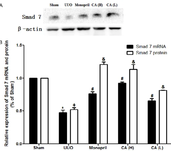
CA increased Smad7 gene and protein expres-sion of the rats in the UUO model
As shown in Table 4 and Figure 5, when com-pared with the model group, the CA treatment was found to promote the expression of the
under tissue injuries and inflammation stimula -tions; and type 3, cancer cells from the epithe-lium lose polarity, which are transformed into cancer cells with EMT ability.
[image:6.612.95.369.70.309.2]In 1995, Strutz et al. showed for the first time that EMT was involved in the formation of renal fibrosis [15]. Rastaldi et al. found that there were different degrees of EMT in all of the path-ological types, and the number of renal tubular epithelial cells with EMT characteristics was found to be closely related to the serum creati-nine concentration and the degree of renal Figure 4. Treatment of CA inhibited Smad3 gene and protein expression in
the renal tissues of the UUO rats. CA or vehicle control was orally adminis-tered in sham- or UUO-operated rats starting on the day of the surgery for 14 days. A. Smad3 was measured by the Western Blot; B. Quantitative data of the Smad3 mRNA and protein expression analyzed for each group. The er-ror bars represent mean ± SD. *P<0.05 compared with Smad3 mRNA level in sham group; #P<0.05 compared with Smad3 mRNA level in UUO group; +P<0.05 compared with Smad3 protein level in sham group; &P<0.05 com-pared with Smad3 protein level in UUO group.
Table 4. Smad7 mRNA and protein expression in each group (mean ± SD)
Groups n Smad7 mRNA Smad7 protein
Sham 10 1.000000±0.0000000 1.000000±0.0000000 UUO 8 0.477183±0.0369971* 0.522132±0.0273262* Monopril 10 0.759414±0.0333397# 1.207967±0.0389462# CA (H) 9 0.924940±0.0160862# 1.135367±0.0729589# CA (L) 10 0.656079±0.0361356# 0.812367±0.0078679#
*P<0.05, compared with the sham group; #P<0.05, compared with the model group.
Smad7 mRNA and protein in the kidney tissues of the UUO rats (P<0.05). Expression in the CA (H) was found to be increased compared with that of the CA (L) group (P<0.05), indicating that the CA treat-ment could regulate the Sm- ad7 expression in a dose de- pendent manner.
Discussion
The characteristic manifesta-tions of renal tubule interstitial fibrosis (RIF) include: tubular basement membrane compo-nents and interstitial ECM ac- cumulation and the mass pr- opagation of the α-SMA posi -tive myofibroblast. Myofibro-blasts are generated by EMT from renal tubular epithelial cells, which leads to massive generation and accumulation of ECM in the interstitial re- gion, and the occurrence of interstitial fibrosis lesions.
[image:6.612.88.371.454.534.2]interstitial damage [16]. Iwano et al. demon-strated that up to 36% of the renal interstitial fibroblasts originated from the local EMT of the kidney tubules. Under normal physiological conditions, the factors that inhibit and promote fibrosis reach a steady state. Meanwhile, renal fibrosis will occur with the onset of homeostatic imbalances.
Smad proteins play a critical role in the TGF-β signal pathway, and also mediate signal trans-duction. The TGF-β signal is transmitted from the cell membrane to the nucleus, and relies on Smads family proteins regulation. The different intracellular regulatory factors in the Smads signal transduction pathway will have com-pletely different results under the same TGF-β stimulation. For example, transcription will be activated when positive regulatory factors are dominant. Conversely, when the negative regu-latory factors are in the dominant position, the transcription will be inhibited. Smad3 is the activated Smad protein, and can promote the formation of kidney fibrosis. The previous
pathway through the ubiquitin ligase-Smad ubiquitination regulatory factors (Smuf1 and Smurf2) [18]. Inhibition of the TGF-β/Smad sig -nal pathway may intervene the normal function-ing of an organism. However, the study results have only involved cellular and animal experi-ments, which still remain at a remote distance from clinical practice.
[image:7.612.93.370.69.313.2]Modern research studies have found that asi-aticoside can inhibit fibroblast proliferation, and inhibit and alleviate the proliferation of fibrous connective tissues, as well as promote the healing of skin wounds. Asiaticoside may provide antioxidant and anti-proliferation ef- fects, as well as immune regulation functions [19, 20]. Pharmacological studies have con-firmed that CA may potentially inhibit fibroblast formation, reduce the aggregation of renal interstitial ECM, inhibit the proliferation of mesangial cells, and inhibit glomerular sclero-sis [21]. These findings indicate that CA plays an important role in the prevention and treat-ment of renal fibrosis. However, its mechanism Figure 5. Treatment of CA inhibited Smad7 gene and protein expression in
renal tissues of the UUO rats. CA or vehicle control was orally administered in sham- or UUO-operated rats starting on the day of the surgery for 14 days. A. Smad7 was measured by the Western Blot; B. Quantitative data of the Smad7 mRNA and protein expression analyzed for each group. The error bars represent mean ± SD. *P<0.05 compared with Smad7 mRNA level in sham group; #P<0.05 compared with Smad7 mRNA level in UUO group; +P<0.05 compared with Smad7 protein level in sham group; &P<0.05 com-pared with Smad7 protein level in UUO group.
remains unclear. Previous studies have shown that [22-25] CA may reduce the expressions of the connective tissue growth factor (CTGF) and the monocyte chemoattractant protein-1 (MCP-1) in the kidney tissues of UUO rates. It has also been found to inhibit renal tubular epithe-lial cells transformed into myofibroblasts and the expression of the bone morphogenetic pro-tein-7 (BMP-7) secreted by the renal tubular epithelial cells cultivated in vitro. It can also regulate the balance of matrix metalloprotein-ase-2/tissue inhibitors of metalloproteinase-2 (MMP-2/TIMP-2) in renal tubular epithelial cells cultivated in vitro, further leading to the anti-TIF effect.
Conclusions
In the present study we investigated the treat-ment effect of CA granula on the UUO rat model. The CA was able to inhibit deposition of lesion kidney collagen fibers and reduce the degree of fibrosis. In addition, the CA could inhibit Smad3 protein expression, and promote Smad7 protein expression, which indicates that CA is able to delay renal tubule interstitial fibrosis after UUO by regulating Smad3 and Smad7 expression. The effects of CA on the fibrosis were found to be dose dependent, and were equivalent to those of fosinopril at the dose of 16.8 mg/100 g/day. This study may potentially provide a theoretical basis for the prevention and treatment of chronic kidney dis-ease with CA granules.
Acknowledgements
This work has been supported by the Nation- al Natural Science Foundation of China (No. 81173409).
Disclosure of conflict of interest
None.
Address correspondence to: Dr. Zhu Zhang, Depar-
tment of Nephrology, The First Affiliated Hospital of
Henan Traditional Chinese Medical University, 19 Renmin Road, Zhengzhou 450000, Henan, China. Tel: +86-371-66231932; Fax: +86-371-66220903; E-mail: zhangzhudoc@126.com
References
[1] Yano Y, Fujimoto S, Asahi K and Watanabe T. Prevalence of chronic kidney disease in China. Lancet 2012; 380: 213-4.
[2] Klahr S and Morrissey J. Obstructive
nephropa-thy and renal fibrosis. Am J Physiol Renal Physi -ol 2002; 283: 861-875.
[3] Kobayashi E, Sasamura H, Mifune M, Shi-mizuhirota R, Kuroda M, Hayashi M and Saruta T. Hepatocyte growth factor regulates
proteo-glycan synthesis in interstitial fibroblasts. Kid -ney Int 2003; 64: 1179-1188.
[4] Eddy AA. Molecular basis of renal fibrosis. Pe -diatr Nephrol 2000; 15: 290-301.
[5] Razzaque MS and Taguchi T. Cellular and mo-lecular events leading to renal
tubulointersti-tial fibrosis. Med Mol Morphol 2002; 35:
68-80.
[6] Meng XM, Huang XR, Xiao J, Chung AC, Qin W, Chen HY and Lan HY. Disruption of Smad4
im-pairs TGF-β/Smad3 and Smad7 transcription
-al regulation during ren-al inflammation and fi -brosis in vivo and in vitro. Kidney Int 2012; 81: 266-279.
[7] Inamdar PK, Yeole RD, Ghogare AB and Souza NJD. Determination of biologically active con-stituents in Centella asiatica. J Chromatogr A 1996; 742: 127-130.
[8] Lee J, Jung E, Kim Y, Park J, Park J, Hong S, Kim J, Hyun C, Kim YS and Park D. Asiaticoside in-duces human collagen I synthesis through TGFbeta receptor I kinase (TbetaRI kinase)-in-dependent Smad signaling. Planta Med 2006; 72: 324-328.
[9] Bylka W, Znajdek-Awiżeń P, Studzińska-Sroka E, Dańczak-Pazdrowska A and Brzezińska M.
Centella asiatica in dermatology: an overview. Phytother Res 2014; 28: 1117-24.
[10] Tang B, Zhu B, Liang Y, Bi L, Hu Z, Chen B, Zhang K and Zhu J. Asiaticoside suppresses
collagen expression and TGF-β/Smad signal -ing through induc-ing Smad7 and inhibit-ing
TGF-βRI and TGF-βRII in keloid fibroblasts. Arch
Dermatol Res 2011; 303: 563-72.
[11] Zeisberg M and Neilson EG. Biomarkers for epithelial-mesenchymal transitions. J Clin In-vest 2009; 119: 1429-1437.
[12] Thiery JP, Acloque H, Huang RY and Nieto MA. Epithelial-mesenchymal transitions in develop-ment and disease. J Clin Invest 2009; 139: 871-90.
[13] Liu Y. New Insights into Epithelial-Mesenchy-mal Transition in Kidney Fibrosis. J Am Soc Nephrol 2010; 21: 212-22.
[14] Sabe H. Cancer early dissemination:
cancer-ous epithelial–mesenchymal transdifferentia
-tion and transforming growth factor β signal -ling. J Biochem 2011; 149: 633-639.
[15] Strutz FM. EMT and proteinuria as progression factors. Kidney Int 2009; 75: 475-481. [16] Rastaldi MP, Ferrario F, Giardino L, Dell’Antonio
transition of tubular epithelial cells in human renal biopsies. Kidney Int 2002; 62: 137-146. [17] Lan HY and Chung CK. TGF-β/Smad signaling
in kidney disease. Semin Nephrol 2012; 32: 236-243.
[18] Ruan Y, Zhang Z, Zhang X, Liu C and Guo M. The expressions of TGF-beta1 and Smad 2
mRNA on diseased glomeruli and their signifi -cance in the development of glomerulosclero-sis. Chin J Pathol 2002; 31: 314-7.
[19] Zhang LN, Zheng JJ, Xiao-Hui LI, Meng-Jiao WU, Zhang L and Wan JY. The protective effect of asiaticoside on sepsis-induced acute liver in-jury in mice. Lishizhen Medicine & Materia Medica Research 2010; 21: 2734-2736. [20] Dipankar Chandra R, Shital Kumar B and Md
Munan S. Current updates on centella asiati-ca: phytochemistry, pharmacology and tradi-tional uses. Med Plant Res 2013; 3.
[21] Dai LB, Shu P, Yan S, Yue H, Rong L, Ning K, Ye-Yang LI, Xiao-Jian LI, Xie YF and Gang LI.
Ef-fects of asiaticoside on dermal fibroblasts in
hypertrophic scar. Chin Pharm J 2010; 45: 1067-1072.
[22] Zhang Z, Wang S, Wang B, Zhao L, Wang L and Tang G. Centella asiatica granule on renal tis-sue in rats with unilateral ureteral obstruction alpha-smooth muscle actin expression. Tradit Chin Med Res 2009; 22: 15-18.
[23] Wang L, Liu P, Ma J, Zhao L, Wang S, Zhang Z, Wang G and Zhang Z. Effect of Centella
asiati-ca granula on TGF-β1-induced expression of
bone morphogenetic protein-7 in renal tubular epithelial cells. Shandong Med J 2009; 49: 13-15.
[24] Zhang Z, Wang G, Jiwei MA, Liu H, Zhang X and Zhu G. Effect of herba centellae on the expres-sion of HGF and MCP-1. Exp Ther Med 2013; 6: 427-434.