Original Article
Increased miR-223 expression promotes proliferation
and migration of retinal endothelial cells and
pathogenesis of diabetic retinopathy by
targeting EIF4E3 and IGF-1R
Yan Zhao1, Dong Li1, Di Chen2, Hongpei Ji1, Qian Hu1, Jianyong Zhang1
1Department of Ophthalmology, Guizhou Provincial People’s Hospital, Guiyang, Guizhou, China; 2Affiliated Hospital of Taishan Medical University, Taian, Shandong, China
Received November 14, 2016; Accepted January 13, 2017; Epub March 1, 2017; Published March 15, 2017
Abstract: This study aimed to investigate the expression of miR-223 microRNA in diabetic retinopathy (DR) and the relationship between miR-223 expression and DR outcomes. Arat DR model was established and retinal en-dothelial cells (RECs) were isolated. The expression of miR-223 was regulated using the miR-223 mimic inhibitor
and scrambled for cell transfection. Furthermore, small interfering RNAs (siRNAs) specific for miR-223 targets were
constructed, and their effects on cell proliferation and apoptosis as well as on cell invasion and migration abilities were examined. We confirmed the upregulation of miR-223 in DR RECs and an association between its mimic trans -fection and increased cell proliferation. Moreover, miR-233 modulated the cell proliferation by negatively regulating eukaryotic translation initiation factor 4E family member 3 (EIF4E3) and the insulin-like growth factor 1 receptor
(IGF1R). Meanwhile, siRNAs specific for EIF4E3 and IGF1R plus the miR-223 inhibitor significantly promoted the cell
proliferation.This study demonstrated the crucial roles of miR-223 and its targets in DR pathogenesis; however, further studies should be conducted to elucidate the underlying mechanism of miR-223 modulation of cell prolifera-tion.
Keywords: miR-223, diabetic retinopathy, EIF4E3, IGF1R, cell proliferation and migration
Introduction
Diabetic retinopathy (DR) is one of the main dia-betes complications and a leading cause of vision impairment and blindness among work-ing age adults [1, 2]. The prevalence of diabe-tes, hyperglycemia, and hypertension in the population accounts for the onset and progres-sion of DR [1]. DR is a multifactorial disease with complex etiology, including systemic abnormalities and alterations such as diabetic microangiopathy [3-6]. The complex etiology and prevalence of diabetes lead to the difficulty of therapy for DR and intensifies this problem.
DR is a neurodegenerative disease [7], thus the amelioration of neurodegenerative changes improves the progression of diabetes-related diseases [8]. A pericyte loss, retinal angiogen-esis, ischemic retinopathy, and retinal pericyte apoptosis observed under high-glucose
modu-lation of DR. Some miRNAs, including miR-223, showed protective effects on optic nerve regen-eration [16]. Dysregulation and other effects of miR-223 have been reported during angiogen-esis in diabetic patients, in osteoarthritis patients with type 2 diabetes, and in a variety of other diseases [17-19]. However, the miR-223 expression and mechanism of action in DR have been unclear until now.
This study aimed to investigate the expression of miR-223 in DR and the relationship between miR-223 expression and DR outcomes. Experi- ments were performed in vitro in cultured RECs and in vivo in an animal model. The targets of miR-223 were predicted and their expression, as well as the role in DR pathogenesis, was analyzed in the present study to reveal novel information on miR-223 effects on DR.
Materials and methods
Animals and model establishment
Animal experiments were approved by the local Animal Care Committee. Eighteen Sprague-Dawley rats (male, 200-250 g) were purchased from the Animal Experiment Center of Shanghai, Chinese Academy of Sciences, and cared for following the Guiding Principle in the Care and Use of Animals. The animals were randomly assigned into two groups, a control group and a diabetic group. Induction of diabetes in rats was performed by a single intravenous injec-tion of streptozotocin (STZ, 65 mg/kg; Sigma-Aldrich, Oakville, ON, Canada). Curcumin and insulin (Sigma-Aldrich) were administered to rats concomitantly [20]. Blood samples were taken from the tail vein to measure glucose lev-els and the animals with glycemia of >16.7 mM were considered diabetic [21]. The animals were euthanized under anesthesia four weeks after the STZ injection.
Isolation of RECs from DR rats
RECs were isolated from the DR rat retina using a magnetic-activated cell sorting (MACS) sys-tem following the manufacturer’s protocol (Mi- ltenyi Biotech, Cologne, Germany) and labeled using platelet endothelial cell adhesion mole-cule 1, also known as cluster of differentiation 31 [22]. Then, fluorescence-activated cell sort-ing (FACS) enrichment of the endothelial cells was performed using a MoFlo high-speed cell
sorter (Becton Dickinson, Franklin Lakes, NJ, USA), together with endothelial-specific gene expression analyses. RECs were cultured in Dulbecco’s modified Eagle’s medium (Sigma) supplemented with 5 mM glucose, 10% fetal bovine serum (FBS), and endothelial cell growth factor (Sigma) according to a previous report [22].
Cell transfection with miRNA mimic and inhibi-tor
RECs were plated on a 60-mm dish and incu-bated for 24 h, followed by transfections with a miRNA inhibitor, mimic, and scramble (Sangon, Shanghai, China) and small interfering RNAs (siRNAs) specific for eukaryotic translation ini-tiation factor 4E family member 3 (EIF4E3; Sangon) and IFG1R (Sangon). Transfections were performed using the Lipofectamine® RNAiMAX transfection reagent (3 μL/mL of the medium; Life Technologies, Carlsbad, CA, USA) in Opti-MEM® I reduced serum medium (100 nM; Life Technologies) for 48 h. The transfect-ed cells were harvesttransfect-ed, and protein and RNA extracts were prepared for further gene expres-sion analysis by western blot and real-time polymerase chain reaction (PCR), respectively. To investigate the effects of the EIF4E3 siRNA, IGF1R siRNA, miR-223 mimic, and miR-223 inhibitor, cell proliferation and migration experi-ments were carried out.
Cell viability assay
REC viability was assessed using a 3-(4, 5-dimethyl-2-thiazolyl)-2,5-diphenyltetrazolium bromide (MTT) assay as previously described [9]. In brief, 2.0 × 10⁴ cells were placed in 96-well plates and incubated for 24 h, followed by the addition of an MTT solution and incuba-tion for an addiincuba-tional 4 h. Cell viability was determined according to the manufacturer’s instructions, and each experiment was repeat-ed three times.
Colony formation assay
crys-tal violet solution, and those with more than 50 cells were counted.
Apoptosis assay
Apoptosis of transfected RECs was measured using a cyanine dye (Cy5)-labeled annexin V apoptosis detection kit and analyzed by flow cytometry [23]. Briefly, cells were harvested, washed, and pelleted after transfection for 24 h. Subsequently, RECs were resuspended in 5 μL of annexin V binding buffer containing annexin V-Cy5 (1:1,000) and 5 μL of buffer con-tained propidium iodide (PI) for 10 min, fol-lowed by analysis using a FACS Calibur flow cytometer (Becton Dickinson, BD, CA, USA). Annexin V-Cy5-positive and PI-negative cells (annexin V+/PI-) were considered early apoptot-ic cells.
Transwell assay
Cell migration and invasion assays were per-formed using Transwell migration chambers (8-μm pore size; Costar Corning, Corning, NY, USA). For the invasion assay, membranes were coated with a diluted extracellular matrix solu-tion (Sigma-Aldrich). The cells (5 × 10⁴ cells/ well) were seeded on the upper surface of the membranes in a serum-free medium. The lower chamber was filled with a medium containing 10% FBS, and the cells were incubated at 37°C for 24 or 48 h. Non-invaded cells were removed from the upper surface of the membranes using cotton swabs, and the invaded cells on the lower surface of the membranes were fixed, stained with a Diff-Quik staining solution (Origgio, Italy), and then counted using light microscopy (Diff-Quik®). All experiments were repeated three times with duplicate wells.
mined using an Eppendorf Mastercycler (Bri- nkman Instruments, Westbury, NY, USA) with the SYBR ExScript qRT-PCR kit (Takara, China). All reactions were run in triplicate and the rela-tive expression levels were calculated using the
2-ΔΔCt method. The glyceraldehyde 3-phosphate
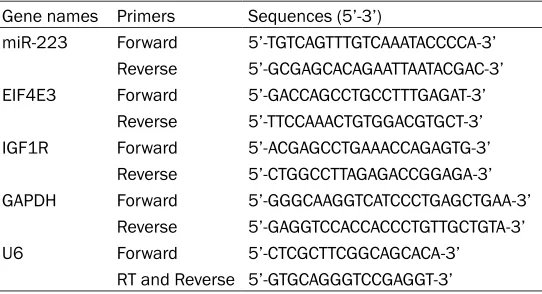
dehydrogenase (GAPDH) and U6 small nuclear RNA genes were used as the internal controls for mRNA and miRNA expression, respectively. The primers used for the amplification of the targets are shown in Table 1. The reaction con-ditions were as follows: 95°C for 10 min, fol-lowed by 40 cycles at 95°C for 30 s and 60°C for 40 s.
Western blot
Cellular proteins were prepared as previously described [24]. Proteins were quantified by a DC protein assay (Bio-Rad) and separated by 10% sodium dodecyl sulfate polyacrylamide gel electrophoresis. Separated proteins were blot-ted onto polyvinylidene difluoride membranes (Millipore, Billerica, MA, USA), which were then blocked in phosphate-buffered saline contain-ing 0.1% Triton X-100. The membranes were probed with primary antibodies against EIF4E3 (1:1,000 dilution; Millipore), IGF1R (1:1,000 dilution; Millipore), and GAPDH (1:2,000 dilu-tion; Millipore) at 4°C overnight. The mem-branes were then incubated with horseradish peroxidase-conjugated secondary antibodies for 1 h, followed by an enhanced chemilumines-cence reaction and the detection using the Quantity One software (Bio-Rad).
Dual-luciferase reporter assay
[image:3.612.92.363.84.231.2]miR-223 targets were predicted using Target Scan Human (http://www.targetscan.org/vert_ 71/) [25], and a dual-luciferase reporter assay Table 1. Primer list used in this study for qRT-PCR detection
Gene names Primers Sequences (5’-3’)
miR-223 Forward 5’-TGTCAGTTTGTCAAATACCCCA-3’
Reverse 5’-GCGAGCACAGAATTAATACGAC-3’
EIF4E3 Forward 5’-GACCAGCCTGCCTTTGAGAT-3’
Reverse 5’-TTCCAAACTGTGGACGTGCT-3’
IGF1R Forward 5’-ACGAGCCTGAAACCAGAGTG-3’
Reverse 5’-CTGGCCTTAGAGACCGGAGA-3’
GAPDH Forward 5’-GGGCAAGGTCATCCCTGAGCTGAA-3’
Reverse 5’-GAGGTCCACCACCCTGTTGCTGTA-3’
U6 Forward 5’-CTCGCTTCGGCAGCACA-3’
RT and Reverse 5’-GTGCAGGGTCCGAGGT-3’
RNA isolation and real-time PCR
deter-was used to confirm the predictions. 3’-Un- translated region (UTR) reporter vectors for EIF4E3 and IGF1R were synthesized by Sangon Biotech (Shanghai, China). The dual (firefly and Renilla)-luciferase reporter plasmids pGL3-EIF4E3/IGF1R-3’-UTR-WT and pGL3-EIF4E3/ IGF1R-3’-UTR-Mut were constructed and trans-fected into cells. Luciferase activities were then measured using the dual-luciferase reporter assay system (Promega) at 48 h
post-transfec-tion. The fold induction of firefly luciferase activ-ity was normalized to that of Renilla luciferase [26].
Statistical analysis
[image:4.612.93.522.68.528.2]groups and those among more than two groups were analyzed using Tukey’s test and analysis of variance, respectively. A p-value of < 0.05 was considered to indicate a statistically signifi-cant difference. GraphPadPrism6.0(GraphPad Software, Inc., La Jolla, CA, USA) was used to draw graphs.
Results
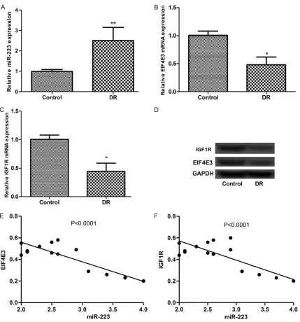
Expression of miR-223, EIF4E3, and IGF1R in RECs isolated from DR rats
[image:5.612.90.526.69.611.2]mine the expression levels of miR-223 and its targets in response to DR using RECs isolated from DR rats. The results showed that the
[image:6.612.90.518.70.615.2]sion of miR-223 and its targets showed nega-tive relationships between miR-223 and EIF4E3 and IGF1R in the rats (Figure 1E and 1F). EIF4E3 and IGF1R are direct targets of miR-223
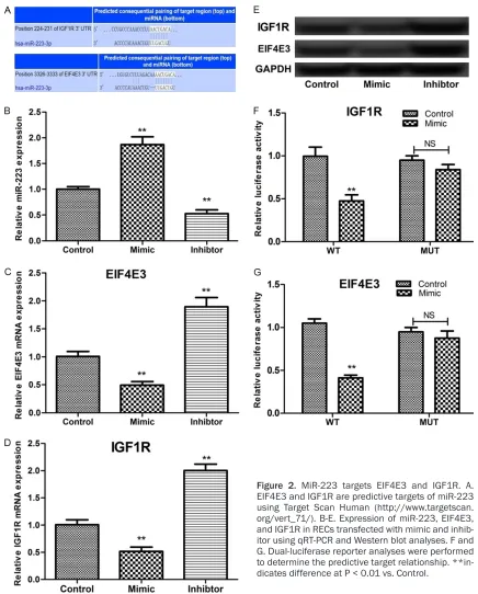
Using Target Scan Human: http://www.tar-getscan.org/vert_71/) [25], we predicted that EIF4E3 and IGF1R were two targets for miR-223 (Figure 2A). Transfection with the miR-223 mimic and inhibitor-induced down- and upregu-lation of EIF4E3 and IGF1R expression, respec-tively (Figure 2B-E). Furthermore, the dual-lucif-erase reporter assay showed that co-transfec-tion of miR-223 and the pGL3-EIF4E3/IGF1R-3’-UTR-WT vector significantly quenched the relative firefly luciferase activity (Figure 2F and 2G), indicating the negative regulation of EIF4E3 and IGF1R expression by miR-223. miR-223 inhibitor inhibited proliferation of RECs
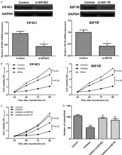
Using the MTT assay, we confirmed that inhibi-tion of miR-223 decreased the cell viability (P < 0.05, Figure 3B). Then, we silenced the expres-sion of the EIF4E3 and IGF1R genes in RECs using siRNAs and detected increased cell via-bility of the siRNA-transfected cells (Figure 3B). By colony formation analysis, we revealed that the miR-223 inhibitor inhibited the cell colony formation, while siRNAs specific against EIF4E3 and IGF1R induced the formation of cell colo-nies (Figure 3D). These results showed that miR-223 expression contributed to cell prolif-eration by targeting EIF4E3 and IGF1R. miR-223 suppression inhibited endothelial cell apoptosis and cell migration
Previous data revealed that cell apoptosis is a major factor of REC damage at high concentra-tions of sugar in a DR model, which accelerates the progression of DR. By the analysis of apop-tosis, we revealed that the miR-223 inhibitor triggered the cell apoptosis activity, and siRNAs specific against EIF4E3 and IGF1R reduced the
cell apoptosis rate and enhanced the migration and invasion activities (Figure 4A-D).
The ultimate result of the DR progress is the formation of new vessels, including the biologi-cal processes of REC migration and prolifera-tion. Therefore, we analyzed the effects of miR-223 expression (mimic and inhibitor transfec-tions) on the REC migration, and the results showed that miR-223 expression significantly suppressed the REC migration (P < 0.05, Figure 4E and 4F). However, this effect was reversed by the silencing of EIF4E3 or IGF1R (P < 0.01, Figure 4G and 4H). Taken together, these results showed that miR-223 modulated cell functions by targeting EIF4E3 and IGF1R. Discussion
Oxidative stress and retinal pericyte apoptosis observed under high-glucose conditions are associated with the pathogenesis of DR [27]. DR is a neurodegenerative disease [7], and the pericyte loss, retinal angiogenesis, ischemic retinopathy, and retinal pericyte apoptosis occur under high-glucose conditions [9, 10]. Numerous studies have shown that DR is asso-ciated with miRNA abnormalities. In our pres-ent study, we confirmed that the miR-223 upregulation was related to DR and REC apop-tosis via targeting EIF4E3 and IGF1R.
It has been reported that the high-glucose-induced proliferation of hRECs and pericyte loss promote the DR process. The expression of miR-223 in the zebrafish retina showed a protective effect on optic nerve regeneration after optic crush [16]. Moreover, miR-223 induction in type 2 diabetes is associated with the upregulation of cell apoptosis [19]. Several studies have suggested downregulation of miR-223 in diabetes [28, 29]. However, an increase of miR-223 and IGF1R was found to be associ-ated with the acute ischemic occurrence [30]. In this study, we confirmed the upregulation of miR-223 in STZ-DR model in vivo and in RECs in vitro. Moreover, REC transduction with a miR-223 inhibitor resulted in the inhibition of cell Figure 4. MiR-223 modulated cell functions of apoptosis and migration by targeting EIF4E3 and IGF1R. A and B. The effects of miR-223 expression on RECs cell apoptosis; C and D. miR-223 regulates RECs apoptosis by targeting EIF4E3 and IGF1R. E and F. The effects of miR-223 expression on RECs cell migration; G and H. miR-223 regulates RECs migration by targeting EIF4E3 and IGF1R. RECs were transfected with miR-223 inhibitor, mimic, si-EIF4E3,
and si-IGF1R. Cell apoptosis was detected using annexin V-Cy5-labeled Apoptosis Detection Kit and analyzed by flow
proliferation, cell viability, and colony forma-tion, as well as in the promotion of cell apopto-sis. These results showed that the upregulation of miR-223 in DR contributed to the high-glu-cose-induced proliferation of RECs.
EIF4E3 and IGF1R were predicted to be nega-tive targets for miR-223. EIF4E3 acts as a tumor suppressor [31], while low levels of IGF1R expression have been detected in type 1 dia-betic patients [32]. In addition, IGF-1 trans-genes showed a protective effect against dia-stolic dysfunction in diabetic cardiomyopathy [33], and expression of IGF1R promoted rat aortic angiogenesis in vitro [34], indicating the promotion of proliferation by IGF1R in diabetes. Moreover, IGF1R is essential for the phos-phoinositide 3-kinase/protein kinase B signal-ing pathway [35, 36]. In the present study, we confirmed that the expression of EIF4E3 and IGF1R was downregulated in DR, and both showed a negative relationship with the miR-223 expression (Figure 1). Moreover, siRNAs specific for EIF4E3 and IGF1R plus a miR-223 inhibitor significantly promoted cell prolifera-tion, indicating the roles of miR-223-mediated EIF4E3 and IGF1R expression in DR REC proliferation.
Conclusions
In the present study, we confirmed the upregu-lation of miR-223 in DR RECs and the associa-tion of its expression with high-glucose-induced cell proliferation. Moreover, miR-223 modulat-ed the cell proliferation by negatively regulating EIF4E3 and IGF1R. This study demonstrated a crucial role of miR-223 in DR pathogenesis. However, the underlying mechanism or path-ways involved in miR-223-mediated cell prolif-eration should be explored in further studies.
Disclosure of conflict of interest
None.
Address correspondence to: Dong Li, Department of Ophthalmology, Guizhou Provincial People’s Hos- pital, Guiyang, 1 South Baoshan Road, Nanming District, Guiyang 550002, Guizhou, China. E-mail: [email protected]
References
[1] Ding J and Wong TY. Current epidemiology of diabetic retinopathy and diabetic macular ede-ma. Curr Diab Rep 2012; 12: 346-354.
[2] Yau JW, Rogers SL, Kawasaki R, Lamoureux EL, Kowalski JW, Bek T, Chen SJ, Dekker JM, Fletcher A and Grauslund J. Global prevalence and major risk factors of diabetic retinopathy. Diabetes Care 2012; 35: 556-564.
[3] Kowluru RA and Mishra M. Oxidative stress, mitochondrial damage and diabetic retinopa-thy. Biochim Biophys Acta 2015; 1852: 2474-2483.
[4] Chen M, Curtis T and Stitt A. Advanced glyca-tion end products and diabetic retinopathy. Curr Med Chem 2013; 20: 3234-3240. [5] Steigerwalt R, Nebbioso M, Appendino G,
Belcaro G, Ciammaichella G, Cornelli U, Luzzi R, Togni S, Dugall M and Cesarone M. Meri-va®, a lecithinized curcumin delivery system, in diabetic microangiopathy and retinopathy. Panminerva Med 2012; 54: 11.
[6] Woerdeman J, van Duinkerken E, Wattjes MP, Barkhof F, Snoek FJ, Moll AC, Klein M, de Boer MP, IJzerman RG and Serné EH. Proliferative retinopathy in type 1 diabetes is associated with cerebral microbleeds, which is part of generalized microangiopathy. Diabetes Care 2014; 37: 1165-1168.
[7] Kadłubowska J, Malaguarnera L, Wąż P, Zore
-na K. Neurodegeneration and neuroinflamma -tion in diabetic retinopathy: potential ap-proaches to delay neuronal loss. Curr Neuropharmacol 2016; 14: 831-839.
[8] Chen S, Liu AR, An FM, Yao WB and Gao XD. Amelioration of neurodegenerative changes in cellular and rat models of diabetes-related Al-zheimer’s disease by exendin-4. Age (Dordr) 2012; 34: 1211-1224.
[9] Park SW, Yun JH, Kim JH, Kim KW, Cho CH and Kim JH. Angiopoietin 2 induces pericyte
apop-tosis via α3β1 integrin signaling in diabetic
retinopathy. Diabetes 2014; 63: 3057-3068. [10] Li SY, Fu ZJ and Lo AC. Hypoxia-induced
oxida-tive stress in ischemic retinopathy. Oxid Med Cell Longev 2012; 2012: 426769.
[11] Zhang X, Lai D, Bao S, Hambly B and Gillies M. Triamcinolone acetonide inhibits p38MAPK ac-tivation and neuronal apoptosis in early dia-betic retinopathy. Curr Mol Med 2013; 13: 946-958.
[12] Zhong Q and Kowluru RA. Regulation of matrix
metalloproteinase-9 by epigenetic modifica -tions and the development of diabetic retinop-athy. Diabetes 2013; 62: 2559-2568. [13] Mortuza R, Feng B and Chakrabarti S.
miR-195 regulates SIRT1-mediated changes in dia-betic retinopathy. Diabetologia 2014; 57: 1037-1046.
[15] Wu JH, Wang YH, Wang W, Shen W, Sang YZ, Liu L and Chen CM. MiR-18b suppresses high-glucose-induced proliferation in HRECs by tar-geting IGF-1/IGF1R signaling pathways. Int J Biochem Cell Biol 2016; 73: 41-52.
[16] Fuller-Carter PI, Carter KW, Anderson D, Har-vey AR, Giles KM and Rodger J. Integrated
analyses of zebrafish miRNA and mRNA ex
-pression profiles identify 29b and
miR-223 as potential regulators of optic nerve re-generation. BMC Genomics 2015; 16: 1. [17] Zhuang G, Meng C, Guo X, Cheruku PS, Shi L,
Xu H, Li H, Wang G, Evans AR, Safe S, Wu C, Zhou B. A novel regulator of macrophage acti-vation: miR-223 in obesity-associated adipose
tissue inflammation. Circulation 2012; 125:
2892-903.
[18] Zhu L, Wang X, Wu J, Yang J, Sun X, Fan X, Zhang M and Zhou S. Effects of miR-223 on cerebral ischemic injury and angiogenesis in diabetic rats. Int J Clin Exp Med 2016; 9. [19] Kim D, Song J, Ahn C, Kang Y, Chun CH and Jin
EJ. Peroxisomal dysfunction is associated with up-regulation of apoptotic cell death via miR-223 induction in knee osteoarthritis patients with type 2 diabetes mellitus. Bone 2014; 64: 124-131.
[20] Chiu J, Khan ZA, Farhangkhoee H and Chakrab-arti S. Curcumin prevents diabetes-associated abnormalities in the kidneys by inhibiting p300 and nuclear factor-kappaB. Nutrition 2009; 25: 964-972.
[21] Jiang T, Chang Q, Cai J, Fan J, Zhang X and Xu G. Protective effects of melatonin on retinal
in-flammation and oxidative stress in experimen -tal diabetic retinopathy. Oxid Med Cell Longev 2016; 2016: 3528274.
[22] Kovacs B, Lumayag S, Cowan C and Xu S. Mi-croRNAs in early diabetic retinopathy in strep-tozotocin-induced diabetic rats. Invest Oph-thalmol Vis Sci 2011; 52: 4402-4409.
[23] Shynkar VV, Klymchenko AS, Kunzelmann C, Duportail G, Muller CD, Demchenko AP, Freys-sinet JM and Mely Y. Fluorescent biomem-brane probe for ratiometric detection of apop-tosis. J Am Chem Soc 2007; 129: 2187-2193. [24] Sun G, Zhou Y, Li H, Guo Y, Shan J, Xia M, Li Y,
Li S, Dan L and Li F. Over-expression of microR-NA-494 up-regulates hypoxia-inducible fac-toR-1 alpha expression via PI3K/Akt pathway and protects against hypoxia-induced apopto-sis. J Biomed Sci 2013; 20: 1-9.
[25] Wei Y, Cui YF, Tong HL, Zhang WW and Yan YQ. MicroRNA-2400 promotes bovine preadipo-cyte proliferation. Biochem Biophys Res Com-mun 2016; 478: 1054-1059.
[26] Laurent-Rolle M, Morrison J, Rajsbaum R, Ma-cleod JL, Pisanelli G, Pham A, Ayllon J, Miorin L, Martínez-Romero C and Tenoever B. The
inter-feron signaling antagonist function of yellow fever virus NS5 protein is activated by type I interferon. Cell Host Microbe 2014; 16: 314-327.
[27] Devi TS, Hosoya KI, Terasaki T and Singh LP. Critical role of TXNIP in oxidative stress, DNA damage and retinal pericyte apoptosis under high glucose: implications for diabetic retinop-athy. Exp Cell Res 2013; 319: 1001-1012. [28] Haneklaus M, Gerlic M, O’Neill L and Masters
S. miR-223: infection, inflammation and can -cer. J Int Med 2013; 274: 215-226.
[29] Duan X, Zhan Q, Song B, Zeng S, Zhou J, Long Y, Lu J, Li Z, Yuan M, Chen X, Yang Q, Xia J. De-tection of platelet microRNA expression in pa-tients with diabetes mellitus with or without ischemic stroke. J Diabetes Complications 2014; 28: 705-710.
[30] Long Y, Zhan Q, Yuan M, Duan X, Zhou J, Lu J, Li Z, Yu F, Zhou X, Yang Q, Xia J. The expression of microRNA-223 and FAM5C in cerebral in-farction patients with diabetes mellitus. Car-diovasc Toxicol 2017; 17: 42-48.
[31] Osborne MJ, Volpon L, Kornblatt JA, Culjkovic-Kraljacic B, Baguet A and Borden KL. eIF4E3 acts as a tumor suppressor by utilizing an atyp-ical mode of methyl-7-guanosine cap recogni-tion. Proc Natl Acad Sci U S A 2013; 110: 3877-3882.
[32] de Souza KS, Ururahy MA, da Costa Oliveira YM, Loureiro MB, da Silva HP, Bortolin RH, Melo Dos Santos F, Luchessi AD, Neto JJ, Arrais RF, Hirata RD, das Graças Almeida M, Hirata MH, de Rezende AA. Low bone mineral density
in Type 1 diabetic patients: influence of re -duced IGF1, IGF1R, and TGFB1 expression. Diabetes Metab Res Rev 2016; 32: 589-95. [33] Huynh K, McMullen JR, Julius TL, Tan JW, Love
JE, Cemerlang N, Kiriazis H, Du XJ and Ritchie
RH. Cardiac-specific IGF-1 receptor transgenic expression protects against cardiac fibrosis
and diastolic dysfunction in a mouse model of diabetic cardiomyopathy. Diabetes 2010; 59: 1512-1520.
[34] Nicosia RF, Nicosia S and Smith M. Vascular endothelial growth factor, platelet-derived growth factor, and insulin-like growth factoR-1 promote rat aortic angiogenesis in vitro. Am J Pathol 1994; 145: 1023.
[35] Wan X, Harkavy B, Shen N, Grohar P and Hel-man L. Rapamycin induces feedback activa-tion of Akt signaling through an IGF-1R-depen-dent mechanism. Oncogene 2007; 26: 1932-1940.